Last Updated on November 26, 2025 by Bilal Hasdemir
Medical imaging has changed how doctors diagnose diseases. Yet, PET CT scans are not used as much as they could be. Every year, over 80 million CT scans are done in the U.S., but PET CT scans are less common.
So, why don’t doctors use PET CT scans more? The reason is in the difference between a CT scan and a PET CT scan. A CT scan shows detailed pictures of the body’s structure. But a PET CT scan adds metabolic activity data, giving a fuller picture of the body’s functions.
Key Takeaways
- PET CT scans offer more detailed diagnostic information than CT scans alone.
- The use of PET CT scans is limited compared to CT scans.
- Understanding the differences between CT and PET CT scans is key for diagnosis.
- PET CT scans provide both anatomical and metabolic data.
- The adoption of PET CT scans is influenced by various factors.
The Current Landscape of Medical Imaging

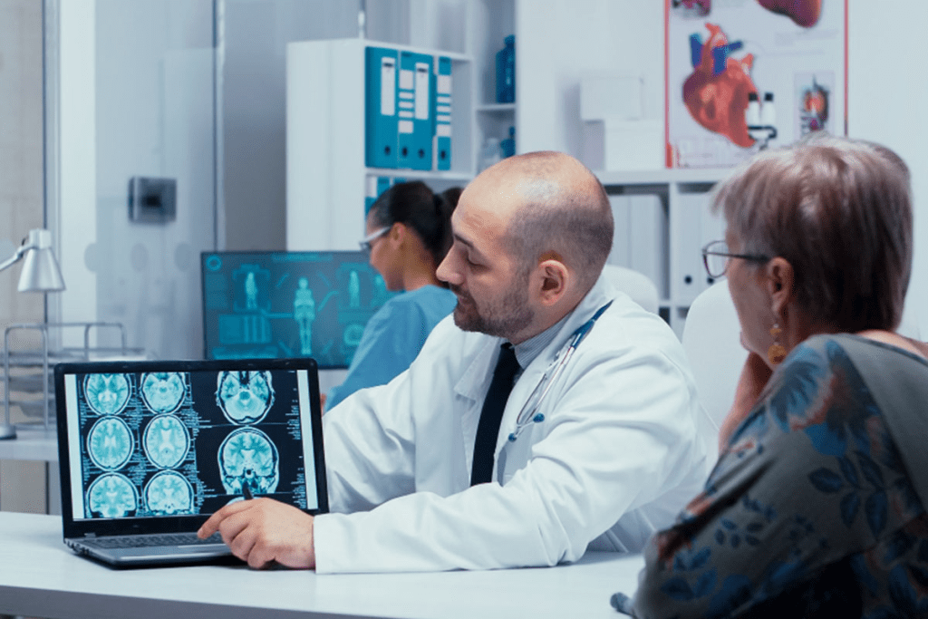
Diagnostic imaging is key in today’s healthcare. It lets doctors see inside the body and find many health issues.
Overview of Common Diagnostic Imaging Techniques
Medical imaging uses many technologies. Each has its own uses and benefits. Here are some common ones:
- Computed Tomography (CT) scans: CT scans show detailed pictures of the body’s inside. They help find injuries, cancers, and guide biopsies.
- Positron Emission Tomography (PET) scans: PET scans show how active the body’s cells are. They help find cancers, brain issues, and heart problems.
- Magnetic Resonance Imaging (MRI): MRI uses magnets and radio waves to see soft tissues. It’s great for brain, spine, and joint problems.
- Ultrasound: Ultrasound uses sound waves to see inside the body. It’s often used for pregnancy and abdominal checks, and to guide some treatments.
Each imaging method has its own strengths. Doctors choose based on what they need to find out
Prevalence and Utilization Statistics
More people are getting imaging tests. This is because of new tech and the need for safe tests. Here are some stats:
- CT scans are very common, with millions done in the U.S. each year.
- PET scans are used less often but are key for cancer and treatment checks.
- More imaging tests are expected as the population ages and health needs grow.
Knowing how often imaging tests are used helps doctors and planners. It helps make sure patients get the right tests for their health issues.
When looking at CT scans and PET CT scans, it’s important to know their differences. CT scans show body structure well, but PET CT scans add functional info. This makes them better for some cases.
Understanding CT Scans: Technology and Applications
CT scans use X-ray technology to create detailed images. These images help doctors diagnose and treat many medical conditions. This tool is crucial in modern medicine, enabling non-invasive examinations of the body’s interior.
How CT Scan Technology Works
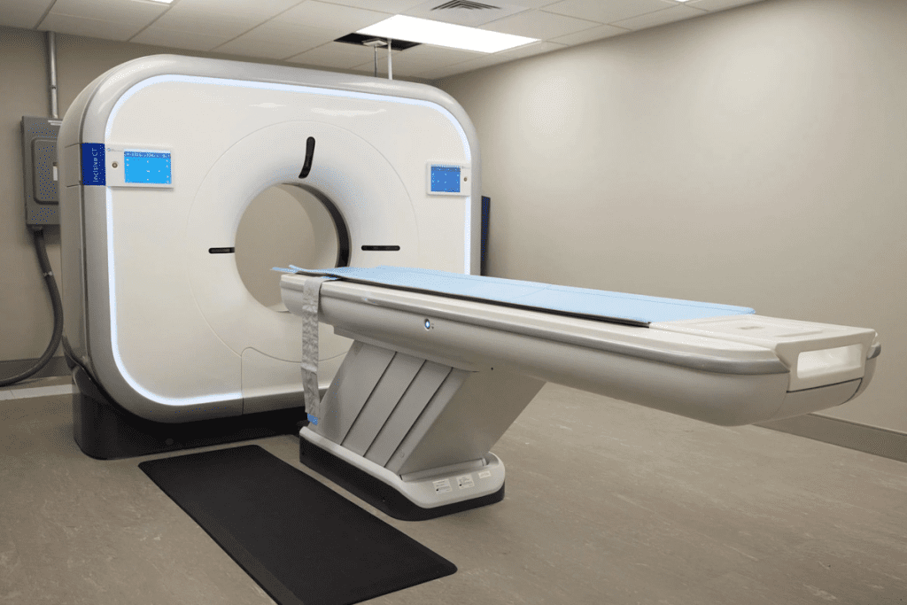
CT scans combine X-rays and computer tech for detailed images. The patient lies on a table that moves into a doughnut-shaped machine. The machine rotates around the body, capturing X-ray images from different angles.
These images are then put together by a computer. It turns them into detailed cross-sectional images or slices of the body’s inside.
Key components of CT scan technology include:
- X-ray tube: Produces X-rays that penetrate the body.
- Detectors: Capture the X-rays that have passed through the body.
- Computer system: Reconstructs the captured X-ray images into detailed cross-sectional images.
Common Medical Uses for CT Scans
CT scans are used in many medical ways. They help diagnose injuries, detect cancers, and more. Here are some common uses:
- Diagnosing injuries, such as internal bleeding or fractures.
- Detecting cancers and monitoring their progression.
- Visualizing vascular diseases, including aneurysms or blockages.
- Guiding interventional procedures, such as biopsies.
Advantages and Limitations of CT Imaging
CT scans have many benefits. They provide detailed images quickly, which is great in emergencies. They’re also good at diagnosing many conditions. But, there are downsides too.
One is the radiation exposure. There’s also a chance of allergic reactions to contrast dyes used to improve image quality.
The benefits of CT scans include:
- Rapid imaging, which is key in emergencies.
- High-resolution images for accurate diagnosis.
- Versatility in diagnosing many medical conditions.
It’s important to consider both the good and bad sides of CT scans. This includes thinking about radiation exposure and the use of contrast agents.
Understanding PET Scans: Technology and Applications
Positron Emission Tomography (PET) scans have changed how we diagnose diseases. They show how the body works, not just what it looks like. This is different from other imaging methods.
How PET Scan Technology Works
PET scans use a special radioactive tracer. This tracer goes into the body and sticks to active cells, like cancer. The scanner picks up the radiation from the tracer to show how the body works.
First, the tracer is made. It’s a sugar molecule with a radioactive atom. When injected, it goes to cells based on how active they are. The scanner then makes images of where the tracer is most active.
Common Medical Uses for PET Scans
PET scans are used in many ways in medicine, including:
- Cancer Diagnosis and Staging: They find cancer, see how far it has spread, and check if treatments are working.
- Neurological Disorders: They help find and manage diseases like Alzheimer’s and Parkinson’s.
- Cardiac Conditions: They check how well blood flows to the heart, helping find heart disease and see if heart tissue is alive.
Advantages and Limitations of PET Imaging
PET scans have big benefits, like finding diseases early and tracking treatment. But, they also have downsides, like radiation exposure and sometimes giving wrong results.
The advantages are:
- They can find diseases early
- They track how well treatments are working
- They show how active cells are
The limitations are:
- They expose you to radiation
- They are expensive
- They might not be available everywhere
In summary, PET scans are a key tool in medicine. They give deep insights into how the body works. Knowing how they work, what they’re used for, and their pros and cons is important for doctors and patients.
The Difference Between CT Scan and PET CT Scan

CT scans and PET CT scans are two different imaging tools. They help doctors see inside the body in different ways. Each tool has its own special use in medical imaging.
Structural vs. Functional Imaging
CT scans show detailed pictures of the body’s inside parts. They are great for looking at organs, bones, and tissues. PET scans, on the other hand, show how the body’s cells work.
PET CT scans do both. They give a complete picture of the body’s structure and how it works.
Resolution and Detail Comparison
CT scans have better detail than PET scans. They show more about the body’s shape. But, PET scans tell us about the body’s activity.
PET CT scans mix both. They show where the body’s activity is happening in its parts. This helps doctors find and understand tumors better.
Diagnostic Capabilities Comparison
CT scans are good at finding problems like injuries and tumors. PET CT scans are better for managing cancer and checking the brain and heart.
- CT scans are ideal for:
- Detecting structural abnormalities
- Guiding biopsies and other interventions
- PET CT scans are ideal for:
- Managing cancer treatment
- Assessing neurological and cardiac conditions
In summary, knowing the difference between CT scans and PET CT scans is key. CT scans give detailed pictures of the body’s shape. PET CT scans show how the body works. Together, they help doctors diagnose and treat better.
Combined PET-CT Technology: The Best of Both Worlds
Healthcare providers now have a better way to understand patient conditions. They use PET and CT technologies together. This mix gives a clearer view of what’s happening inside the body.
How PET-CT Integration Works
PET and CT scans are combined in one session. PET scans show how tissues work, while CT scans give detailed pictures. Together, they paint a full picture of the body’s health.
PET-CT integration works by using one device for both scans. The images are then merged using special software. This helps doctors see how different parts of the body work together.
Clinical Benefits of Combined Imaging
PET-CT scans offer big advantages. They give doctors a clearer view of what’s going on inside the body. This is key in fighting cancer, where PET-CT scans help find and track tumors.
- Enhanced diagnostic accuracy
- Better localization of abnormalities
- Reduced need for extra tests
In short, PET-CT scans are a big step forward in medical imaging. They combine two types of scans to give doctors a complete view. This leads to better diagnosis and treatment plans.
Availability and Accessibility Challenges
PET scanners are not easy to find everywhere, making it hard for patients to get them. These scans are very useful for diagnosing diseases. But, many people can’t get them because of several reasons.
Geographic Distribution of PET Scanners
PET scanners are mostly found in big cities and major hospitals. This means people in rural areas have limited access to them.
Several things affect where PET scanners are located:
- The high cost of buying and keeping PET scanners
- The need for a steady supply of special medicines
- The need for skilled people to run the scanners
Staffing and Expertise Requirements
To use a PET scanner, you need specialized training. People like radiologists, nuclear medicine technologists, and medical physicists are needed. Not having enough of these experts can make PET scans hard to get.
Here are some key skills needed:
- Radiologists who know about nuclear medicine
- Nuclear medicine technologists with PET scanning experience
- Medical physicists who make sure the scanners are safe
Patient Access Barriers
Patients face many obstacles when trying to get PET scans. These include:
- Geographic barriers, like having to travel far to get a scan
- Financial barriers, like the high cost of scans and insurance issues
- Logistical barriers, like preparing for the scan and scheduling delays
It’s important to work on these issues. This way, more people can get PET scans when they need them.
Radiation Exposure Concerns and Safety Considerations
CT and PET CT scans are used in medical diagnostics. They both involve radiation, but they have different safety levels. It’s important to understand their safety profiles.
Radiation Doses: CT vs. PET CT Scans
It’s key to know the radiation difference between CT and PET CT scans. CT scans use X-rays to show body details. The dose depends on the scan and body part.
PET CT scans mix PET’s function with CT’s anatomy. They use small radioactive tracers, adding to the radiation exposure.
- CT scans: The dose can be from 2 to 10 millisieverts (mSv), based on the scan and protocol.
- PET CT scans: The dose includes CT and PET tracer doses. It can be from 4 to 25 mSv or more, depending on the tracer and CT protocol.
Risk-Benefit Assessment for Different Patient Populations
The risk of radiation from CT and PET CT scans varies by patient group. For oncological applications, the benefits often outweigh the risks.
For pediatric patients or those needing repeated scans, the long-term risks must be weighed against the diagnostic benefits.
- For cancer patients, the benefits of PET CT scans justify the radiation exposure.
- Pediatric patients need careful consideration of alternative imaging to reduce radiation.
- Patients with chronic conditions may benefit from protocols that reduce cumulative radiation dose over time.
Safety Protocols and Minimizing Exposure
Healthcare providers use safety protocols to address radiation concerns. They optimize scan protocols, use dose-reduction technologies, and ensure scans are only done when necessary.
Patient education is also key in radiation safety. Informing patients about risks and benefits helps them make informed decisions.
- Optimizing scan protocols for the lowest effective dose.
- Using dose-reduction technologies in newer CT scanners.
- Limiting the use of CT and PET CT scans to medically justified cases.
Clinical Decision-Making: When Doctors Choose PET Over CT
Doctors often face tough choices when it comes to imaging tests. They must decide between PET and CT scans for the best results. This decision is based on the patient’s condition and what the doctors need to see.
For cancer diagnosis, PET scans are often the top choice. They show how cancer cells are working and where they are in the body. This helps doctors plan the best treatment.
But, for other conditions like neurological disorders, CT scans might be better. They are great for looking at the brain and nervous system. This is because they can show detailed images of soft tissues.
When it comes to contrast, PET CT scans are a good option. They combine the strengths of both PET and CT scans. This gives doctors a clearer picture of the body’s structures and how they are working.
So, doctors choose PET scans for cancer and CT scans for neurological issues. They also pick PET CT scans for a detailed look at the body. This helps them make the best decisions for their patients.
Future Developments in Medical Imaging Technology
New technologies in medical imaging are making diagnoses more precise and effective. These advancements are changing how we diagnose and treat diseases. They offer exciting new ways to help patients.
Advancements in PET and CT Technologies
PET and CT technologies have seen big improvements. New PET technologies give us more detailed views of the body’s metabolic processes. Advanced CT scanners help us see complex body structures better.
The creation of hybrid imaging systems like PET-CT is a major breakthrough. They combine PET’s functional info with CT’s anatomical details. This combo leads to more accurate disease diagnoses, mainly in cancer cases.
Role of Artificial Intelligence in Image Analysis
Artificial intelligence (AI) is becoming key in medical imaging, focusing on image analysis. AI algorithms spot abnormalities, track disease progress, and aid radiologists in making better diagnoses.
AI’s role in medical imaging goes beyond analysis. It’s also being used for image reconstruction and enhancement. AI boosts image quality, reducing the need for extra scans and boosting confidence in diagnoses.
- Improved diagnostic accuracy through advanced image analysis
- Enhanced image quality using AI-driven reconstruction techniques
- Increased efficiency in radiology workflows
As medical imaging tech keeps advancing, we’ll see big changes in patient care. The mix of new PET technologies, advanced CT scanners, and AI in image analysis will lead to better diagnoses and treatment plans. This will improve patient outcomes significantly.
Patient Perspectives: Understanding Your Imaging Options
Knowing about imaging options can improve your health care. When you need a test, knowing the differences is key.
Questions to Ask Your Doctor About Imaging Tests
Talking to your doctor before a test is important. Here are some questions to ask:
- What is the purpose of the recommended imaging test?
- How does the test work, and what will it diagnose?
- Are there any alternative imaging options available?
- What are the risks and benefits associated with the test?
- How should I prepare for the test?
These questions help you understand your options and make good choices for your care.
Preparing for Different Types of Scans
Getting ready for a scan is important for a good test. Here are some tips for different scans:
- CT Scans: You might need to not eat or drink for hours before. Also, remove metal objects or jewelry.
- PET Scans: You might need to fast before the test. Avoid hard exercise too.
- PET-CT Scans: You’ll need to prepare like you would for both a PET and a CT scan.
Knowing what you need to do can make you feel less anxious and more ready.
Conclusion: The Evolving Role of PET Scans in Modern Medicine
PET scans are changing with new technology and medical practices. Knowing the difference between CT scans and PET CT scans is key. PET scans give functional info that CT scans don’t, making PET CT scans a top choice for doctors.
Both CT scans and PET CT scans show detailed body images. But, PET scans are better at spotting metabolic activities. This makes them essential in fighting cancer, studying the brain, and heart health. As tech gets better, PET scans will help doctors more, leading to better care for patients.
PET scans will be even more important in making medicine personal. They help doctors find the right treatment for each patient. By knowing what PET scans can do, doctors can make better choices. This leads to better health for everyone.
FAQ
What is the main difference between a CT scan and a PET CT scan?
CT scans show the body’s structure. PET CT scans, on the other hand, show how the body’s tissues and organs work. They combine CT’s structural images with PET’s functional information.
How do CT scans and PET CT scans compare in terms of diagnostic capabilities?
CT scans are great for finding structural problems like injuries and tumors. PET CT scans are better for seeing how tissues work. They’re key for diagnosing cancer, neurological issues, and infections.
Why are PET scans less common than CT scans?
PET scans are less common because they cost more and are harder to find. They also use radioactive tracers, which can be a problem.
What are the benefits of combining PET and CT technologies?
Combining PET and CT gives a full view of the body’s health. It helps doctors make better treatment plans by knowing both anatomy and function.
How do radiation exposure concerns compare between CT and PET CT scans?
Both scans use radiation. CT scans use X-rays, while PET scans use radioactive tracers. PET CT scans have a higher radiation dose because of the PET part.
In what clinical scenarios are PET scans preferred over CT scans?
PET scans are best for cancer, brain function, heart health, and finding infections. They offer detailed information that CT scans can’t.
What are the regulatory hurdles for PET imaging?
PET imaging must meet FDA rules and guidelines. These ensure PET scans are used safely and effectively.
How can patients prepare for different types of scans?
Patients should follow their doctor’s or imaging center’s instructions. This might include fasting or avoiding certain meds.
What questions should patients ask their doctors about imaging tests?
Ask about the scan’s purpose, what to expect, risks, and benefits. Also, ask about other imaging options and how they might affect treatment.
What is the future of medical imaging technology?
New PET and CT technologies, and AI for image analysis, will improve diagnosis. They’ll help make treatments more personalized and effective.
What is the difference between CT scan and PET CT scan in terms of resolution and detail?
CT scans have better spatial resolution and detailed anatomy. But, PET CT scans combine anatomy with functional information. This is key for some diagnoses.
How do doctors decide between CT scans and PET CT scans for patient diagnosis?
Doctors choose based on the patient’s condition and what they need to know. They consider the disease type, stage, and the patient’s health.


