Last Updated on November 27, 2025 by Bilal Hasdemir

Diagnosing bone fractures requires high precision. CT scans are now considered one of the most effective tools for detecting fractures. At Liv Hospital, we use advanced CT technology to capture detailed body images, helping doctors identify even the most complex breaks with accuracy.
Studies show that CT scans can detect fractures in nearly 98% of cases, making them a vital diagnostic tool. So, if you’re wondering “will a CT scan show broken bones?” — the answer is yes. CT scans provide clear, detailed images that allow doctors to confirm and assess fractures effectively.
Key Takeaways
- CT scans provide detailed images that are key for spotting complex fractures.
- They are very good at finding fractures, with a success rate of up to 98%.
- Advanced CT scans help plan treatments by showing how bad the fracture is.
- CT scans are great for finding fractures in hard-to-reach areas.
- Three-dimensional views of fracture sites help doctors plan surgeries better.
The Advanced Technology Behind CT Bone Imaging

Advanced CT scanning technology is key in finding and diagnosing fractures today. We use computed tomography (CT) scans to get detailed images of the body. These images help us spot fractures and other bone issues accurately.
How CT Scans Create Detailed Cross-Sectional Images
CT scans combine X-rays and computer tech to show the body’s inside. They rotate an X-ray source and detectors around the patient. This captures data from many angles.
This data is then turned into detailed images using advanced algorithms. The result is a highly detailed image that can spot even the smallest fractures or bone problems. This makes CT scans a vital tool for diagnosing complex injuries.
Evolution of CT Technology for Fracture Detection
CT technology has improved a lot over the years. Modern CT scanners give clearer images and scan faster. This reduces the chance of motion artifacts and makes patients more comfortable.
The development of multi-detector CT (MDCT) scanners is a big step forward. They can take multiple slices at once. This boosts their ability to diagnose complex fractures.
Comparing CT Scans to Traditional Radiography
CT scans and traditional X-rays have some key differences. Here’s a comparison:
| Feature | CT Scans | Traditional X-Rays |
| Image Detail | High-resolution cross-sectional images | 2D images with limited detail |
| Fracture Detection | Highly sensitive for detecting complex and subtle fractures | Limited in detecting complex or non-displaced fractures |
| Scanning Time | Fast scanning times, typically under a minute | Quick, usually a few seconds |
As the table shows, CT scans have big advantages over traditional X-rays. They are better at finding complex fractures and give detailed images of bones.
Will a CT Scan Show Broken Bones? The Definitive Answer

CT scans are key in diagnosing broken bones today. They give us detailed info on fractures, which is vital for treatment.
CT scans use advanced tech to show body parts inside. They’re great for finding fractures X-rays miss.
Detection Capabilities for Various Fracture Types
CT scans can spot many fracture types. They help doctors treat bone injuries well.
Some fractures CT scans can find include:
- Stress fractures
- Avulsion fractures
- Compression fractures
- Comminuted fractures
Subtle Fractures Only Visible on CT Imaging
Some fractures are too small for X-rays to see. But CT scans can spot them. They’re very good at finding these hard-to-see fractures.
Case Studies Demonstrating CT Scan Effectiveness
Many studies show CT scans are great for finding fractures. For example, a study found CT scans found fractures X-rays missed.
| Fracture Type | Detection Method | Accuracy Rate |
| Simple Fracture | X-ray | 85% |
| Complex Fracture | CT Scan | 98% |
| Subtle Fracture | CT Scan | 95% |
These studies highlight CT scans’ role in fracture care. They’re a reliable tool for doctors.
The Gold Standard: CT Scan Accuracy Rates for Fracture Detection
CT scans are the top choice for finding fractures. They give detailed pictures of the body’s inside. This makes them key in spotting fractures.
Statistical Evidence: The 98% Detection Rate
Research shows CT scans are very good at finding fractures. They have a 98% success rate. This makes them a trusted tool in hospitals.
A study in a well-known medical journal found CT scans right 98% of the time. This shows how important CT scans are in treating fractures.
Factors Influencing Diagnostic Precision
Several things can change how well CT scans work. The quality of the equipment, the radiologist’s skill, and the patient’s state during the scan matter a lot. It’s key to use top-notch equipment and have skilled radiologists.
Also, how the patient moves during the scan can impact the results. So, it’s important for patients to listen to the instructions before the scan.
Radiologist Expertise and Interpretation
The radiologist’s skill is vital in reading CT scans. They can spot small fractures and tell different injuries apart. This is important for planning the right treatment.
Radiologists use their experience to find complex fractures. Their knowledge is essential for accurate diagnoses and care.
CT Scans vs. X-Rays: When and Why CT Is Superior
CT scans are now a top choice for finding complex fractures in orthopedics. They offer more detail than X-rays, which are often used first. But sometimes, X-rays don’t give enough info for a correct diagnosis and treatment plan.
Limitations of Traditional X-Rays in Complex Fractures
X-rays have been key in finding fractures for years. But they have big limits with complex or hard-to-spot fractures. They show a 2D view of a 3D body part, making it hard to see some fractures.
Many times, X-rays miss fractures that CT scans find. This is common in tricky areas like the spine, pelvis, or joints.
Key limitations of X-rays include:
- Two-dimensional representation of 3D structures
- Limited detail of soft tissues and bone marrow
- Potential for overlapping structures to obscure fractures
Occult Fractures: When X-Rays Miss What CT Detects
Occult fractures are hard to spot on first X-rays. They often need more tests, like CT scans, to find.
“Occult fractures are a significant clinical concern as they can lead to delayed diagnosis and treatment, potentially resulting in complications such as nonunion or malunion.”
CT scans are great at finding these hidden fractures. They show more detail than X-rays, helping spot fractures that are hard to see.
Cost-Benefit Analysis of CT vs. X-Ray
CT scans cost more than X-rays. But, they are more accurate, which can save money in the long run. Better diagnosis means better care and fewer complications.
| Imaging Modality | Diagnostic Accuracy | Cost |
| X-ray | Lower for complex fractures | Lower |
| CT Scan | Higher for complex and subtle fractures | Higher |
It’s important to think about the cost of each test. Misdiagnosis or delayed diagnosis can lead to more tests, longer treatment, and complications. So, the initial cost of a CT scan might be worth it.
3D Reconstruction: How CT Enhances Fracture Treatment Planning
3D reconstruction in CT scans has changed how we plan fracture treatments. It gives a detailed, three-dimensional view of the fracture. This helps in making accurate diagnoses and treatments.
Benefits of Three-Dimensional Visualization
Three-dimensional visualization has many benefits for fracture treatment planning. It lets surgeons assess the fracture from multiple angles. They can see how bone fragments relate to each other and spot any complications.
This detail is key for complex fractures. Traditional 2D images often can’t give enough information.
Seeing fractures in 3D helps us understand their severity and complexity better. This lets us create treatment plans that meet each patient’s specific needs.
Impact on Surgical Decision-Making
3D images greatly influence surgical decisions. Surgeons use these images to plan the optimal surgical approach. They choose the best incision site and technique for each patient.
This planning can lead to reduced surgery times and improved outcomes.
3D models also help surgeons simulate surgeries. They can prepare for challenges ahead. This can lead to more successful surgeries and faster recovery times for patients.
Patient Education Through 3D Models
3D reconstruction is also key in patient education. It provides patients with a clear, visual representation of their fracture and treatment plan. This helps them understand their injury and the treatment better.
This understanding can increase patient satisfaction and cooperation. It also helps patients make informed decisions about their care. This makes treatment planning more collaborative.
CT Scans for Specific Body Regions and Fracture Types
CT scans are key in diagnosing fractures in certain body parts. They offer detailed images, which is great for complex areas.
Diagnosing Fractures with Precision
CT scans are used to find fractures in the ankle, leg, and complex joints. They give doctors clear images to see how bad the fracture is.
Broken Ankle CT Scan: Procedure and Benefits
A broken ankle CT scan gives detailed views of the ankle and nearby areas. It’s helpful when X-rays aren’t enough, like for tricky or small fractures.
The scan moves the patient through the scanner, taking pictures from all sides. The benefits are:
- Clear images of bones
- Can spot small or hidden fractures
- Shows the ankle and soft tissues well
CT Scan of the Leg: Detection Capabilities
A CT scan of the leg helps find fractures in the femur, tibia, fibula, and more. CT scans are better than X-rays for complex fractures.
The detailed images help doctors:
- Know how bad the fracture is
- See where the pieces are
- Check for damage to soft tissues
Complex Joint Fractures and CT Imaging
Complex joint fractures are hard to deal with because of their detailed anatomy. CT imaging helps by showing the joint and its parts clearly.
Using CT scans for complex joint fractures helps:
- Understand the fracture better
- Plan surgery well
- Get better results from treatment
CT Scan vs. MRI: Choosing the Right Imaging for Bone Fractures
CT scans and MRI are both great for finding bone fractures. But they do different things. CT scans are better at showing bone details. MRI is better at seeing soft tissues. Knowing what each can do helps pick the best test.
Does MRI Show Bone Fractures? Understanding Capabilities
MRI can show bone fractures, even if CT scans or X-rays can’t. It’s great for finding occult fractures that are hard to see. MRI shows both bone and soft tissue, helping to understand the injury fully.
When MRI Might Be Preferred Over CT
There are times when MRI is better than CT for diagnosing fractures. This is true when soft tissue injuries, like sprains or tears, are suspected. MRI is also good for checking the spinal cord and finding bone marrow edema, signs of a fracture.
| Imaging Modality | Bone Detail | Soft Tissue Visualization | Radiation Exposure |
| CT Scan | Excellent | Fair | Yes |
| MRI | Good | Excellent | No |
Complementary Use of Both Imaging Methods
Often, both CT scans and MRI are used together. A CT scan might first check the bone fracture. Then, an MRI looks at the soft tissues. This way, both bone and soft tissue injuries are well understood and treated.
Healthcare providers use CT scans and MRI based on what each can do. This helps them choose the best test for each patient.
The CT Scan Procedure for Suspected Fractures
Learning about the CT scan procedure can ease worries for those with suspected fractures. We’ll explain what happens during the scan, how to prepare, and the safety steps involved.
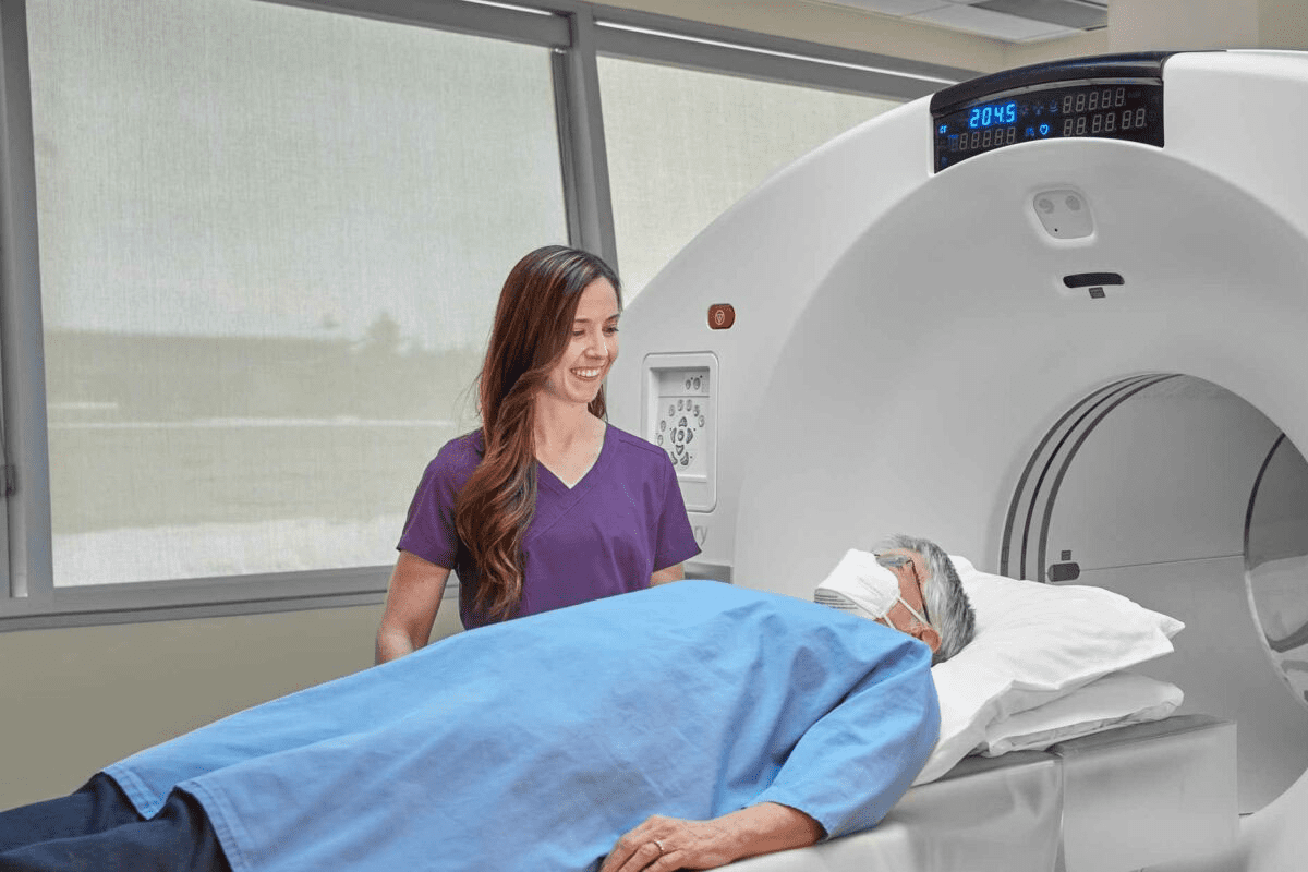
What Patients Can Expect During the Scan
A CT scan for suspected fractures is done in hospitals or imaging centers. You’ll be asked to wear a hospital gown and remove metal objects or jewelry.
During the scan, you’ll lie on a table that moves into a large machine. The machine takes pictures of your bones from different angles. The whole process takes 10 to 30 minutes, depending on the scan’s complexity.
Preparation and Safety Considerations
To have a safe and effective CT scan, follow the preparation steps carefully. You might need to avoid eating or drinking for a few hours before, if contrast dye is used.
Tell your healthcare provider about any allergies, like iodine or contrast dye, and medical conditions. Pregnant women or those who might be pregnant should also inform their healthcare provider, as the scan involves radiation.
Radiation Exposure: Risks in Context
CT scans expose you to a small amount of radiation. The risk of harm from radiation is low, but it’s important to consider the benefits of getting a precise diagnosis.
We take steps to reduce radiation exposure, like using the lowest dose needed and tailoring the scan to your needs. The table below shows important details about radiation exposure during a CT scan.
| Aspect | Description | Precaution |
| Radiation Dose | Typically measured in millisieverts (mSv) | Adjusted based on patient size and scan type |
| Risk to Pregnant Women | Potential risk to the fetus | Inform healthcare provider if pregnant or suspecting pregnancy |
| Contrast Dye | Used to enhance image clarity | Assess for allergies and kidney function before use |
By understanding the CT scan procedure and taking precautions, we can make sure it’s safe and effective. If you have any concerns or questions, talk to your healthcare provider.
Conclusion: The Critical Role of CT Scans in Modern Fracture Management
CT scans are key in diagnosing and managing fractures. They provide detailed images needed for treatment plans. This technology helps doctors spot fractures, even the small ones missed by regular X-rays.
Research shows CT scans are very good at finding fractures. A study on PubMed Central proves their high sensitivity and specificity. This makes CT scans essential in modern fracture care.
As medical tech advances, CT scans will become even more vital. They help us make better treatment choices and improve patient care. By using CT scans, we can offer more effective treatment for fracture patients, leading to better health results.
FAQ
Will a CT scan show broken bones?
Yes, CT scans are very good at finding broken bones. They show detailed images that help doctors diagnose fractures.
Does a CT scan show bones?
Yes, CT scans are made to see bones. They can spot fractures, bone pieces, and other bone problems.
Can a CT scan detect subtle fractures?
Yes, CT scans are great at finding small fractures. They can see things that X-rays or other tests can’t.
How accurate are CT scans in detecting fractures?
CT scans are very accurate. Studies show they can find about 98% of certain fractures.
What is the difference between a CT scan and an X-ray?
CT scans give more detailed images than X-rays. This makes them better for finding complex or small fractures.
When is MRI preferred over CT for bone fractures?
MRI is often chosen over CT if there’s a soft tissue injury with the fracture. MRI is better at seeing soft tissues.
Can MRI detect broken bones?
Yes, MRI can find broken bones. But it’s not usually the first choice for bone imaging because it’s better at soft tissue.
What can patients expect during a CT scan for a suspected fracture?
Patients can expect a quick and painless test. The CT scanner will take detailed pictures of the area.
Are there any risks associated with CT scans?
The main risk of CT scans is radiation. But, the benefits of accurate diagnosis are usually worth it.
How does 3D reconstruction from CT scans aid in fracture treatment?
3D reconstruction helps doctors and patients understand the fracture. It helps with planning surgery and educating patients.
Reference
- Si, C., Zhu, J., & Zheng, Y. (2025). Value of CT-based 3D reconstruction in analyzing pelvic fractures. Journal of Orthopaedic Surgery and Research. https://pmc.ncbi.nlm.nih.gov/articles/PMC12135960/
- Kang, J., et al. (2022). Application value of CT scan 3D reconstruction technology in clinical diagnosis and treatment of maxillofacial fracture patients. Journal of Craniofacial Surgery. https://pmc.ncbi.nlm.nih.gov/articles/PMC9283051/
- Haydel, A. L., et al. (2024). Preoperative computed tomography scan in distal radius fractures: impact on treatment planning. Journal of Orthopaedic Trauma. https://www.sciencedirect.com/science/article/pii/S258951412300186X


