Last Updated on November 27, 2025 by Bilal Hasdemir

Would a CT scan show a brain tumor? At Liv Hospital, we use CT scans and MRI for precise imaging needed to diagnose and treat brain tumors. These methods provide detailed views to guide effective care.
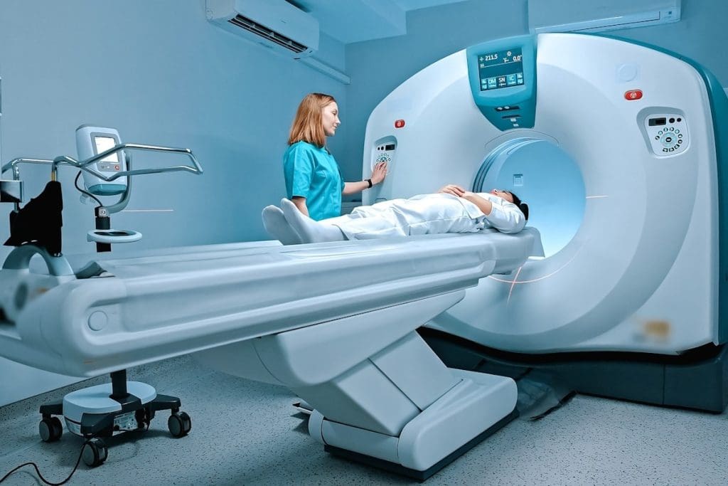
CT scans make detailed images with X-rays. MRI uses strong magnets and radio waves. This tech difference impacts how they find brain tumors.
CT scans are quick and used in emergencies. They give fast diagnoses. MRI, though, shows soft tissues better. It’s great for seeing brain tumor details.
Key Takeaways
- CT scans use X-rays, while MRI uses strong magnets and radio waves.
- CT scans are faster and often used in emergency situations.
- MRI provides more detailed images of soft tissues.
- The choice between CT scans and MRI depends on the specific needs of the diagnosis.
- Liv Hospital employs both CT scans and MRI for accurate brain tumor detection.
Understanding Brain Imaging Technologies

To accurately diagnose brain tumors, it’s essential to comprehend the principles behind CT scan and MRI technologies. These imaging modalities are key in detecting and diagnosing brain tumors. Each has its unique strengths and limitations.
Basic Principles of CT Scanning
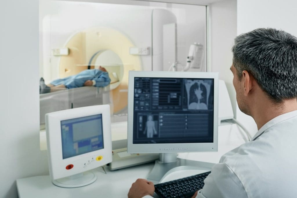
CT scans use X-rays to create detailed images of the brain. A CT scanner rotates around the patient, capturing X-ray measurements from multiple angles. These measurements are then reconstructed into images using computer algorithms.
CT scans are great for quick information in emergencies, like detecting hemorrhage or fractures. But, they might not show soft tissue details as well as MRI does.
Basic Principles of MRI Technology
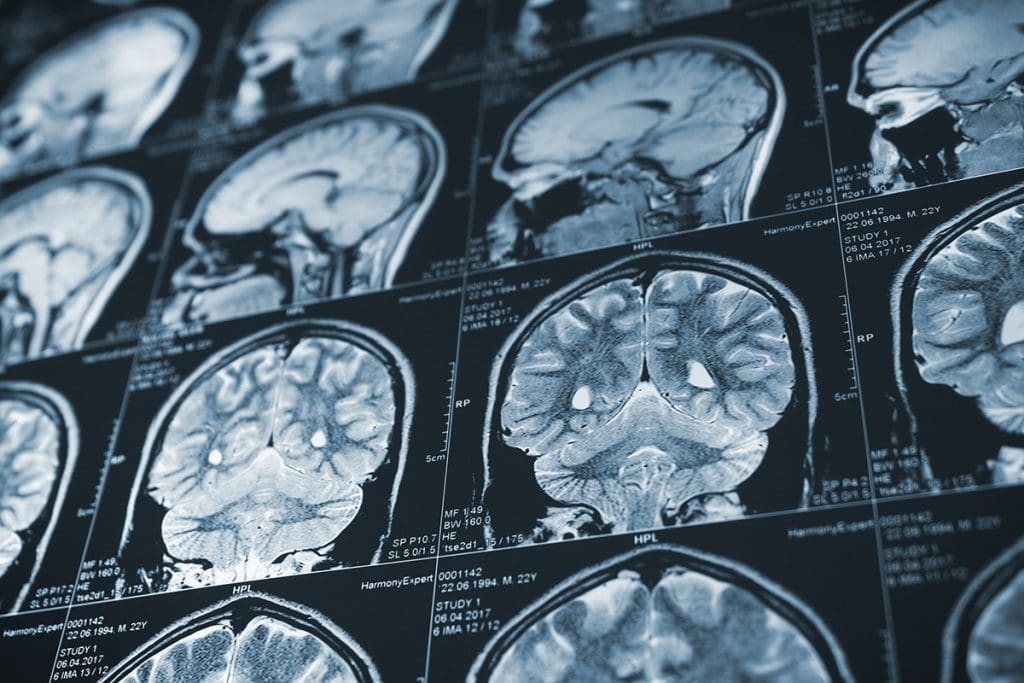
MRI technology uses a strong magnetic field and radio waves to create images of the brain’s soft tissues. It aligns hydrogen nuclei in the body with the magnetic field and then disturbs this alignment with radio waves. As the nuclei return to their aligned state, they emit signals that create detailed images.
MRI excels at showing soft tissue details, making it great for diagnosing conditions like certain brain tumors. Its superior soft tissue contrast helps in better defining tumor boundaries and internal structures.
Key Differences in Imaging Techniques
The main differences between CT and MRI lie in their technologies and the information they provide. CT scans use X-rays and are faster, while MRI uses magnetic fields and radio waves, providing more detailed soft tissue images.
| Characteristics | CT Scan | MRI |
| Imaging Technology | X-rays | Magnetic field and radio waves |
| Speed | Faster, ideal for emergencies | Slower, more detailed |
| Soft Tissue Detail | Limited | High resolution |
Understanding these differences is key for choosing the right imaging modality for diagnosing brain tumors. By selecting the right technology, healthcare providers can ensure accurate diagnoses and effective treatment plans.
Would a CT Scan Show a Brain Tumor? Capabilities and Limitations

CT scans can find brain tumors, but it depends on the tumor’s type and size. They are a key tool for doctors, but they don’t work for all tumors.
Types of Brain Tumors Detectable by CT
CT scans work well for some brain tumors. For example, meningiomas and oligodendrogliomas have calcifications that show up on scans. Also, big tumors that push brain structures around can be seen.
But, not all tumors are easy to spot with CT scans. Small, isodense, or spreading tumors are harder to find.
Sensitivity and Specificity of CT for Brain Tumors
The accuracy of CT scans is measured by sensitivity and specificity. Sensitivity is how well they find tumors. Specificity is how well they don’t find tumors when they’re not there.
| Tumor Type | Sensitivity of CT | Specificity of CT |
| Calcified Tumors | High | High |
| Soft Tissue Tumors | Moderate | Moderate |
| Small/Infiltrating Tumors | Low | High |
When CT Might Miss a Brain Tumor
CT scans might not catch every tumor. This is true for small or isodense tumors. Also, bone or other structures can hide tumors.
In summary, CT scans are helpful for finding brain tumors, but they have limits. Knowing these limits helps doctors give better care.
How Brain Tumors Appear on CT Scans
Brain tumors on CT scans look different, making it key to know their signs. Radiologists look for special signs on CT scans to tell tumors apart.
Characteristic Features of Primary Brain Tumors
Primary brain tumors show unique signs on CT scans. Some common signs include:
- Heterogeneous density: Many primary brain tumors look like mixed masses with different densities.
- Calcifications: Some tumors, like oligodendrogliomas, have calcifications seen on CT scans.
- Necrosis: Bigger tumors might have necrosis, showing up as low-density areas.
- Mass effect: Tumors can push brain structures aside.
For example, glioblastomas look like irregular, ring-shaped lesions with necrosis in the middle. Meningiomas, on the other hand, are well-defined, homogeneously enhanced masses near the meninges.
Metastatic Brain Lesions on CT
Metastatic brain lesions have their own signs. Key signs include:
- Multiple lesions: Metastases are often many and found at the gray-white junction.
- Ring enhancement: Many metastases enhance in a ring shape after contrast.
- Surrounding edema: Metastases usually have a lot of vasogenic edema.
On CT scans, metastatic lesions can look like solid or ring-enhancing masses. Seeing many lesions at different enhancement stages can hint at metastatic disease.
Enhancement Patterns with Contrast
Contrast enhancement is key in looking at brain tumors on CT scans. Different tumors show different enhancement patterns:
- Homogeneous enhancement: Some tumors, like certain meningiomas, enhance evenly.
- Ring enhancement: Glioblastomas and some metastases show ring enhancement around a necrotic center.
- Heterogeneous enhancement: Many high-grade gliomas and some metastases enhance unevenly.
Knowing these patterns helps radiologists tell tumor types apart and see how far the disease has spread.
MRI Brain Tumor Detection: Gold Standard Approach
Advanced MRI techniques have changed how we find and understand brain tumors. MRI’s ability to show soft tissues clearly makes it key in neuro-oncology.
Superior Soft Tissue Contrast in MRI
MRI gives exceptional soft tissue contrast. This lets us see brain tumors clearly, even when other images can’t. It’s vital for spotting brain details and telling different soft tissues apart.
Advanced MRI Techniques for Tumor Characterization
We use many MRI methods to better understand brain tumors. These include:
- Functional MRI (fMRI) for checking how tumors affect brain function
- Magnetic Resonance Spectroscopy (MRS) for studying tumor metabolism
- Diffusion-weighted imaging (DWI) for looking at tumor cell density
- Perfusion-weighted imaging (PWI) for seeing tumor blood flow
Visualization of Tumor Boundaries and Invasion
MRI is great at showing tumor boundaries and if tumors are spreading. This info is essential for planning surgery and choosing the best treatment.
| Feature | CT Scan | MRI |
| Soft Tissue Contrast | Limited | Excellent |
| Tumor Boundary Visualization | Poor | Excellent |
| Advanced Imaging Techniques | Limited | Multiple options available |
Comparative Analysis: CT vs. MRI for Brain Tumor Diagnosis
Knowing the differences between CT and MRI is key for accurate brain tumor diagnosis. Both have their strengths and weaknesses. The choice between them depends on several factors.
Diagnostic Accuracy Comparison
MRI is generally better than CT for brain tumor detection. MRI’s high soft tissue contrast helps see tumor boundaries and edema better. Experts say MRI is more sensitive than CT, even in early stages.
“MRI has become the gold standard for brain tumor imaging due to its ability to provide detailed information about tumor size, location, and characteristics.”
But CT scans have their own benefits. They are quick, making them great for emergencies. They can spot larger tumors or those causing a lot of mass effect.
Speed and Accessibility Factors
CT scans are fast, which is a big plus. They’re perfect for emergencies or when patients can’t stay in one place for long. Plus, CT scanners are more common than MRI machines, even in rural areas.
MRI scans take longer and are less common in some places. But MRI’s better accuracy makes it worth using when possible.
Patient Considerations: Claustrophobia and Implants
Choosing between CT and MRI depends on the patient. For example, those with claustrophobia might prefer CT or open MRI. MRI’s enclosed space can be tough for them.
Also, patients with metal implants, like pacemakers, can’t have MRI. CT scans are safer for them because they don’t have strong magnetic fields.
In summary, MRI is better for brain tumor diagnosis, but CT scans have their own benefits. Speed, accessibility, and patient needs like claustrophobia and implants also play a role in choosing between them.
Detecting Metastatic Brain Tumors: Special Considerations
Metastatic brain tumors are hard to diagnose. They start in other parts of the body and move to the brain. This makes finding them tricky.
CT vs. MRI Sensitivity for Metastases
When it comes to finding metastatic brain tumors, MRI is better. It can spot smaller tumors than CT scans. MRI shows the brain’s soft tissues clearly, helping to see where tumors are.
MRI is the top choice for finding these tumors because it shows the brain’s details well. But, CT scans are faster and easier to get. They’re good for quick checks in emergencies.
Multiple Metastases Detection Rates
How well CT and MRI find multiple tumors is different. MRI finds more tumors, small or many, than CT. This is because MRI is better at showing the brain’s details.
| Imaging Modality | Sensitivity for Single Metastasis | Sensitivity for Multiple Metastases |
| CT | 70-80% | 40-60% |
| MRI | 90-95% | 80-90% |
Follow-up Imaging Protocols
For people with cancer, checking for new tumors is key. They might need CT and MRI scans to watch how tumors change. The plan depends on the patient’s cancer type and how far it has spread.
High-risk patients should get MRI scans often. How often depends on their cancer type, how far it has spread, and any treatments they’ve had.
Beyond the Brain: CT and Bone Metastases Detection
Cancer often spreads to the bones, making it key to detect bone metastases for treatment. Advanced imaging like CT scans helps find and measure bone involvement.
Metastatic Osseous Lesions in Spine CT Non-Con Imaging
CT scans are great for spotting bone metastases in the spine. Non-contrast CT imaging shows changes in bone density and structure. These changes can be lytic lesions, where bone is destroyed, or sclerotic lesions, where bone gets denser.
Key Features of Metastatic Osseous Lesions on CT:
- Lytic lesions: Areas where bone is destroyed, appearing as darker regions on CT images.
- Sclerotic lesions: Areas where bone becomes denser, appearing as brighter or whiter regions.
- Mixed lesions: Combination of lytic and sclerotic features.
How CT Reveals Bone Involvement in Cancer
CT scans give detailed views of bone anatomy, helping us see how much bone is involved in cancer. We can spot fractures, cortical destruction, and soft tissue masses linked to bone metastases.
| Feature | Description | Clinical Significance |
| Lytic Lesions | Destruction of bone appearing as darker areas | Indicates bone metastasis, possible fracture risk |
| Sclerotic Lesions | Increased bone density appearing as brighter areas | Often seen in prostate cancer metastases |
| Cortical Destruction | Breakdown of the outer layer of bone | Increases risk of fracture |
Limitations of CT in Bone Metastasis Detection
CT scans are useful for finding bone metastases but have limits. Small or early metastatic lesions might not show up, if they haven’t changed bone density or structure much. Also, CT scans don’t tell us much about the metabolic activity of bone metastases.
We must keep these limits in mind when looking at CT scans. Often, we use other imaging like bone scans or MRI to fully understand the disease.
Bone Scan Technology: Complementary to CT Imaging
Bone scan technology is key in finding bone metastases. It’s often paired with CT imaging. This tool helps doctors understand bone health, aiding in diagnosis and treatment.
How Bone Scans Work
Bone scans use a small amount of radioactive material, like technetium-99m. It’s injected into the blood. The material goes to active bone areas, like cancer spots.
A special camera picks up the radiation. This creates images showing where the bone is most active.
The scan is simple and done outside the hospital. Patients wait a few hours after the injection. This lets the tracer spread throughout the body.
Interpreting Uptake on Bone Scans
Doctors look for high uptake areas on bone scans. This can mean cancer, arthritis, or trauma. The uptake’s intensity and pattern give clues about the cause.
A strong focal uptake might mean cancer. But a spread-out uptake could be arthritis. It’s important to look at all the patient’s information when reading these scans.
Does Uptake on a Bone Scan Mean Cancer?
Uptake on a bone scan can hint at cancer, but it’s not always cancer. Other things like fractures, infections, or inflammation can also show up.
Here’s a table showing different reasons for uptake:
| Cause | Characteristics |
| Cancer | Focal or multifocal uptake, often in a pattern consistent with metastatic disease |
| Arthritis | Diffuse uptake in joints, often with a characteristic pattern (e.g., symmetric involvement) |
| Fracture | Focal uptake at the site of the fracture, which may be linear or irregular |
| Infection | Diffuse or focal uptake, often with associated soft tissue swelling or other signs of inflammation |
As the table shows, many conditions can cause uptake. So, it’s vital to look at the whole patient picture when reading bone scan results.
Differential Diagnosis in Cancer Imaging
When we look at cancer imaging results, figuring out what they mean is key. We use different imaging methods to spot and measure cancer. But, it’s hard to tell if something is cancer or not.
Bone scans can show changes in bone that might mean cancer. Yet, these changes can also happen for other reasons. This makes it tough to tell what’s going on.
Bright White Spots on Bone Scan: Cancer vs. Other Conditions
Bright spots on a bone scan might mean cancer has spread. But, they could also show other issues like broken bones, infections, or wear and tear.
To figure out if it’s cancer, we look at the patient’s history, symptoms, and other scan results. For example, if someone with a cancer history has many bright spots, it’s likely cancer has spread.
Bone Scan Arthritis vs. Cancer
Arthritis can also show up as bright spots on scans, making it look like cancer. We need to look closely at where and how these spots appear to tell them apart.
Arthritis spots usually show up in joints and are on both sides. Cancer spots might look different, but not always. Sometimes, we need more scans to be sure.
When Additional Imaging Is Necessary
If we’re not sure what’s going on, we might suggest more scans like CT or MRI. These scans give us more details.
They can help us understand what the bone scan spots mean. For example, a CT scan can show if there’s damage to the bone or soft tissue, which is often a sign of cancer.
By using different scans and looking at the patient’s whole situation, we can make a better diagnosis. This helps us plan the best treatment.
Advanced and Integrated Imaging Approaches
Cancer diagnosis and treatment are evolving fast. SPECT and PET/CT are key in this change. We’re moving towards more detailed imaging that helps us understand cancer better.
SPECT and PET/CT Advantages
SPECT and PET/CT are changing cancer imaging. SPECT shows how tumors work, helping us see if treatments are working. PET/CT combines this with detailed images, making it easier to find tumors.
Using SPECT and PET/CT has many benefits. These include:
- They help find small tumors or spread early
- They give us a better look at how tumors work
- They help us see how well treatments are working
Combination Approaches: CT, MRI, and Bone Scan
Using different imaging methods together gives a clearer picture of cancer. CT, MRI, and bone scans each offer unique insights. Together, they help us understand cancer better.
For example, CT scans show tumor size and location well. MRI is great for soft tissue details. Bone scans spot bone metastases. Combining these, we get a detailed view of the disease.
Future Directions in Cancer Imaging
The future of cancer imaging looks bright. New tech and AI will improve it. We’ll see better images, faster scans, and smarter analysis tools.
New trends include:
- New contrast agents and tracers for better images
- More hybrid imaging, like PET/MRI
- AI and machine learning for better image analysis
These changes will lead to better cancer care and outcomes.
Conclusion
Choosing between a CT scan and MRI for brain tumor diagnosis depends on several factors. These include the type of tumor, its location, and patient considerations. We have looked at the capabilities and limitations of both imaging modalities in detecting brain tumors.
CT scans are often used first because they are fast and easy to get, even in emergencies. But MRI is usually the best choice for brain tumor diagnosis. This is because MRI shows soft tissues better and can tell more about tumor boundaries and invasion.
In cancer imaging, picking the right imaging modality is key for accurate diagnosis and treatment planning. Techniques like CT, MRI, and bone scans are important for finding and managing cancer. Knowing the strengths and weaknesses of each helps healthcare providers make better choices and care for their patients well.
FAQ
Does uptake on a bone scan mean cancer?
What does cancer look like on a bone scan?
How does a bone scan differ from a CT scan?
Can a bone scan detect bone cancer?
What is the role of CT in detecting bone metastases?
How do MRI and CT compare for brain tumor diagnosis?
Can CT scans show brain tumors?
What are the advantages of using SPECT or PET/CT in cancer imaging?
How do bright white spots on a bone scan relate to cancer?
What is the difference between a bone scan and a CT scan for detecting bone metastases?
References
- National Health Service (NHS). (2023). CT Scan. NHS.uk. https://www.nhs.uk/conditions/ct-scan/


