
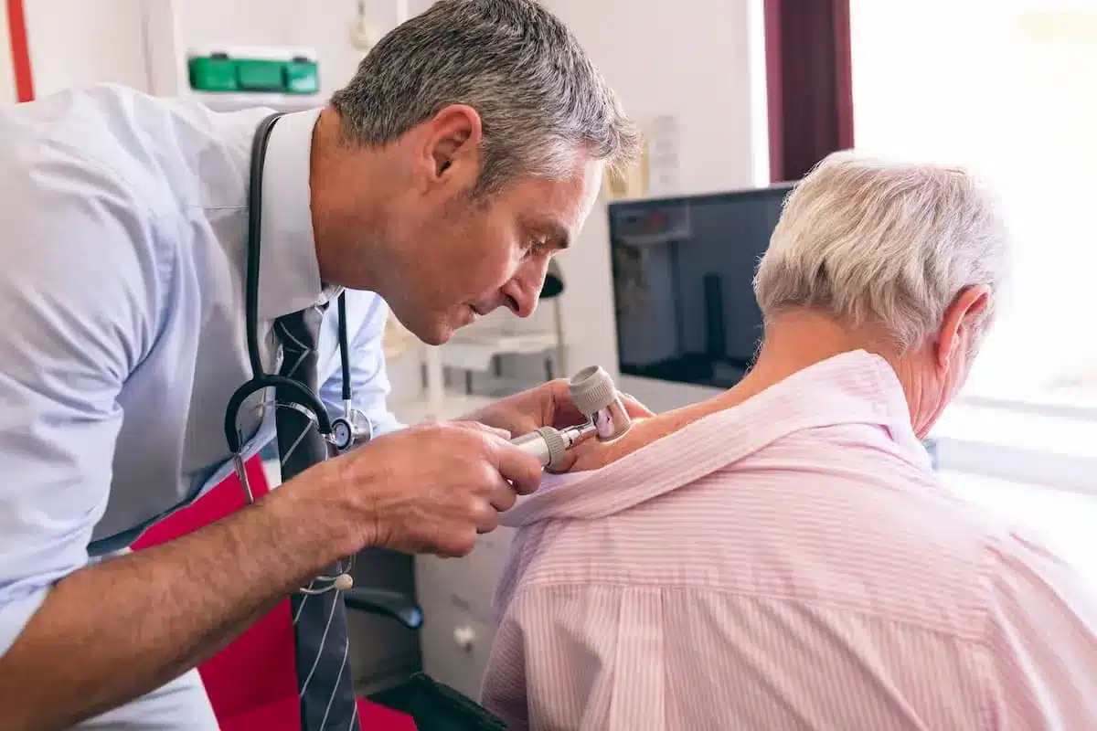
At Liv Hospital, we know how key an abdominal aorta ultrasound is. It helps spot abdominal aortic aneurysms and check aorta size. Ultrasound is becoming more important in medicine, used more for prenatal checks and baby monitoring, as the U.S. ultrasound market report shows.
We’ll show you the essential steps for a detailed aorta sonogram. This ensures you get top-notch care. Getting accurate images is vital for diagnosing and treating blood vessel issues. Our guide is made to help doctors get the best results.
Key Takeaways
- Understanding the importance of accurate abdominal aorta ultrasound
- Learning the key steps for a precise aorta sonogram
- Recognizing the role of ultrasound technology in medical imaging
- Appreciating the significance of detecting abdominal aortic aneurysms
- Assessing aorta size with high precision
The Clinical Significance of Abdominal Aorta Ultrasound

Abdominal aorta ultrasound is key in today’s vascular diagnostics. It’s non-invasive and reliable. Abdominal aortic ultrasound helps check vascular health and spot problems early.
This diagnostic method is vital in clinical practice. It helps find and manage vascular diseases early. Ultrasound for abdominal aorta is great for both diagnosis and monitoring.
Diagnostic Value in Vascular Assessment
Ultrasound of the abdominal aorta is very useful. It gives detailed images of the aorta. Doctors can see the aorta’s structure and function, find problems, and track disease.
A study showed ultrasound screening for aortic aneurysms cuts down on deaths. Early detection is key.
“Screening for abdominal aortic aneurysm (AAA) with ultrasonography is effective in reducing mortality, and is recommended for men aged 65 to 75 years who have ever smoked.”
Screening Applications and Guidelines
Ultrasound screening for abdominal aortic aneurysms is common in high-risk groups. Guidelines suggest aortic US for men aged 65-75 who smoked. This early detection and management can lower rupture risk and death.
Mortality Reduction Through Early Detection
Early ultrasound screening for aortic aneurysms cuts down on deaths. Finding aneurysms early lets doctors take action. This can include watching the aneurysm or surgery if needed.
We stress following screening guidelines and using ultrasound for abdominal aorta first. This can lead to better patient outcomes and lower aneurysm-related deaths.
Essential Equipment and Patient Preparation

Getting a clear view of the abdominal aorta with ultrasound needs the right equipment and patient prep. We’ll cover the must-have tools for ultrasound and how to get the patient ready for the best images.
Ultrasound Technology Selection
The type of ultrasound tech used is key for good images. New portable and handheld ultrasound devices are popular, thanks to recent tech advances. They’re great for quick checks in emergencies or at the bedside.
When picking an ultrasound for the aorta, look for these important features:
- A transducer that can go deep and show details well
- Advanced image tech for clearer views
- Doppler imaging to see blood flow
- An easy-to-use interface for quick setup
Patient Positioning and Preparation
Getting the patient set up right is essential for clear ultrasound images. It’s best if patients don’t eat for 8-12 hours before. This helps reduce gas in the bowels that can block ultrasound waves.
The patient should lie on their back with arms up. This makes it easier to see the belly. Sometimes, lying on the side helps get a better view. Keeping the patient comfy is key to avoid moving during the scan.
Important steps in getting the patient ready include:
- Telling the patient what to expect to calm them down
- Making sure they’ve followed any pre-exam rules (like fasting)
- Putting the patient in the right spot on the table
- Adjusting the ultrasound machine for the patient’s body
Step 1: Selecting the Appropriate Transducer and Settings
Getting a good abdominal aorta ultrasound starts with picking the right transducer and settings. This first step is key to getting clear images for accurate diagnosis.
Frequency Selection Based on Patient Body Habitus
The transducer’s frequency depends on the patient’s body type and the aorta’s depth. For most adults, a curvilinear transducer with a 3-5 MHz frequency works best. This range balances depth and image quality well.
For thinner patients, a 5-7 MHz frequency can improve image sharpness. But for obese patients, a 2-3 MHz frequency is needed for better penetration.
Depth and Focus Optimization Techniques
Setting the right depth is vital to keep the aorta in the center of the image. Adjust the depth so the aorta is clearly seen in the middle of the screen.
Adjust the focus to the aorta’s level for better side detail. Many ultrasound machines have dynamic focusing to automatically adjust focus.
Gain and Time Gain Compensation Adjustments
Adjusting the gain is key for image quality. Set the gain so the aorta’s lumen is black, and the surrounding tissues are clear.
Time Gain Compensation (TGC) helps adjust gain at different depths. Use TGC sliders to keep the image bright and even, compensating for signal loss with depth.
| Parameter | Adjustment | Purpose |
|---|---|---|
| Frequency | 3-5 MHz | Balance between penetration and resolution |
| Depth | Center aorta on screen | Ensure clear visualization of aorta and surrounding structures |
| Focus | At the level of aorta | Optimize lateral resolution |
| Gain | Anoechoic lumen | Optimize image quality |
| TGC | Uniform brightness | Compensate for signal attenuation with depth |
Choosing the right transducer and settings is the first step to a successful aortic sonogram. These steps are essential for getting high-quality images for accurate diagnosis and care.
Step 2: Identifying Key Anatomical Landmarks
To do a good abdominal aorta ultrasound, finding key landmarks first is key. This step makes sure the exam is both right and complete.
Using the Spine and Great Vessels as Reference Points
The spine is a main reference because of its clear shadow on ultrasound. The great vessels, like the aorta and inferior vena cava, are also key landmarks. They help us find the abdominal aorta. We use these to guide our scan, making sure we get all the views needed.
Locating the Celiac Trunk Origin
Finding where the celiac trunk starts is another big step. The celiac trunk is a big branch of the abdominal aorta. Knowing where it starts helps us make sure we’re in the right spot. This is really helpful for patients with complex anatomy or aortic problems. We adjust our scan based on the patient’s body and aorta shape.
Differentiating Aorta from Inferior Vena Cava
Telling the aorta from the inferior vena cava (IVC) is critical for correct diagnosis. The aorta is smaller and rounder than the IVC. The aorta also looks more detailed and has thicker walls than the IVC. For more info on aortic sizes, check out 5 techniques for mastering aortic measurements. This can help make your ultrasound more accurate.
By carefully finding these landmarks, we make sure our abdominal aorta ultrasound is precise. This sets the stage for accurate diagnosis and the right care for patients.
Step 3: Mastering Transverse Scanning Protocol
Ultrasound scans of the abdominal aorta need a good grasp of the transverse scanning protocol. This method is key for getting clear images and accurate diagnoses.
Systematic Approach from Diaphragm to Bifurcation
We start by finding the diaphragm as our first landmark. Then, we scan the aorta down to its bifurcation. This method makes sure we see the whole aorta.
We focus on the aorta’s size, thickness, and any issues during the scan. A systematic scan helps spot small changes that might mean trouble.
Proper Probe Orientation and Pressure
Getting the probe right is essential for quality images. We make sure it’s straight to the aorta to cut down on bad images. We use light pressure to avoid squishing the aorta or moving gas around.
The table below shows how to use the probe correctly:
| Probe Orientation | Pressure Application | Benefits |
|---|---|---|
| Perpendicular to the aorta | Gentle pressure | Minimizes artifacts, optimizes visualization |
| Adjusted for patient anatomy | Avoids compressing the aorta | Reduces interference from bowel gas |
Visualization Techniques for Challenging Patients
Some patients can be tough to scan, like those with a lot of gas or obesity. We use special techniques to get around these problems. For example, we might ask the patient to breathe deeply to move gas or use harmonic imaging for obese patients.
With these techniques, we can get great images even when it’s hard. The trick is to stay patient and adjust our approach as needed for each patient.
Step 4: Performing Effective Longitudinal Scanning
Longitudinal scanning is key for seeing the whole aorta during an ultrasound. It checks the aorta’s health, finds aneurysms, and looks at blood flow.
Sagittal Plane Orientation Methods
To scan well, we align the probe in the sagittal plane. This means lining up the probe with the aorta’s long axis. This way, we see the whole aorta in one view. “Proper alignment is key to accurate diagnosis,” as many ultrasound guides say.
We find the aorta first in the transverse plane. Then, we turn the probe 90 degrees for the sagittal view. Making small changes to the probe’s position and angle is vital to keep the aorta in the middle of the image.
Capturing the Complete Aortic Length
To get the whole aortic length, we carefully move the ultrasound probe. We slowly sweep the probe laterally to see the whole aorta, from top to bottom. This helps spot any issues along the aorta.
Choosing the right depth settings on the ultrasound machine is also key. This makes sure we see the whole aorta as we scan.
Techniques for Continuous Visualization
To keep seeing the aorta, we keep the probe aligned with its long axis. We make gentle pressure adjustments to keep the aorta in view as we scan. “Maintaining continuous visualization is key for spotting small problems,” say vascular ultrasound experts.
Color Doppler also helps by showing blood flow in the aorta. This gives us important info about its health.
Learning these scanning techniques improves how well we can diagnose issues with the abdominal aorta.
Step 5: Precise Measurement Techniques for Aorta Sonogram
Aorta sonogram measurements need to be precise for accurate diagnoses. We will cover the exact methods for measuring the aorta. This includes where and how to measure.
Standard Measurement Locations and Protocols
To get accurate results, we must stick to established protocols. We measure the aorta at three key spots: the start, middle, and end. These measurements help us spot any issues.
Outer Wall to Outer Wall Measurement Method
This method is widely used to measure the aorta’s diameter. It measures from one outer edge of the aortic wall to the other.
Normal Diameter Reference Values by Age and Gender
Knowing the normal diameter ranges is key for spotting problems. The aorta’s size changes with age and gender. Below are the normal diameter ranges for different ages and genders.
| Age Group | Gender | Normal Diameter Range (mm) |
|---|---|---|
| 20-39 | Male | 20-24 |
| 20-39 | Female | 18-22 |
| 40-59 | Male | 22-26 |
| 40-59 | Female | 20-24 |
| 60+ | Male | 24-28 |
| 60+ | Female | 22-26 |
By using these methods and reference values, we can make accurate diagnoses. This helps us provide better care for our patients.
Step 6: Evaluating Aortic Wall Integrity and Blood Flow
The sixth step in our guide focuses on checking the aortic wall and blood flow. This step is key to spotting any vascular problems and checking the aorta’s health.
Wall Thickness and Echogenicity Assessment
Checking the aortic wall’s thickness and how it looks is very important. We look for any thickening or odd shapes that might mean there’s a problem. The wall’s look can tell us if there’s inflammation or other issues.
Normally, the aortic wall is thin and even. If it looks different, we need to look closer. We’re watching for any spots that are more reflective, which could mean there’s plaque or atherosclerosis.
Color and Spectral Doppler Applications
To see how blood flows in the aorta, we use color and spectral Doppler. Color Doppler shows us where the blood is moving. It helps us spot any unusual flow.
Spectral Doppler gives us more details about blood flow. By focusing on the aorta, we can measure blood speed and look at waveforms. This helps us find any blockages or problems with blood flow.
Identifying Atherosclerotic Plaque and Calcifications
Looking for plaque and calcifications in the aortic wall is a big part of this step. We search for any signs of plaque buildup or calcification, as they can show atherosclerosis.
Plaque might look like bright spots in the aortic wall, sometimes with shadows if it’s calcified. We check how much plaque there is and what it looks like. This info is key for figuring out the risk and planning treatment.
| Feature | Normal | Abnormal |
|---|---|---|
| Aortic Wall Thickness | Uniform, thin layer | Thickened, irregular |
| Echogenicity | Homogeneous | Heterogeneous, increased |
| Blood Flow | Laminar, normal velocity | Turbulent, abnormal velocity |
| Plaque/Calcifications | Absent | Present, varying characteristics |
By carefully checking the aortic wall and blood flow, we get a full picture of the aorta’s health. This info is essential for making the right diagnosis, understanding the risk, and planning the best treatment.
Step 7: Detecting and Characterizing Abdominal Aortic Aneurysms
Abdominal aortic aneurysms need careful detection and characterization. This helps guide the right treatment. We will explain how to find and understand these aneurysms. This includes how to measure them and follow up on their growth.
Aneurysm Definition and Classification
An abdominal aortic aneurysm (AAA) is a big bulge in the aorta. It’s bigger than 1.5 times its normal size or 3 cm or more. AAAs can be divided into types based on where they are and how they look.
Critical Measurement Techniques
Measuring the aneurysm size accurately is key. We use a specific method to measure it. This method is important for tracking the aneurysm’s growth.
Risk Stratification and Follow-up Protocols
The risk of rupture depends on the aneurysm size. We categorize aneurysms by their size:
- Less than 3 cm: Low risk
- 3-4 cm: Moderate risk, check every 12 months
- 4-5.4 cm: Moderate to high risk, check every 6-12 months
- 5.5 cm or larger: High risk, might need surgery
Patients with AAAs need regular ultrasound checks. This helps track the aneurysm’s growth and decide if surgery is needed.
Step 8: Comprehensive Evaluation of Aortic Branches
The aorta’s branches are vital for blood flow. They are key in an abdominal aorta ultrasound. We check the celiac trunk, superior mesenteric artery, renal arteries, and iliac arteries for a full review.
Celiac Trunk and Superior Mesenteric Artery Assessment
The celiac trunk and superior mesenteric artery are important. They start from the abdominal aorta. We use ultrasound to see these vessels.
- Find where the celiac trunk starts and splits into the common hepatic and splenic arteries.
- Look at where the superior mesenteric artery starts and how it goes.
- Check blood flow with color Doppler.
Looking at these branches helps us spot problems that could cause mesenteric ischemia.
Renal Arteries Visualization Techniques
Seeing the renal arteries is key for checking kidney blood flow. We use special ways to see them:
- Look at the renal artery starts from the side or back.
- Use color Doppler to find the renal arteries and check their flow.
- Make sure the Doppler angle is right for accurate measurements.
By checking the renal arteries, we can find problems that might hurt kidney function.
Iliac Arteries Examination at Bifurcation
The aorta splits into the common iliac arteries at a key point. We look at the iliac arteries this way:
- Follow the common iliac arteries to where they split into internal and external iliac arteries.
- Use color Doppler to see blood flow in the iliac arteries.
- Look for any signs of blockage or swelling.
By fully checking the aortic branches, we get a detailed look at the abdominal aorta. This helps us find any issues that could affect patient care.
Step 9: Documentation and Standardized Reporting
Good documentation and reporting are key to a successful abdominal aorta ultrasound. We must make sure our records are detailed, correct, and follow a standard. This helps everyone in healthcare talk clearly and share information with patients.
Essential Images for Complete Documentation
Getting the right images is vital for a full aorta sonogram. We need pictures that clearly show the aorta from different angles. These should include measurements and any unusual findings.
When we document images, we must label them properly. This includes the patient’s info, the date, and the type of view. This focus on detail is important for top-notch patient care.
Measurement Recording Standards
Keeping measurements the same is important for comparing different exams. We should follow set rules for measuring the aorta’s size. This means using outer wall to outer wall measurements.
| Measurement Location | Standard Protocol | Notes |
|---|---|---|
| Proximal Aorta | Outer wall to outer wall | Include in all reports |
| Mid Aorta | Outer wall to outer wall | Note any abnormalities |
| Distal Aorta | Outer wall to outer wall | Include bifurcation measurements |
Structured Reporting Templates
Using structured reporting templates can make our reports better and more consistent. These templates help us include all important details. This makes our documentation complete and standard.
A good template should have sections for patient info, exam details, findings, and recommendations. Using these templates helps us report more efficiently. It also improves how we talk to other healthcare providers.
Critical Findings Communication Protocols
Telling others about important findings is critical for good patient care. We need clear ways to share urgent or significant info with doctors or other healthcare teams.
Our protocols should cover both regular and emergency reports. This ensures that important findings are shared quickly and correctly. This might be through phone calls, secure messages, or other methods.
By following these steps, we can improve patient care. We can also lower the chance of mistakes in communication. Our work in documenting and reporting will meet the highest standards in abdominal aorta ultrasound.
Step 10: Overcoming Technical Challenges in Aortic Ultrasound
Aortic ultrasound faces many technical challenges. These obstacles can affect image quality and accuracy. We will explore effective solutions for these issues.
Solutions for Bowel Gas Interference
Bowel gas can make ultrasound images poor. To improve this, we focus on patient preparation. We tell patients to fast or avoid gas foods before the scan. This helps reduce bowel gas.
We also use gentle compression with the probe. This moves gas-filled loops, making the aorta clearer. Another strategy is alternative scanning windows. By finding different windows, we can avoid gas and get better images.
Techniques for Imaging Obese Patients
Ultrasound in obese patients is tough due to deep tissue and loss of signal. We use lower frequency transducers for deeper penetration. But, this means some loss of detail.
Adjusting gain and depth settings is also key. This improves image quality in obese patients. Tissue harmonic imaging also helps by reducing artifacts and making walls clearer.
Approaches for Tortuous Aortic Anatomy
Tortuous aortas are hard to image. We use multiple scanning planes to fully see the aorta’s shape. This ensures accurate measurements and a full view of the wall.
3D ultrasound techniques are also helpful. They give a detailed view of the aorta and its surroundings.
Managing Post-Surgical Anatomy Challenges
Post-surgical changes, like grafting, add to the challenge. Knowing typical post-surgical appearances is key. We must also be aware of complications.
It’s vital to correlate with the patient’s surgical history. This ensures accurate ultrasound findings. We also watch out for artifacts from surgery or changed anatomy.
Conclusion: Advancing Abdominal Aortic Ultrasound Practice
Ultrasound technology keeps getting better, helping us diagnose and manage aortic diseases more effectively. We’ve covered the key steps for a good aorta sonogram. This includes choosing the right transducer and settings, and how to document and report findings.
Healthcare professionals can improve their skills by following the 10 key steps for abdominal aortic ultrasound. This leads to better care for patients. Keeping up with new education and technology is key to improving ultrasound practice.
It’s important to stay current with new ultrasound of aorta and aortic sonogram developments. This boosts our ability to diagnose accurately. It also helps us care for patients better by catching diseases early.
By improving our skills in abdominal aortic ultrasound, we can give patients better care. This improves their quality of life and health outcomes.
FAQ
What is an aorta sonogram, and why is it important?
An aorta sonogram is a test that uses sound waves to see the aorta. This artery carries blood from the heart to the body. It helps find problems like aneurysms and checks the aorta’s health.
How do I prepare for an abdominal aorta ultrasound?
Before the test, you might need to fast and avoid certain foods. Wear loose clothes that let you move easily. Your doctor will tell you what to do.
What is the role of ultrasound technology in abdominal aorta imaging?
Ultrasound is key for seeing the aorta without harm. It’s safe, cheap, and doesn’t use radiation. It helps find problems like aneurysms.
How is the appropriate transducer selected for an aortic sonogram?
Choosing the right transducer depends on the patient’s body and the aorta’s depth. Lower frequency transducers go deeper, while higher ones are better for thinner patients.
What are the key anatomical landmarks to identify during an abdominal aorta ultrasound?
Important landmarks include the spine and great vessels. Also, the celiac trunk origin and inferior vena cava are key. They help the sonographer get the right view of the aorta.
How do you differentiate the aorta from the inferior vena cava during an ultrasound?
Sonographers look at size and wall thickness. The aorta is smaller and thicker, with pulsatility. The inferior vena cava is larger and thinner, with respiratory changes.
What is the systematic approach for transverse scanning of the abdominal aorta?
Start at the diaphragm and move down to the bifurcation. Use a consistent method and pressure. Techniques like bowel gas management help overcome challenges.
How do you measure the diameter of the aorta accurately during an ultrasound?
Measure from outer wall to outer wall at specific points. Make sure the probe is straight and avoid slanted measurements.
What are the normal diameter reference values for the aorta by age and gender?
Normal sizes vary by age and gender. For men, it’s less than 3 cm, and for women, less than 2.7 cm. These can change slightly based on the source and patient.
How is an abdominal aortic aneurysm detected and characterized during an ultrasound?
An aneurysm is seen as a dilation of the aorta that’s at least 1.5 times normal size. Size, shape, and complications like rupture are evaluated.
What is the significance of evaluating aortic branches during an abdominal aorta ultrasound?
Checking aortic branches is vital. It ensures blood flow to organs and spots stenosis or other issues that could harm the patient.
How do you overcome technical challenges, such as bowel gas interference, during an aortic ultrasound?
Try repositioning the patient or using gentle pressure. Adjust the probe settings or use harmonic imaging or contrast agents for tough cases.
What are the essential elements of documentation and reporting for an abdominal aorta ultrasound?
Capture key images and precise measurements. Use reporting templates and share findings quickly with the doctor. This ensures timely care.
References
- Devin Tooma, Vi Dinh; Co-authors Jessica Ahn, Jade Deschamps, Satchel Genobaga, Annalise Lang, Victor Lee, Reed Krause, Seth White. Aorta Ultrasound Made Easy: Step-by-Step Guide. POCUS 101. Available from: https://www.pocus101.com/aorta-ultrasound-made-easy-step-by-step-guide/ (POCUS 101)
- Risler Z, Dean AJ, Ku BS. Abdominal Aortic Aneurysm | Basic Sonoguide. American College of Emergency Physicians (ACEP). August 17, 2020. Available from: https://www.acep.org/sonoguide/basic/aorta/ (acep.org)
- [Author(s) not specified]. Abdominal aorta – protocol (PDF). University of California San Francisco (UCSF). Available from: https://edus.ucsf.edu/sites/edus.ucsf.edu/files/wysiwyg/UCSF20ED20US20Protocol20Abdominal20Aorta_Final.pdf
- [Author(s) not specified]. Aorta — POCUS Protocols / Tutorials. POCUS 101. Available from: https://www.pocus101.com/category/aorta/ (POCUS 101)
- [Author(s) not specified]. Abdominal aortic aneurysm screening: ultrasound equipment guidelines. GOV.UK. Available from: https://www.gov.uk/government/publications/abdominal-aortic-aneurysm-screening-ultrasound-equipment-guidance (GOV.UK)


