Last Updated on November 27, 2025 by Bilal Hasdemir

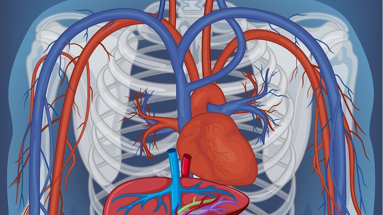
Knowing how veins and arteries work is key for doctors and everyone else. A detailed artery body map shows how blood moves around the body.
At Liv Hospital, we know how important clear pictures are. Our diagrams of veins and arteries help you understand blood flow. They guide your health choices.
It’s important to know how our body works. The circulatory system, or cardiovascular system, is a complex network. It carries blood all over the body.
Arteries and veins are different. Arteries are thick and carry oxygenated blood to the body. Veins are thinner and have valves to stop blood from flowing backward. They return deoxygenated blood to the heart.
Knowing these differences helps us understand artery and vein diagrams on humans. It also helps us see the map of veins in the body.
Oxygen transport is vital. Oxygen goes from the lungs to the body’s tissues through the blood. The arteries of human body diagram shows how oxygenated blood spreads out.
Oxygen then moves into the tissues. There, it helps with metabolic processes.
The circulatory system is organized in a way that makes it efficient. It starts with the heart, pumping blood into big vessels like the aorta. Blood then moves through smaller vessels, from arteries to arterioles, and then to capillaries.
In capillaries, oxygen, nutrients, and waste are exchanged. Then, deoxygenated blood goes back to the heart through venules and veins. This completes the cycle.
Important parts of this system include:
Diagrams of veins and arteries are key in modern medicine. They help doctors diagnose, treat, and teach. These images show the blood vessel network in our bodies.
Accurate diagrams are essential for spotting heart diseases. They help doctors see blood flow issues, blockages, and vascular health. Research shows these diagrams boost diagnosis and patient care.
In surgeries, diagrams guide doctors through complex tasks. They show blood vessel locations and conditions. This info lowers surgery risks, like in vascular surgery.
Diagrams are also great for teaching. They help students learn the circulatory system’s anatomy. This knowledge is key for their studies.
| Application | Description | Benefits |
|---|---|---|
| Diagnostic | Identifying vascular abnormalities | Improved diagnostic accuracy |
| Surgical Planning | Planning complex vascular procedures | Reduced risk of complications |
| Education | Teaching vascular anatomy | Enhanced understanding of circulatory system |
Healthcare uses diagrams to better care for patients, improve surgery results, and grow medical knowledge.
Understanding the arterial body map is key for doctors to work with the circulatory system. The arterial system is a network of blood vessels. It’s vital for the body’s health by bringing oxygen to organs.
The aorta is the biggest artery, starting from the heart’s left ventricle. It has parts like the ascending aorta and the descending aorta. The aortic arch splits into major branches.
These include the brachiocephalic trunk, left common carotid artery, and left subclavian artery. They carry blood to the head, neck, and arms.
The carotid and vertebral systems are key for the brain. The common carotid arteries split into internal and external carotid arteries. The internal carotid artery then splits into the anterior and middle cerebral arteries.
The vertebral arteries join to form the basilar artery. This artery supplies blood to the brain’s back side.
Peripheral arterial networks supply the limbs and other tissues. The femoral artery and brachial artery are major ones for the lower and upper limbs. Collateral circulation helps by providing backup blood flow when main arteries are blocked.
Critical anastomoses are vital connections between blood vessels. They offer alternative paths for blood flow. For example, the circle of Willis connects the brain’s blood flow paths.
Knowing about these connections is key for diagnosing and treating vascular diseases.
| Arterial Pathway | Origin | Major Branches |
|---|---|---|
| Aorta | Left ventricle | Brachiocephalic trunk, left common carotid artery, left subclavian artery |
| Carotid Arterial System | Common carotid artery | Internal carotid artery, external carotid artery |
| Vertebral Arterial System | Subclavian artery | Basilar artery |
It’s important to know how blood gets back to the heart. The venous system is a network of veins. It returns deoxygenated blood to the heart for oxygen.
The superior and inferior vena cava are key veins. They carry deoxygenated blood from the body to the heart. The superior vena cava handles the upper body, and the inferior vena cava handles the lower body.
These veins empty into the right atrium. This helps blood return to the heart.
The jugular venous system drains blood from the head and neck. It includes the internal and external jugular veins. Together, they ensure blood gets back to the heart efficiently.
Good function of this system is key for healthy brain circulation.
Veins have one-way valves to stop backflow. This ensures blood flows only towards the heart. These valves are vital, more so in the legs where gravity can cause blood to pool.
Venous insufficiency happens when vein valves are damaged. This leads to poor circulation and symptoms like varicose veins and swelling. Knowing the venous return system is key for diagnosing and treating this condition.
Healthcare professionals use detailed diagrams to understand the venous return system. An accurate artery and vein map helps them see the whole circulatory system. This improves patient care.
The nervous system and blood vessels work together closely. This teamwork is key to keeping blood pressure and flow steady. It helps us understand how our body controls blood through arteries and veins.
The sympathetic nervous system controls blood vessels. It changes their size to adjust blood pressure. This is important for quick changes in blood flow during stress or activity.
Vasomotor control manages blood vessel size. It’s vital for keeping blood pressure normal. The brainstem’s vasomotor center plays a big role, using signals to adjust vessel size.
Baroreceptors and chemoreceptors sense blood pressure and chemical changes. They send signals to the vasomotor center. This helps control blood vessel size and keeps blood flow steady. Knowing about these receptors helps us understand how our circulatory system works, as shown in a map of veins and arteries or diagram of the veins and arteries.
Learning about neurovascular integration helps us see how our circulatory health is managed. It shows how a diagram veins arteries human body is useful for both doctors and patients.
The head and neck have a complex network of arteries and veins. This network is key to keeping the brain working well. We’ll look at the circle of Willis, venous sinuses, and important vascular spots.
The circle of Willis is at the brain’s base. It helps protect against brain problems by allowing blood to flow around blocked arteries. This circle is vital for keeping brain blood flowing when an artery is blocked.
Venous sinuses are between the dura mater’s layers, the brain’s outer covering. They help drain blood from the brain to the heart. Knowing about venous sinus anatomy is key for managing brain pressure and conditions like cerebral venous sinus thrombosis.
Knowing vascular landmarks in the face and neck is critical for medical procedures. Understanding these landmarks helps avoid problems and makes medical procedures successful.
In summary, knowing about cephalic circulation is essential for medical care and diagnosing brain diseases. The complex network of arteries and veins in the head and neck needs precise knowledge for good patient care.
It’s key to know the veins and arteries in the thoracic area for heart health. The thoracic space has many blood vessels. These vessels are vital for blood flow.
The heart gets its blood from the coronary circulation. The coronary arteries branch from the aorta. They carry oxygen-rich blood to the heart muscle. Blockages here can cause heart problems.
The coronary veins take deoxygenated blood from the heart back to the right atrium. This happens through the coronary sinus.
The lungs and blood exchange gases through the pulmonary vasculature. The pulmonary arteries carry blood to the lungs. The pulmonary veins bring oxygen-rich blood back to the heart.
| Vessel | Function |
|---|---|
| Pulmonary Arteries | Carry deoxygenated blood to the lungs |
| Pulmonary Veins | Return oxygenated blood to the heart |
The azygos system is a backup for blood flow to the heart. It includes the azygos, hemiazygos, and accessory hemiazygos veins. This system helps when main veins are blocked.
Knowing where to access the heart for procedures is important. Common access points are the femoral vein and artery, and the radial artery. This knowledge helps cardiologists with procedures like angioplasty.
The abdominal vascular network is a complex system. It plays a key role in blood circulation across the body. It includes arteries and veins that supply blood to important organs like the liver, kidneys, and digestive tract.
The hepatic portal system is a special venous system. It directs blood from the gastrointestinal tract and spleen to the liver. The liver gets blood from the hepatic artery and the hepatic portal vein, helping it work right.
“The hepatic portal system is key for the liver’s role in metabolism and detoxification,” as medical literature says. This dual blood supply is essential for the liver’s health and overall well-being.
The renal vasculature supplies blood to the kidneys for filtration. The renal arteries branch from the aorta and split into smaller vessels. These vessels reach the nephrons, the kidneys’ functional units.
Knowing about renal vasculature is vital for diagnosing and treating kidney diseases. The complex network of blood vessels in the kidneys is key to their function.
Splanchnic circulation is the blood flow to the digestive organs. This includes the stomach, small intestine, and large intestine. It’s essential for nutrient absorption and the digestive process.
Knowing vascular landmarks is critical for surgeons in abdominal operations. Identifying key vessels helps avoid complications and ensures successful surgeries.
We stress the need for detailed vascular mapping in surgical planning. Understanding the abdominal vascular network layout helps surgeons navigate complex anatomical structures.
Mapping veins and arteries in limbs is key for medical treatments and surgeries. We explore the vascular anatomy in upper and lower limbs. This includes both arterial supply and venous drainage.
The upper limb gets its blood from the brachial artery. It splits into radial and ulnar arteries. These then branch into smaller vessels, reaching the forearm, wrist, and hand.
The upper limb’s venous drainage uses both superficial and deep systems. The superficial veins are the cephalic and basilic veins. The deep veins follow the arteries.
| Vein | Location | Drainage Area |
|---|---|---|
| Cephalic Vein | Superficial, lateral aspect | Radial side of forearm and hand |
| Basilic Vein | Superficial, medial aspect | Ulnar side of forearm and hand |
| Brachial Veins | Deep, accompanying brachial artery | Upper arm |
The lower limb’s blood supply starts with the femoral artery. It turns into the popliteal artery and splits into the anterior and posterior tibial arteries. These eventually form the pedal circulation.
The lower limb’s venous drainage is split into superficial and deep systems. The superficial veins are the great and small saphenous veins. The deep veins run with the arteries.
Vascular access sites in limbs are vital for medical procedures. These include dialysis, chemotherapy, and angiography. Common sites are the radial artery for arterial lines and the femoral vein for central venous access.
We’ve looked at how diagrams of veins and arteries are key in medicine. They help doctors diagnose and plan treatments. They also teach students about the circulatory system.
Research shows vascular diagrams make care better. They help doctors make accurate diagnoses and plan treatments well. Using these diagrams helps healthcare teams understand complex blood systems.
A detailed map of veins and arteries is very important. It’s a key tool for learning about blood vessels. It helps students and doctors see how different parts of the system work together.
As medical technology gets better, vascular diagrams will become even more important. We need to make sure these tools help improve patient care and results.
Knowing the vascular system is key for spotting heart diseases and planning surgeries. It helps doctors give better care to patients. It’s all about veins and arteries.
Diagrams of veins and arteries help doctors spot problems and plan treatments. They are vital for understanding the heart and planning surgeries.
The main paths include the aorta, carotid, and vertebral arteries. There’s also the peripheral network. Knowing these paths is vital for treating heart diseases.
Arterial nerves control blood pressure through nerves and sensors. This complex system is key for keeping blood flowing right to our organs.
The venous system, like the vena cava, helps blood get back to the heart. It’s important for treating blood flow problems.
Diagrams teach doctors about blood vessels and heart diseases. They’re key for learning and staying updated in the field.
The head’s blood flow includes the circle of Willis and sinuses. Knowing this is vital for treating brain and neck issues.
Diagrams help surgeons plan and do complex surgeries safely. They’re essential for success in the operating room.
Knowing the blood flow in the belly is key for treating diseases there. It helps doctors tackle problems with organs like the liver and kidneys.
Maps of limbs help doctors diagnose and treat blood flow issues. They’re vital for planning treatments and checking how patients do.
Subscribe to our e-newsletter to stay informed about the latest innovations in the world of health and exclusive offers!