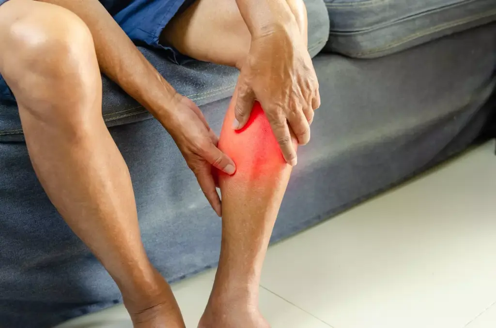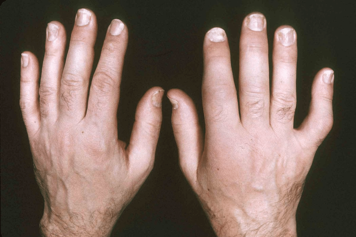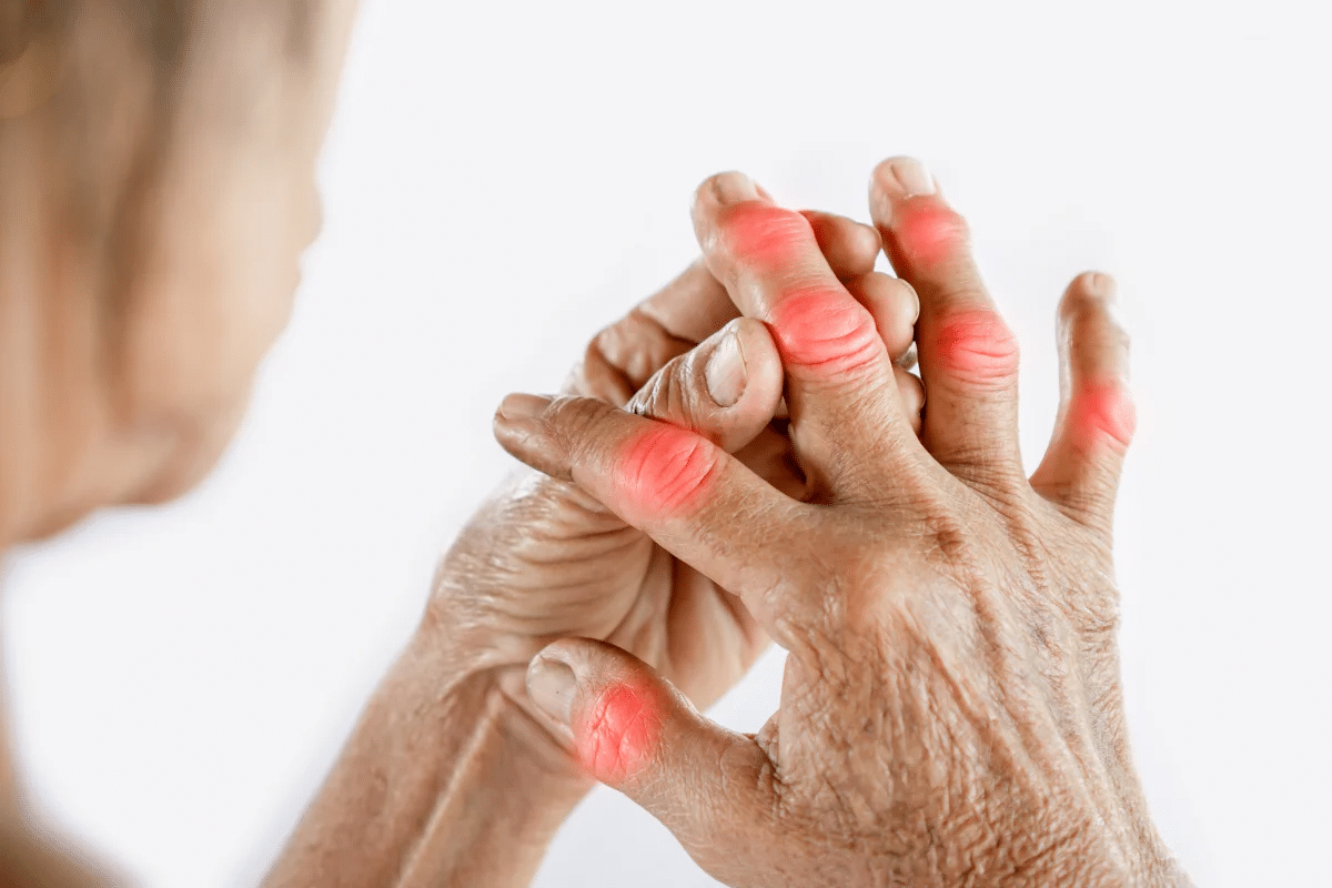
Arteriovenous malformations (AVMs) are unusual connections between arteries and veins. They skip the capillary system. AVMs in the skin and legs can cause swelling, warmth, pulsation, discoloration, and pain. Sometimes, they can lead to bleeding or ulcers.
At Liv Hospital, our teams are skilled in diagnosing and treating AVMs. We know these malformations are caused by birth defects. They affect the arterial and venous origins, creating direct connections between vessels.
We focus on patient-centered care. We address the unique needs of each person with AVMs in the skin and legs.
Key Takeaways
- AVMs are abnormal connections between arteries and veins.
- Symptoms of AVMs in the skin and legs include swelling, warmth, and pain.
- Liv Hospital offers expert diagnosis and treatment for AVMs.
- AVMs are congenital vascular malformations.
- Patient-centered care is our priority at Liv Hospital.
Understanding Arteriovenous Malformations (AVMs): Definition and Pathophysiology

Arteriovenous malformations (AVMs) are abnormal connections between arteries and veins. They skip the capillary system. This condition is a congenital disease, often caused by vascular dysplasia in early pregnancy.
Studies show AVMs are present at birth. They form during a baby’s growth in the womb.
What Does AVM Stand for in Medical Terms?
In medical terms, AVM stands for Arteriovenous Malformation. It’s a tangle of blood vessels that connects arteries to veins abnormally. This disrupts blood flow and oxygenation, leading to health issues.
The Abnormal Vascular Connection: Bypassing the Capillary System
AVMs have an abnormal vascular connection that skips the capillary system. Normally, arteries carry oxygenated blood to capillaries for oxygen exchange. In AVMs, blood goes straight from arteries to veins, causing tissue damage due to poor oxygen delivery.
Common Causes and Risk Factors of Arteriovenous Malformations
The exact causes of AVMs are not fully understood. Research suggests they are present at birth and linked to vascular dysplasia in fetal development. Genetic conditions like hereditary hemorrhagic telangiectasia (HHT) increase the risk. Other risk factors include genetic mutations and environmental influences.
To better understand the risk factors associated with AVMs, let’s examine the following table:
| Risk Factor | Description |
|---|---|
| Genetic Predisposition | Family history of AVMs or genetic syndromes like HHT |
| Vascular Dysplasia | Abnormal development of blood vessels during fetal growth |
| Genetic Mutations | Specific genetic mutations that may contribute to AVM formation |
Understanding these risk factors is key for early detection and management of AVMs. We will explore symptoms, diagnosis, and treatment options in the next sections.
The 7 Key Arteriovenous Malformation Symptoms to Recognize

It’s important to know the symptoms of arteriovenous malformations (AVMs) early. This helps in getting the right treatment. AVMs can show different signs that both patients and doctors need to be aware of.
We will look at the seven main symptoms of AVMs, focusing on those that affect the skin and legs. These signs can really affect someone’s life and need quick medical check-ups.
1. Visible Swelling and Enlargement
One key symptom of AVMs is swelling or getting bigger in the affected area. This happens because of the abnormal blood flow, which can make tissues grow more.
In the leg, this swelling is easy to see. It can also cause pain or discomfort, mainly when standing or walking.
2. Skin Discoloration: Reddish or Purple Appearance
Another common symptom is skin discoloration. The area might look reddish or purple because of the blood vessels near the skin.
This change in color can worry patients a lot. It’s because it’s visible and might upset them.
3. Increased Warmth in the Affected Area
AVMs can make the affected area feel warmer than usual. This is because of the high blood flow, which raises the temperature in the limb or area.
| Symptom | Description | Common Location |
|---|---|---|
| Visible Swelling | Enlargement due to abnormal blood flow | Leg, Skin |
| Skin Discoloration | Reddish or purple appearance | Skin |
| Increased Warmth | Elevated temperature due to high-flow AVM | Leg, Skin |
4. Pulsation or Throbbing Sensation
People with AVMs might feel a pulsation or throbbing in the affected area. This is usually because of the abnormal blood flow through the malformation.
This feeling is a big sign of an AVM, even when other symptoms are not clear.
Knowing these symptoms helps patients and doctors work together to manage AVMs well.
Skin AVMs: Characteristics and Clinical Presentation
Skin AVMs, or arteriovenous malformations on the skin, are tricky to diagnose and treat. They have a complex vascular structure. This means an abnormal connection between arteries and veins, skipping the capillary system.
This direct connection can cause various symptoms. It’s important to recognize these symptoms early for timely treatment.
Birthmark-Like Appearance of Skin AVMs
Skin AVMs often look like birthmarks, with reddish or purple spots. This is because of the abnormal vascular connection under the skin. It’s key to tell them apart from simple birthmarks for the right care.
If you notice such a mark, get a healthcare professional to check it out.
Thermal Differences: Why Skin AVMs Feel Warmer
Skin AVMs are warmer than the skin around them. This is because of the high blood flow from the arteriovenous shunt. This increased blood flow makes the area warmer.
Patients might feel this warmth, which could be a sign of an AVM. For more on AVMs, check out this resource.
Progressive Changes and Complications in Skin AVMs
Skin AVMs can change over time, leading to complications. As they grow, they can cause pain, ulcers, and bleeding. If not treated, they can lead to serious issues like disfigurement and loss of function.
Early diagnosis and treatment are key to avoid these problems. This helps improve patient outcomes.
It’s important for healthcare providers to understand skin AVMs. This knowledge helps in accurate diagnosis and effective treatment. Recognizing symptoms early can prevent serious issues, improving patients’ lives.
AV Malformation in Leg: Specific Symptoms and Complications
Leg AVMs can cause swelling, pain, and other issues. They can affect a person’s quality of life and how well they can move.
Leg Swelling and Venous Hypertension
Swelling is a common symptom of AVM in the leg. It happens because of high pressure in the veins. This is due to the malformation not using capillaries.
Venous hypertension can also cause varicose veins and other changes in blood vessels.
“The swelling and pain from leg AVMs can really limit a person’s movement and comfort,” says a vascular specialist. “It’s important to catch and treat these problems early to help manage symptoms.”
Chronic Skin Changes: Hyperpigmentation and Ulcers
AVMs in the leg can also cause skin changes. Patients might see hyperpigmentation, where the skin turns color because of bad blood flow. In serious cases, this can lead to painful ulcers that are hard to heal.
- Hyperpigmentation due to chronic venous insufficiency
- Development of venous ulcers
- Increased risk of skin infections
Advanced Complications: Gangrenous Changes and Mobility Issues
In severe cases, AVMs in the leg can cause tissue death. This is because of poor blood supply. It can lead to serious problems and might even need amputation if not treated quickly. The pain and swelling can also make it hard for patients to move around.
Dr. [Last Name] says, “It’s key to treat AVMs in the leg early to avoid these serious problems and improve health outcomes.” Treatment might include blocking off the malformation, using special treatments, or surgery, depending on the AVM’s size and location.
Diagnosing AVMs: The Essential Role of MRI and Other Imaging
Advanced imaging, like MRI, has changed how we diagnose arteriovenous malformations. Accurate diagnosis is key to finding the best treatment and improving patient care.
MRI AVM Detection: Gold Standard for Diagnosis
MRI is the top choice for diagnosing AVMs. It gives clear images of the malformation and the tissues around it. MRI AVM detection is very good at showing the complex blood vessels involved.
Complementary Diagnostic Tools: Ultrasound and Angiography
While MRI is the main tool, other methods help too. Ultrasound is used for quick checks, showing blood flow and vascular details. Angiography gives detailed views of blood vessels, helping with treatment planning.
| Imaging Modality | Key Features | Clinical Use |
|---|---|---|
| MRI | High-resolution images of soft tissues and vascular structures | Primary diagnostic tool for AVMs |
| Ultrasound | Real-time imaging, Doppler flow assessment | Initial assessment, bedside evaluation |
| Angiography | Detailed vascular anatomy, flow dynamics | Pre-interventional planning, embolization guidance |
The Diagnostic Process: What Patients Should Expect
The first step is a detailed medical history and physical check. Then, imaging tests are done to confirm the diagnosis and see how big the malformation is. Patients might need to have several tests as part of their diagnosis.
Knowing what to expect can help reduce stress. Our team is here to support patients every step of the way, from diagnosis to treatment and after.
AVM Embolization: Leading Minimally Invasive Treatment Option
Embolization has changed how we treat AVMs. AVM embolization is a main treatment. It aims to reduce venous pressure by targeting the malformation’s core.
Procedure Step by Step
The embolization process starts with a detailed check of the AVM. Then, we use a small, non-invasive method to guide a catheter to the AVM. Next, we use special agents to block the malformation’s core.
This careful process needs skill to block the AVM well and keep the patient safe.
Targeting the Nidus: Glue, Onyx, and Other Embolization Agents
Choosing the right agent is key for success. We often use glue, Onyx, and other liquid embolics. Each agent has its own benefits, chosen based on the AVM and patient’s health.
For example, Onyx is great because it doesn’t stick, making it easier to control. Picking the right agent is important for blocking the malformation’s core well.
Recovery Timeline and Follow-up Protocol After Embolization
After the procedure, patients need time to recover. They are watched closely for any problems. The recovery timeline depends on the AVM’s complexity and the patient’s health.
It’s important to follow up to see if the treatment worked. We check for any new problems or symptoms. Regular check-ups and scans are part of our care plan.
Knowing about AVM embolization helps patients understand their treatment. This includes the procedure, the agents used, and recovery.
Surgical Approaches for AVM Treatment
Surgery is a key part of treating arteriovenous malformations (AVMs). It can be a cure for some patients. We look at each case to see if surgery is right.
Indications for Surgical Intervention
Surgery is often chosen for AVMs that are easy to reach and not too big. We check the AVM’s size, where it is, and the patient’s health. This helps us decide the best treatment.
- AVMs that are easily accessible and pose a significant risk if left untreated
- Cases where embolization or other treatments are not feasible or have failed
- Patients experiencing significant symptoms or complications due to the AVM
Modern Surgical Techniques for AVM Removal
New surgical methods have made AVM treatment better. We use modern techniques to lower risks and help patients heal faster. These include:
- Microsurgical techniques that allow for precise removal of the AVM
- Intraoperative angiography to ensure complete removal of the malformation
- Minimally invasive approaches to reduce tissue damage and promote faster healing
Weighing Risks and Benefits of Surgical Treatment
Surgery to remove an AVM has risks, like bleeding during surgery and not getting all of the AVM. We think about these risks and the benefits. We look at how likely the AVM is to rupture, how bad the symptoms are, and how surgery might improve life.
Key considerations include:
- The risk of surgical complications versus the risk of AVM rupture
- The chance for symptom improvement or resolution
- The impact of surgery on the patient’s quality of life
We look at each patient’s situation carefully and use the latest surgery methods. This helps us get the best results for AVM treatment.
Rupture AVM and Bleeding Complications: Risk Assessment
AVMs are complex vascular anomalies that can rupture. This can lead to serious health issues if not managed well. The risk of rupture or bleeding depends on several factors, like the size and location of the AVM.
Factors Influencing AVM Rupture Risk
Several factors can increase the risk of an AVM rupturing. These include:
- The size of the AVM: Larger AVMs are generally considered to be at higher risk of rupture.
- Location of the AVM: AVMs located in certain areas, such as deep brain structures, may have a higher risk of rupture.
- Venous drainage pattern: The pattern of venous drainage can influence rupture risk.
- Presence of associated aneurysms: AVMs with associated aneurysms may have an increased risk of rupture.
Recognizing Signs of AVM Hemorrhage
It’s important to recognize the signs of AVM hemorrhage early. Symptoms may include:
- Sudden and severe headache, often described as “the worst headache of my life”
- Seizures
- Nausea and vomiting
- Altered mental status or loss of consciousness
- Focal neurological deficits, depending on the location of the hemorrhage
AVMs can cause bleeding in various locations, including the brain, known as a hemorrhage. This can damage surrounding tissue.
Emergency Management of Bleeding AVMs
Emergency management of bleeding AVMs involves a team effort. Initial steps include:
- Stabilizing the patient: Ensuring airway, breathing, and circulation (ABCs) are maintained.
- Imaging: Urgent imaging, typically CT or MRI, to assess the extent of hemorrhage.
- Intervention: Depending on the severity and location, interventions may include embolization, surgery, or other treatments to control bleeding and manage complications.
We stress the importance of quick and effective management. This can help minimize long-term damage and improve patient outcomes.
AVMs Beyond Skin and Legs: Arm AVM, Heart AVM, and Other Locations
Arteriovenous malformations (AVMs) can happen in many parts of the body, not just the skin and legs. They can also appear in the arms and heart. These vascular issues can be tricky to diagnose and treat, depending on where they are.
Arm AVMs: Unique Considerations and Treatment Approaches
AVMs in the arm can cause a lot of problems if not treated right. Symptoms include swelling, pain, and trouble moving. Doctors usually use a mix of treatments like embolization and surgery.
- Embolization to reduce blood flow to the AVM
- Surgical excision to remove the malformation
- Post-operative care to minimize complications
Heart AVMs: Diagnosis and Specialized Treatment
Heart AVMs are rare but very serious. Doctors use advanced imaging like MRI and angiography to find them. Treatment needs a team effort, including cardiologists and surgeons.
Key diagnostic tools for Heart AVMs include:
- Echocardiography to assess cardiac function
- Cardiac MRI to visualize the AVM
- Angiography to detail the vascular anatomy
AVMs in Other Body Locations: Brain, Spine, and Visceral Organs
AVMs can also show up in the brain, spine, and organs inside the body. Each one has its own set of challenges. For example, brain AVMs can cause bleeding or seizures, while spinal AVMs can lead to nerve problems.
Treatment strategies vary widely depending on the location and size of the AVM. For instance, brain AVMs might be treated with radiosurgery or embolization. Spinal AVMs might need surgery.
It’s important to understand the different ways AVMs can affect the body and how to treat them. Every patient is different, so a tailored approach is key.
Are AVMs Genetic? Understanding Hereditary Factors
Studies have shown that some AVMs are linked to hereditary conditions. The exact cause of AVMs is not known, but genetics seem to play a part.
Some people might inherit conditions that raise their risk for brain AVMs. For example, hereditary hemorrhagic telangiectasia (HHT) can lead to multiple AVMs in the brain, lungs, and liver.
Genetic Syndromes Associated with Arteriovenous Malformations
Several genetic syndromes increase the risk of AVMs. These include:
- Hereditary Hemorrhagic Telangiectasia (HHT): Also known as Osler-Weber-Rendu syndrome, HHT is characterized by multiple AVMs in various organs.
- Capillary Malformation-Arteriovenous Malformation (CM-AVM): This syndrome is associated with an increased risk of developing AVMs and capillary malformations.
Family Screening Recommendations for At-Risk Individuals
For those with a family history of AVMs or related syndromes, screening is key. We suggest that family members get checked by a healthcare professional to see their risk.
Screening might include imaging like MRI or ultrasound to spot any AVMs.
Genetic Counseling for Patients with AVMs
Genetic counseling is vital for AVM patients, even more so for those with a family history. Counselors can explain the risks and benefits of genetic tests. They help patients understand their condition and make informed choices about their care.
By grasping the genetic aspects of AVMs, we can better spot those at risk. This targeted approach can lead to better care and outcomes.
Living with AVMs: Long-term Management and Quality of Life
Living with AVMs is a big challenge. It needs a detailed plan to improve life quality. We focus on both medical care and how it affects daily life.
Comprehensive Pain Management Strategies
Managing pain is key when living with AVMs. We use many methods, like medicine and other therapies. Each plan is made just for the patient, based on their AVM and health.
For many, mixing treatments works best. This might include:
- Medicine to control pain and discomfort
- Physical therapy to keep moving and strong
- Other therapies like acupuncture or mindfulness
Preventing Complications Through Vigilant Monitoring
Watching AVMs closely is very important. We help patients set up regular check-ups. We use new imaging to spot any changes.
By watching closely, we can:
- Catch problems early
- Change treatment plans as needed
- Act fast to stop big problems
Support Resources and Patient Communities
AVMs affect not just the body but also the mind. We know how important support is. We help patients find groups and resources for help.
Patients get:
- Groups to share and get advice
- Info to understand their condition
- Counseling for emotional and mental health
Specialized Care at Centers Like Liv Hospital
At Liv Hospital, we aim for top-notch care for AVM patients. Our team works together to cover all AVM needs.
Choosing Liv Hospital means patients get:
- Latest treatments and tech
- Experts with lots of AVM experience
- Focus on improving life quality
Conclusion: Advances in AVM Care and Hope for the Future
Research has greatly improved how we treat AVMs. This has led to better results for those with AVMs. New studies and medical tech are helping doctors understand AVMs better. This means they can give more tailored and effective care.
Now, we’re seeing more use of less invasive treatments like AVM embolization. These methods have shown great promise in helping patients feel better. Places like Liv Hospital offer complete care, from start to finish, tailored to each patient’s needs.
The outlook for AVM care is bright. With ongoing research, we can expect even better treatments soon. Patients will likely see a better quality of life, fewer complications, and overall well-being.
FAQ
What does AVM stand for in medical terms?
AVM stands for Arteriovenous Malformation. It’s an abnormal connection between arteries and veins. This connection skips the capillary system.
What are the common symptoms of an AVM in the skin?
Symptoms include visible swelling and skin discoloration. You might see red or purple spots. The skin can also feel warm and pulsating.
How is an AVM diagnosed?
MRI is the main tool for diagnosis. It’s often used with ultrasound and angiography for more details.
What is AVM embolization?
AVM embolization is a treatment that uses agents to block the AVM. This reduces blood flow and relieves symptoms.
Are AVMs genetic?
Most AVMs are not genetic. But, some are linked to genetic syndromes. Family screening and genetic counseling might be suggested for those at risk.
What are the risks associated with AVM rupture?
Rupture can cause severe bleeding, which is an emergency. The risk depends on the AVM’s size, location, and past bleeding.
Can AVMs occur in locations other than the skin and legs?
Yes, AVMs can happen in many places. This includes the arm, heart, brain, spine, and organs. Each location has its own treatment.
How are AVMs in the leg managed?
Treatment focuses on symptoms like swelling and skin changes. It includes embolization and surgery to manage these issues.
What is the role of surgery in AVM treatment?
Surgery is used for AVMs that can be removed. It’s an option when embolization doesn’t work or has failed. Modern surgery aims to reduce risks and improve results.
How can patients with AVMs manage their condition long-term?
Long-term care includes managing pain and watching for complications. Patients should also seek support and specialized care at places like Liv Hospital.
What are the latest advances in AVM care?
New advances include better diagnostic tools and treatments like embolization. Surgery has also improved. These changes help patients live better lives.


