At Liv Hospital, we know how vital accurate heart checks are. Heart disease is a big worry worldwide. It’s key to diagnose it right for treatment. The coronary angiogram, or dye test for heart, is a major tool.
This test is quite detailed. It uses X-rays and dye to see the heart’s arteries. This helps doctors spot any problems or blockages. It’s vital for finding out if you have heart disease and what treatment you need.
We’ll walk you through nine important heart tests. We’ll compare their uses and benefits. This will help you figure out which one might be best for you.
Key Takeaways
- Coronary angiogram, or dye test, is key for finding heart disease.
- The test uses X-rays and dye to see the heart’s arteries.
- Knowing about different heart tests helps patients make better choices.
- Liv Hospital offers care that focuses on the patient and is innovative.
- We’ll explain nine heart tests, including how dye tests and echocardiograms compare.
The Importance of Cardiac Diagnostic Testing
Heart disease is a big problem worldwide. Cardiac diagnostic testing is key to solving it. These tests check how well the heart works.
Why Regular Heart Monitoring Matters
Keeping an eye on your heart is vital. It helps find problems early. This way, doctors can act fast and avoid big issues.
Regular heart checks have many benefits. They help spot heart disease early. They also keep an eye on heart conditions and see if treatments work.
Risk Factors That Necessitate Heart Testing
Some things make you need heart tests more. These include family heart disease history, high blood pressure, diabetes, and smoking. If you have these, you should get heart tests often.
A wide range of heart tests can help with heart disease. These include:
- Echocardiograms
- Stress tests
- Electrocardiograms (ECG)
- Cardiac catheterization
The Evolution of Cardiac Diagnostics
Cardiac diagnostics have come a long way. New tech makes tests better and less scary. Now, we have things like cardiac MRI and CT angiography.
These new tools help doctors give better care. They can make treatment plans that really work. And, new heart test units make tests more accurate.
Dye Test for Heart: Understanding Coronary Angiography
Coronary angiography, also known as a dye test for the heart, is a key test. It helps see the coronary arteries.
This test is used to find and check how bad coronary artery disease is. It works by putting a dye into the arteries. This makes them show up on X-ray images.
What Happens During a Cardiac Angiogram
A cardiac angiogram starts with a catheter going through an artery in the leg or arm. It then reaches the coronary arteries. Next, a dye is injected, and X-rays are taken to see the arteries.
This test happens in a special lab and takes 30-60 minutes. Patients are awake but a bit sleepy to feel less pain.
The Role of Contrast Dye in Visualization
The dye used in coronary angiography is key for seeing the arteries. It makes the arteries stand out on X-ray images. This helps doctors spot any problems.
The dye is made of iodine and is safe for most people. But, those with kidney issues or iodine allergies need special care before the test.
Detecting Blockages and Arterial Narrowing
Coronary angiography is great at finding blockages and narrow spots in arteries. Doctors can see how bad the disease is. This helps them decide the best treatment.
The info from this test is very important. It tells doctors if they need to do angioplasty, stenting, or other treatments to fix the heart’s blood flow.
| Procedure | Description | Benefits |
|---|---|---|
| Coronary Angiography | Injection of contrast dye into coronary arteries to visualize them on X-ray | Accurate diagnosis of coronary artery disease, assessment of blockages and narrowing |
| Cardiac Catheterization | Insertion of a catheter into an artery to guide it to the coronary arteries | Minimally invasive, allows for simultaneous treatment of blockages |
Echocardiogram: Assessing Heart Structure and Function
The echocardiogram is a non-invasive test that gives detailed insights into the heart. It uses ultrasound technology to check the heart without dye or invasive methods.
Ultrasound Technology for Heart Imaging
Echocardiograms use high-frequency sound waves to make detailed heart images. This non-invasive technique lets doctors see the heart’s structure and function in real-time. It provides important info about the heart’s health.
Types of Echocardiograms
There are several echocardiogram types, each for a specific purpose:
- Transthoracic Echocardiogram (TTE): The most common, where the probe is on the chest.
- Transesophageal Echocardiogram (TEE): A probe goes through the esophagus for closer heart images.
- Stress Echocardiogram: Done before and after stressing the heart, usually through exercise or meds, to check heart function under stress.
Insights into Heart Valves and Pumping Ability
Echocardiograms show key info about heart valves and pumping. They can spot valve problems and check the heart’s pumping. This info is key for treating heart conditions.
Understanding echocardiograms helps patients and doctors make better heart care choices. Whether comparing echocardiogram vs angiogram or picking the right test, echocardiograms are key in heart diagnostics.
Angiography vs Echocardiogram: Comparative Analysis
Two important tests are often used to diagnose heart conditions: angiography and echocardiogram. Both are key tools, but they offer different views into heart health.
Invasive vs Non-Invasive Approaches
Angiography is an invasive test. It involves putting a catheter into a blood vessel to inject dye. This dye helps show the inside of the arteries on X-rays, spotting blockages.
Echocardiography, on the other hand, is non-invasive. It uses ultrasound to create heart images. This test is safer and more comfortable for patients.
Diagnostic Capabilities and Limitations
Angiography gives detailed views of the arteries. It’s great for finding blockages and deciding if more treatments are needed. But, it’s risky because it’s invasive.
Echocardiography checks the heart’s structure and function. It’s good for seeing how the heart works overall. But, it might not show as much about artery blockages as angiography does.
Comparison of Diagnostic Tests
| Test | Invasiveness | Primary Use | Key Benefits |
|---|---|---|---|
| Angiography | Invasive | Coronary artery imaging | Detailed blockage assessment |
| Echocardiography | Non-invasive | Heart structure and function | Comprehensive heart health assessment |
When Doctors Choose One Test Over the Other
Doctors pick between angiography and echocardiography based on the patient’s needs. For example, if someone might have big artery problems, angiography is often chosen. It shows artery details well.
In summary, both angiography and echocardiography are important for diagnosing heart issues. Knowing their differences helps both patients and doctors make better choices for heart care.
Comprehensive List of Heart Tests and Their Names
Cardiac diagnostic testing includes many procedures, each with its own purpose. It’s important for both doctors and patients to know about these tests. This helps them understand heart health better.
Standard Diagnostic Procedures
Standard tests are the base of cardiac diagnostics. They include:
- Electrocardiogram (ECG): A test that measures the heart’s electrical activity.
- Echocardiogram: An ultrasound test that shows the heart’s structure and function.
- Stress Test: A test that checks how the heart works under stress.
Advanced Imaging Techniques
Advanced tests give detailed views of the heart. Some of these are:
- Cardiac MRI: A magnetic resonance imaging test that shows detailed heart images.
- CT Angiography: A computed tomography test that shows the coronary arteries.
- Coronary Angiography: An invasive test that uses dye to see blockages in the coronary arteries.
Specialized Cardiac Assessments
Specialized tests check specific heart health aspects. These include:
- Holter Monitor: A portable device that records the heart’s electrical activity for 24 to 48 hours.
- Event Recorder: A device that records the heart’s electrical activity for longer than a Holter monitor.
- Cardiac Catheterization: A procedure that involves inserting a catheter into the heart to diagnose and treat certain conditions.
These heart tests are key in diagnosing and managing heart conditions. Knowing about the different tests helps patients navigate their cardiac care journey.
Electrocardiogram (ECG): Tracking Heart’s Electrical Activity
An ECG is a non-invasive test that records the heart’s electrical activity. It helps doctors diagnose heart conditions. This tool is key to understanding how the heart works electrically.
Recording Heart Rhythms
An ECG attaches electrodes to the skin to detect heart signals. These signals are then shown on a monitor or printed out. This gives a clear view of the heart’s rhythm and electrical activity.
To do this, we place electrodes on the chest, arms, and legs. The ECG machine records the heart’s electrical activity from different angles. This info is key for spotting irregular heartbeats or arrhythmias.
Interpreting ECG Patterns and Abnormalities
Reading an ECG needs skill, as it involves analyzing patterns and spotting abnormalities. We look at the P wave, QRS complex, and T wave to check the heart’s electrical activity.
Abnormalities can show different heart conditions, like arrhythmias or coronary artery disease. For example, an abnormal QRS complex might mean a blockage in the heart’s electrical system.
| ECG Component | Normal Finding | Abnormal Finding |
|---|---|---|
| P Wave | Upright in lead II | Inverted or absent |
| QRS Complex | Narrow (<120 ms) | Wide (≥120 ms) |
| T Wave | Upright in lead II | Inverted |
When an ECG Is the First-Line Diagnostic Tool
An ECG is often the first test for heart disease symptoms like chest pain. It’s quick, non-invasive, and affordable. It gives vital info about the heart’s electrical activity.
In emergencies, an ECG is very useful. It quickly shows the heart’s condition and helps make urgent treatment decisions.
Understanding what an ECG shows helps us make the right decisions for heart care. This ensures patients get the best treatment for their heart health.
Stress Tests: Evaluating Cardiac Performance Under Exertion
Stress tests are key for checking heart health. They see how the heart works when it’s under stress. This is usually through exercise or medicine.
Exercise vs Pharmacological Stress Testing
There are two main stress tests: exercise and pharmacological. Exercise stress tests use physical activity, like on a treadmill. It’s the preferred method because it’s more natural.
Pharmacological stress tests use medicine to mimic exercise. They’re for those who can’t exercise due to health issues.
The choice between tests depends on the patient’s health and ability to exercise. For example, those with muscle problems or trouble reaching heart rate goals might need pharmacological tests.
Monitoring Parameters During Testing
During a stress test, several things are watched closely. These include:
- Heart rate and rhythm
- Blood pressure
- Electrocardiogram (ECG) readings
- Symptoms such as chest pain or shortness of breath
Watching these helps doctors see how the heart handles stress. It helps spot any problems.
Identifying Coronary Artery Disease and Functional Capacity
Stress tests are great for finding coronary artery disease (CAD) and checking how well you can function. They show how the heart handles stress. This helps find blockages in the heart’s arteries.
They also show how well you can do daily tasks and exercise. This info is key for creating good exercise plans for heart patients.
In short, stress tests are very important for checking heart health. Knowing about the different tests and what’s watched during them helps doctors care for patients better.
Advanced Cardiac Imaging: CT Angiography and MRI
Advanced cardiac imaging, like CT angiography and MRI, has changed cardiology a lot. They give detailed pictures of the heart’s inside and how it works. This helps doctors diagnose and treat heart diseases better.
Dye Usage in CT Angiography Compared to Traditional Angiograms
CT angiography uses dye to see the heart’s arteries, just like old angiograms. But, it’s less invasive. It uses a CT scanner to take detailed pictures after dye is given. This gives a clearer view of the arteries and nearby areas.
Key differences in dye usage:
- CT angiography needs less dye than traditional angiography.
- The dye goes through a vein, making it less invasive.
- CT scans show cross-sections, giving a detailed look at the heart and its vessels.
Cardiac MRI Applications and Advantages
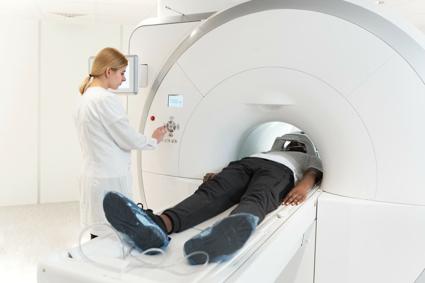
Cardiac MRI is a non-invasive way to see the heart’s inside and how it works. It’s great for checking the heart’s structure, valve work, and how well the heart muscle is doing.
The advantages of cardiac MRI include:
- No radiation, making it safer for repeated checks.
- It’s good at showing soft tissues, helping diagnose heart issues.
- It can check heart function and blood flow without radiation.
Radiation Considerations and Safety Profiles
Radiation is a big deal in heart imaging. CT angiography uses radiation, but newer scanners use less. MRI doesn’t use radiation, which is better for some patients.
| Imaging Modality | Radiation Exposure | Safety Profile |
|---|---|---|
| CT Angiography | Yes | Generally safe, but radiation dose should be minimized. |
| Cardiac MRI | No | Safe for patients without MRI contraindications. |
It’s important to know the good and bad of these imaging methods. This helps doctors pick the best tool for each patient.
What Do Doctors Use to Check Your Heart: Decision Factors
Doctors pick from many heart tests based on symptoms and risk. The right test depends on the patient’s history, symptoms, and health. This careful choice helps find heart problems accurately.
Symptom-Based Test Selection
The test type often matches the symptoms. For chest pain or shortness of breath, a stress test is common. It checks how the heart works when active. For suspected valve problems, an echocardiogram is used to look at the heart’s structure.
| Symptom | Common Test(s) Ordered | Purpose |
|---|---|---|
| Chest Pain | Stress Test, Coronary Angiography | Evaluate heart function under stress, detect blockages |
| Shortness of Breath | Echocardiogram, Electrocardiogram (ECG) | Assess heart structure and function, evaluate electrical activity |
| Palpitations | Electrocardiogram (ECG), Holter Monitor | Monitor heart rhythm, detect arrhythmias |
Risk Profile Considerations
A patient’s risk level also guides test choice. Those with heart disease history, diabetes, or high blood pressure are at higher risk. They might get tests like CT angiography or cardiac MRI.
Risk factors that may need detailed testing include:
- Family history of heart disease
- Diabetes
- High blood pressure
- High cholesterol
- Smoking history
Sequential Testing Strategies
Doctors might start with a simple test and add more tests as needed. This method reduces risk and improves accuracy.
Healthcare providers choose the best test for each patient. This ensures a correct diagnosis and the right treatment plan.
Conclusion: Navigating Your Cardiac Testing Journey
It’s key to know about different heart tests and what they do. We’ve looked at tests like the Electrocardiogram (EKG/ECG), Echocardiogram (Echo), Stress Test, Cardiac Catheterisation, and Angioplasty. Each test helps check your heart’s health and find any problems.
Learning about these tests helps you understand your cardiac journey better. For more info on heart health and diagnostics, check out Apollo 247 Health Topics. This site offers detailed advice on cardiology and cardiologists, aiding your heart health choices.
Knowing your options and talking to your doctor is vital for heart health. Even though cardiac testing can seem tough, with the right info and support, you can achieve great results for your heart.
What is a dye test for the heart, and how does it work?
A dye test for the heart, also known as coronary angiography, is a way to see the heart’s arteries. It uses a contrast dye to spot blockages or narrowing. The dye is injected through a catheter, and X-ray images show the dye’s flow.
What is the difference between angiography and echocardiography?
Angiography is an invasive test that uses dye to see the heart’s arteries. Echocardiography is non-invasive, using ultrasound to check the heart’s structure and function. Angiography finds blockages, while echocardiography looks at heart valves and pumping.
What are the different types of heart tests available?
There are many heart tests, like electrocardiograms (ECG), stress tests, CT angiography, echocardiography, and MRI. Each test gives unique insights into heart health and helps diagnose different conditions.
How does an ECG work, and what does it measure?
An ECG records the heart’s electrical activity. It checks the heart’s rhythm and finds any issues. It’s a non-invasive test often used first to check heart health.
What is a stress test, and how is it used to diagnose coronary artery disease?
A stress test checks how the heart works when exerted, usually through exercise or medicine. It looks at heart rate, blood pressure, and ECG readings to spot coronary artery disease and check how well the heart functions.
How does CT angiography use dye, and what are its advantages?
CT angiography uses dye to see the heart’s arteries, like traditional angiography. But it’s non-invasive, giving detailed images of the heart and its blood vessels. It’s great for spotting blockages and narrowing because of its high-resolution images.
What are the benefits and limitations of cardiac MRI?
Cardiac MRI is a non-invasive test that gives detailed images of the heart and its blood vessels. It’s good for checking heart function and structure. But, it might not work for people with certain metal implants or those who are claustrophobic.
How do doctors choose the right heart test for a patient?
Doctors look at symptoms, risk profile, and medical history to pick a heart test. They might use more than one test to diagnose and monitor heart conditions. The choice depends on what’s best for each patient.
What are the risk factors that necessitate heart testing?
High blood pressure, high cholesterol, diabetes, smoking, and family history of heart disease are risk factors that might need heart testing. Regular monitoring is key for those with these risks to catch and manage heart conditions early.
How often should I have my heart tested?
How often to test your heart depends on your risk factors and medical history. It’s best to talk to your doctor to figure out the right testing schedule for you.
References
- Coronary angiography. Retrieved from: https://www.mountsinai.org/health-library/tests/coronary-angiography
- Coronary angiogram. Retrieved from: https://www.betterhealth.vic.gov.au/health/conditionsandtreatments/coronary-angiogram
- Coronary angiography. Retrieved from: https://www.pennmedicine.org/treatments/coronary-angiography
- CT angiography. Retrieved from: https://www.radiologyinfo.org/en/info/angioct?PdfExport=1


