Knowing about vascular health is key for good medical care. At Liv Hospital, we focus on advanced care and patient needs. This ensures we diagnose and treat well.
An arteriogram is a tool used to find vascular problems. New tech in arteriograms has made diagnosis and treatment better.
We’ll look into the definitions, procedures, and key differences of arteriograms. This will help you understand this important diagnostic tool.
Key Takeaways
- Arteriograms are key for finding vascular issues.
- New tech has made diagnosis more accurate.
- Liv Hospital focuses on patient care and advanced methods.
- Knowing about arteriograms helps make smart health choices.
- Arteriograms are essential for spotting vascular health problems.
Arteriogram Definition: Understanding This Vital Diagnostic Test
An arteriogram is a key medical imaging method. It helps diagnose and treat artery diseases. The arteriogram definition involves detailed images of arteries. This is vital for checking vascular health.
Medical Purpose and Clinical Applications
Arteriograms show the inside of arteries. Doctors use them to spot blockages, aneurysms, or other issues. The vascular arteriogram is great for checking blood vessel health all over the body.
Arteriograms are used in many ways:
- Diagnosing coronary artery disease
- Evaluating peripheral vascular disease
- Planning surgical interventions or angioplasty
- Monitoring the effectiveness of vascular treatments
| Clinical Application | Description |
|---|---|
| Coronary Artery Disease | Arteriograms help visualize blockages in heart arteries. |
| Peripheral Vascular Disease | Arteriograms assess blood flow in limb arteries. |
| Surgical Planning | Detailed arteriogram images guide surgeons during procedures. |
Historical Development of Arterial Imaging
The history of arteriography started in the early 20th century. The first attempts used X-ray technology to see arteries. Over time, better catheter tech and imaging have made arteriograms safer and clearer.
The CathWorks FFRangio System is a recent big step. It adds vital info to routine angiograms. This marks a big leap in vascular imaging.
Knowing about the arteriography definition and its history shows its importance in medicine. As tech keeps improving, arteriograms will keep being key in treating vascular diseases. This offers hope for better patient care.
The Science Behind Arteriograms: How They Visualize Arteries
Arteriograms use contrast dye and X-ray technology to see arteries. They are key in modern medicine. Doctors use them to check artery health.
To get how arteriograms work, we must look at contrast dye and X-ray tech.
Contrast Dye Mechanisms and Properties
Contrast dye makes body structures visible in medical images. In arteriograms, it’s injected into the blood to show arteries.
Its useful properties include:
- High atomic number elements (like iodine or barium) that absorb X-rays, making it visible on X-ray images.
- Low toxicity to keep reactions down.
- Right viscosity to move through blood vessels.
Researchers keep improving contrast dye. For example, new sensors help monitor blood leakage after repairs, as seen in studies from Hanyang University.
“The development of novel sensors for continuous endoleak monitoring represents a significant advancement in the field of vascular diagnostics.”
X-ray Imaging Technology in Vascular Visualization
X-ray tech is key for arteriograms. It lets us see arteries. Modern systems give clear images with little radiation.
The process is:
- X-ray emission: X-rays come from an X-ray tube.
- X-ray absorption: The dye absorbs X-rays, showing arteries against tissues.
- Image capture: Images are captured and made clearer.
| Technology | Description | Benefits |
|---|---|---|
| Digital Subtraction Angiography (DSA) | A technique that subtracts images before and after dye injection. | Shows blood vessels better. |
| 3D Reconstruction | Software makes 3D images from 2D data. | Helps understand complex vascular anatomy. |
Arteriograms combine advanced contrast dye and X-ray tech. They give doctors vital info for vascular medicine.
The Complete Arteriogram Procedure: A Complete Guide
An arteriogram is a detailed imaging test that shows the arteries. Knowing what to expect can make you feel less anxious. At Liv Hospital, we use the latest methods for vascular imaging. This ensures our patients get the best care.
Pre-Procedure Preparation and Requirements
Before an arteriogram, several steps are taken to make the procedure smooth. Patients should tell their doctor about any medications, like blood thinners. It’s also important to mention any allergies, like to contrast dye used during the procedure.
- Stop eating and drinking for a set time before the procedure.
- Arrange for someone to drive you home after the procedure.
- Wear comfortable, loose-fitting clothing.
Our medical team will give you detailed instructions on how to prepare. This includes any changes to your medication.
During the Procedure: What to Expect
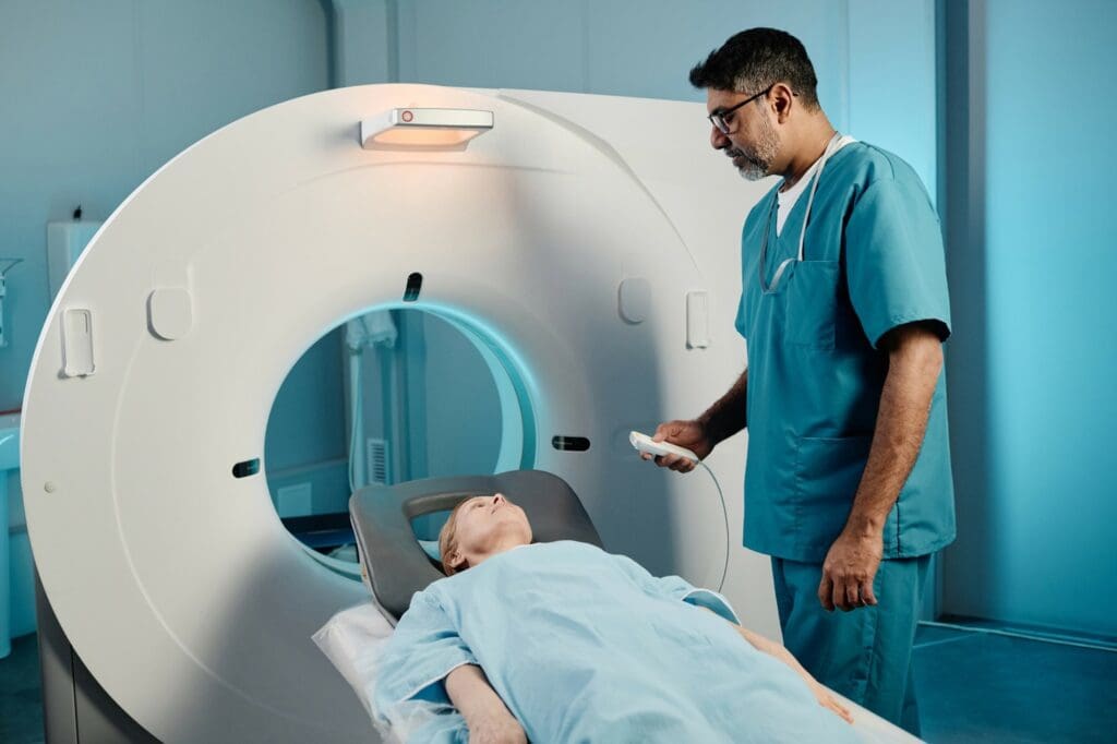
During the arteriogram, you’ll be on an X-ray table. The area where the catheter will be inserted will be cleaned and numbed. The procedure is usually done under local anesthesia to reduce discomfort.
The catheter is then guided to the area of interest using X-ray imaging. Once in place, a contrast dye is injected to see the arteries. You might feel a slight warmth or flushing as the dye is given.
| Procedure Step | Description | Patient Experience |
|---|---|---|
| Preparation | Local anesthesia, catheter preparation | Mild discomfort during anesthesia administration |
| Catheter Insertion | Guiding the catheter to the area of interest | Minimal discomfort due to local anesthesia |
| Contrast Dye Injection | Injecting dye to visualize arteries | Sensation of warmth or flushing |
Post-Procedure Care and Recovery Timeline
After the procedure, the catheter is removed, and pressure is applied to the site to prevent bleeding. You will be monitored for a period to check for any immediate complications. Most patients can go back to normal activities in a few days.
Recovery tips include:
- Avoid heavy lifting and strenuous activities for a few days.
- Keep the insertion site clean and dry.
- Follow the medication regimen as advised by your doctor.
At Liv Hospital, we are committed to providing complete care throughout your treatment. We ensure a smooth and successful recovery from your arteriogram procedure.
Types of Arteriograms and Their Specialized Applications
Arteriograms come in many types, each for a specific use. They help doctors diagnose and treat different vascular conditions. Thanks to new technology, these tests are key in many medical areas.
Cardiac Arteriograms for Heart Vessel Assessment
Cardiac arteriograms, or coronary angiograms, look at the heart’s blood supply. They’re vital for spotting coronary artery disease, a big heart attack cause. “The CathWorks FFRangio System has changed how we treat heart disease,” showing tech’s role in heart care.
During the test, a dye is put into the heart’s arteries. This lets doctors see blockages clearly. Knowing this helps decide the best treatment, like opening blocked arteries.
Cerebral Arteriograms for Brain Vessel Imaging
Cerebral arteriograms check the brain’s blood vessels. They help find problems like aneurysms and stenosis. A dye is injected into brain arteries to get clear X-ray images.
These tests are key for planning brain surgeries or treatments. They help doctors avoid risks during these complex procedures.
Renal Arteriograms for Kidney Vessel Evaluation
Renal arteriograms look at the kidneys’ blood vessels. They help spot issues like stenosis and aneurysms. A dye is used to see the kidney’s blood supply.
This info is vital for treatments like angioplasty. It helps improve blood flow to the kidneys, which is key for those with high blood pressure or kidney disease.
Extremity Arteriograms for Limb Circulation Assessment
Extremity arteriograms check the blood flow in arms or legs. They help find problems like PAD. Doctors can see blockages or narrowing in the arteries.
These tests are important for planning treatments. They help restore blood flow and improve symptoms in affected limbs.
In summary, arteriograms have many types, each for a specific use. They give doctors the info they need to make treatment plans. As tech gets better, these tests will become even more accurate and helpful.
Arteriogram vs. Arteriography: Understanding Terminology Differences
In medical diagnostics, arteriogram and arteriography are not just words. They are about different parts of vascular imaging.
Defining the Image vs. the Technique
An arteriogram is the image of arteries from a medical test. Arteriography is the method to make this image. It’s like saying arteriography is how you get an arteriogram.
Arteriography uses contrast dye and X-ray to see the arteries. This helps doctors find and check vascular problems.
Clinical Usage and Documentation Contexts
In clinics, arteriogram and arteriography have different uses. An arteriogram is what you see in patient records. Arteriography talks about the procedure, its risks, and benefits.
Knowing the difference helps doctors talk clearly. It makes sure patient care and plans are right.
Even though they’re different, arteriogram and arteriography are linked. Better arteriography means better arteriograms. This helps doctors diagnose better.
Medical Conditions Diagnosed Through Arterial Imaging
Healthcare experts use arterial imaging to spot and check on many important vascular issues. Arteriograms are key in today’s medicine. They help see the arteries clearly and find different vascular diseases.
Coronary Artery Disease Detection and Assessment
Coronary artery disease (CAD) is a big cause of sickness and death. Arteriograms help find CAD by showing the coronary arteries in detail. This lets doctors see how blocked or narrow they are.
Arteriograms help in CAD detection by:
- Seeing how narrow or blocked the coronary arteries are
- Figuring out how bad the blockages are
- Helping decide on treatments like angioplasty and stenting
| Condition | Description | Arteriogram Findings |
|---|---|---|
| Coronary Artery Disease | Narrowing or blockage of coronary arteries | Stenosis, occlusion, or plaque buildup in coronary arteries |
| Peripheral Vascular Disease | Narrowing or blockage of peripheral arteries | Stenosis or occlusion in peripheral arteries |
| Aneurysm | Abnormal dilation of an artery | Dilated arterial segment, possible thrombus formation |
Peripheral Vascular Disease Evaluation
Peripheral vascular disease (PVD) is when blood vessels outside the heart get narrow or blocked. Arteriograms help see how bad PVD is. This helps doctors decide on treatments.
Arteriograms offer many benefits in PVD evaluation:
- They accurately show how narrow or blocked the blood vessels are
- They help find if there are other paths for blood to flow
- They help plan for treatments like angioplasty or surgery
Identifying Aneurysms and Vascular Malformations
Arteriograms are also key in finding aneurysms and vascular malformations. These are abnormal blood vessel formations that can be dangerous if not treated.
Arteriograms help by:
- Showing exactly where and how big an aneurysm is
- Looking at how blood flows in the aneurysm
- Helping plan for treatments like endovascular or surgery
Arteriograms give detailed views of blood vessels. This helps doctors diagnose and treat many vascular conditions well.
Risks, Complications, and Safety Protocols in Modern Arteriography
Modern arteriography has made big strides in reducing risks. Yet, it’s key to know and tackle the possible complications. As we keep improving, knowing the risks and following safety steps is essential.
Common Side Effects and Their Management
Arteriography is usually safe, but some side effects can happen. These include:
- Bruising or discomfort at the catheter insertion site
- Allergic reactions to the contrast dye
- Mild to moderate pain during or after the procedure
We handle these side effects by closely watching patients and providing good care after the procedure. For example, we use antihistamines or corticosteroids to fight allergic reactions.
Potential Serious Complications and Prevention
Though rare, serious issues can happen during or after arteriography. These include:
- Vascular injury or bleeding
- Stroke or transient ischemic attack
- Renal complications due to contrast-induced nephropathy
To avoid these problems, we use careful techniques, the right catheter sizes, and check patients’ kidney health before dye use. New research on sensors for monitoring during endovascular repair shows promise, as seen in BMC Cardiovascular Disorders.
Advanced Safety Measures and Monitoring
We use top-notch monitoring during arteriography to boost safety. This includes live imaging, constant vital sign checks, and emergency plans. These steps help lower the chance of issues.
We also focus on teaching patients and preparing them before the procedure. Knowing the risks and being ready can make arteriography safer for everyone.
Recent Advances in Arteriogram Technology and Techniques
Arteriogram technology has changed a lot, making vascular diagnosis better and safer. Healthcare pros now have more precise tools to check and treat blood vessel issues. We see big steps forward in catheter tech, digital images, and safety during procedures.
Evolution of Catheter Technology and Design
Catheters for arteriography have gotten a lot better. They’re now more flexible, making it easier to move through blood vessels. This makes procedures safer and more comfortable for patients.
- Improved Materials: New materials make catheters last longer and work better.
- Advanced Tip Designs: Catheter tips are now better for moving around and less likely to hurt blood vessels.
- Hydrophilic Coatings: Many catheters now have special coatings that help them slide through blood vessels smoothly.
Digital Imaging Enhancements and 3D Reconstruction
Digital imaging has made huge leaps, making arteriograms much better. One big step is being able to see 3D images of blood vessels.
Key Benefits of Digital Imaging Enhancements:
- Images are clearer, helping doctors make better diagnoses.
- 3D views help plan complex procedures better.
- Everyone gets less radiation, making procedures safer.
Research Findings on Improved Accuracy and Safety
Studies show these new techs make arteriograms more accurate and safe. Advanced catheters and digital images lead to better diagnoses and fewer problems.
A study at TCT 2025 showed 3D arteriography improves results in tough vascular cases.
Arteriogram tech keeps getting better, with more research on safety and accuracy. As these advancements grow, patients will get even better care for their vascular needs.
Patient Preparation and Guidelines for Undergoing an Arteriogram
Getting ready for an arteriogram is key to getting good results and feeling better fast. At Liv Hospital, we help international patients get ready for arteriograms. We guide them through every step.
Pre-Procedure Instructions and Considerations
Before your arteriogram, follow important steps to stay safe and get the best results. These steps include:
- Tell your doctor about any medicines you take, like blood thinners.
- Let them know if you have any allergies, like to contrast dye.
- Don’t eat or drink for a while before the test.
- Make sure someone can drive you home after.
It’s very important to listen to your doctor’s advice. This helps avoid risks and get the best results.
| Pre-Procedure Requirement | Description |
|---|---|
| Medication Disclosure | Tell your doctor about all medicines, including supplements and blood thinners. |
| Allergy Disclosure | Report any allergies, like to iodine or contrast dye. |
| Fasting | Avoid eating and drinking for a while before the test. |
Essential Questions to Discuss With Your Doctor
Talk openly with your doctor before the arteriogram. Ask about the procedure and your health. Some questions to ask include:
“What are the risks associated with this procedure, and how will they be managed?”
Your doctor can give you advice tailored to you. They can also answer any worries you have.
- What are the expected outcomes of the arteriogram?
- Are there any other tests I could have instead?
- How will you tell me the results?
What to Expect During Recovery and Follow-up
After the arteriogram, you’ll be watched for any problems. We tell patients to:
- Rest for the rest of the day.
- Watch the puncture site for bleeding or infection.
- Follow any medicine or follow-up instructions from your team.
At Liv Hospital, we aim to give top-notch healthcare to international patients. Knowing what to expect and how to prepare helps you feel confident during your arteriogram.
Alternative Diagnostic Procedures to Arterial Imaging
New ways to check vascular health are being developed. These new methods are safer and more advanced. They help doctors diagnose vascular conditions better.
Non-Invasive Vascular Assessment Options
Non-invasive tests are changing how we check blood vessels. Ultrasound technology uses sound waves to see inside without harm. It’s great for watching how blood vessels change over time.
Magnetic Resonance Angiography (MRA) uses magnetic fields and radio waves. It gives clear pictures of blood vessels. MRA is good for seeing complex blood vessel structures and planning surgeries.
Comparative Analysis of Effectiveness and Applications
Looking at different tests, we see their strengths and weaknesses. Arteriograms show detailed images but are invasive and risky.
- Ultrasound and MRA are safer but might not show as much detail as arteriograms.
- The right test depends on the condition and the patient’s health.
- Computed Tomography Angiography (CTA) has gotten better, making it a good choice for some cases.
Research at places like Hanyang University is making big strides. They’re working on new sensors for checking blood vessels. These advancements give doctors more tools for precise and less invasive tests.
As we learn more about vascular diseases, new tests will play a bigger role. By understanding the good and bad of these new tools, we can give patients the best care.
Conclusion: The Evolving Role of Arteriograms in Vascular Medicine
Arteriograms are key in diagnosing and treating vascular conditions. They are used a lot in modern medicine. We’ve looked at what arteriograms are, how they work, and why they’re important for seeing blood vessels.
New technology in arteriograms has made them more accurate and safer for patients. As vascular medicine grows, arteriograms will keep being a vital tool. They help doctors understand and manage many vascular diseases.
It’s important for both patients and doctors to know about arteriograms. They help us find better ways to treat patients. This leads to better health outcomes for everyone.
As medical tech gets better, arteriograms will too. We expect them to help us diagnose and treat vascular diseases even better. Arteriograms are essential for top-notch patient care.
References
- Diagnostic Arteriogram or Aortogram with or without intervention. Retrieved from: https://navicenthealth.org/service-center/atrium-health-navicent-heart-vascular-care/diagnostic-arteriogram-or-aortogram-with-or-without-intervention
- Arteriography. Retrieved from: https://www.cancer.gov/publications/dictionaries/cancer-terms/def/arteriography
- What is an arteriogram? Retrieved from: https://www.chop.edu/treatments/arteriogram


