Last Updated on November 27, 2025 by Bilal Hasdemir

Knowing about the aortic arch arteries is key to understanding how blood gets to the head, neck, and arms. At Liv Hospital, we focus on our patients and the health of their blood vessels.
The aortic arch has three main branches: the brachiocephalic trunk, left common carotid artery, and left subclavian artery. These branches are important for bringing oxygen-rich blood to the upper body.
We know how important the anatomy of aortic arch arteries is for diagnosing and treating blood vessel problems. Our goal is to offer top-notch care for our patients’ vascular health.
The aorta is the biggest artery and key to the heart’s blood flow. It starts at the left ventricle and goes to the belly, splitting into the common iliac arteries. This path is vital for bringing oxygen-rich blood to the body.
The heart and aorta are closely linked. The aorta comes from the left ventricle, the heart’s main pump. The aortic valve lets blood flow into the aorta but stops it from going back. The aortic root is the start of the aorta, where the heart’s blood supply begins.
Knowing how the heart and aorta connect is key for health. Problems here can cause serious issues, like aortic valve problems and heart disease.
The aorta has several parts as it travels through the body. These are:
Each part of the aorta has branches for different body areas. The aortic arch has three main branches. These supply blood to the head, neck, and arms.
Knowing the anatomy of the aortic arch arteries is key for treating heart problems. These arteries are vital for getting oxygen-rich blood to the body. Their complex design is essential for blood flow.
We will look at where these arteries are, their structure, and how they develop.
The aortic arch is in the chest. It splits into three main branches. These branches supply blood to the head and arms. The branches are the brachiocephalic trunk, the left common carotid artery, and the left subclavian artery.
These arteries are near important structures like the trachea, esophagus, and spine.
The aortic arch arteries have three layers. The innermost layer is the tunica intima, lined with endothelial cells. The middle layer, the tunica media, has smooth muscle cells and elastic fibers. These provide flexibility and contraction.
The outermost layer, the tunica externa, is made of connective tissue. It supports the artery.
| Layer | Composition | Function |
|---|---|---|
| Tunica Intima | Endothelial cells | Lining the lumen, regulating blood flow |
| Tunica Media | Smooth muscle cells, elastic fibers | Providing elasticity and contractility |
| Tunica Externa | Connective tissue | Supporting the artery |
For more details on arterial walls, check out NCBI’s resource on the topic.
The aortic arch forms from the left fourth pharyngeal arch in the embryo. This process creates the aortic sac, which becomes the aortic arch and its branches. Knowing how the aortic arch develops helps us understand its adult structure and variations.
The development of the aortic arch is a complex process. It involves many genetic and environmental factors. Problems during this time can cause congenital anomalies, like a bovine arch or unusual branching patterns.
The aortic arch arteries branch in a typical way in most people. This pattern shows three main branches coming from the aortic arch.
Studies show that about 75 percent of people have this three-branch pattern. Knowing this is key for doctors and students of anatomy.
This branching pattern has been around for a long time, shared by many mammals. It helps blood get to the brain, arms, and body efficiently.
While humans mostly have three branches, other mammals can have different numbers. This shows how animals adapt to their environments and needs.
| Species | Aortic Arch Branching Pattern |
|---|---|
| Human | Tripartite (3 branches) |
| Canine | Bipartite (2 branches) |
| Feline | Variable, often bipartite |
In summary, the three-branch pattern is common in humans and has deep evolutionary roots. Studying this, along with variations in other mammals, helps us understand the complex heart and blood system.
The brachiocephalic trunk is the first big branch of the aortic arch. It’s key for blood flow to the right arm, head, and neck. It starts the blood supply to these areas.
The brachiocephalic trunk goes up and back to the right, in front of the trachea. It’s near the left brachiocephalic vein in front and the trachea and thymus gland in back. Knowing its path helps in treating vascular issues.
At the right sternoclavicular joint, it splits into two branches. The right subclavian artery goes to the right arm. The right common carotid artery goes to the right head and neck.
| Branch | Supply Region |
|---|---|
| Right Subclavian Artery | Right Arm |
| Right Common Carotid Artery | Right Head and Neck |
The brachiocephalic trunk’s branches supply the right upper limb, head, and neck. This includes muscles, bones, and vital structures like the thyroid gland and brain parts.
The brachiocephalic trunk is vital for vascular health in its areas. Its role in the heart system shows the need for accurate diagnosis and treatment of any issues.
The left common carotid artery is a key part of the aortic arch. It supplies blood to the left side of the head and neck. Knowing its anatomy helps in diagnosing and treating vascular conditions.
The left common carotid artery starts from the aortic arch, as its second major branch. It goes up the neck, inside the carotid sheath. Here, it’s with the internal jugular vein and the vagus nerve.
This relationship is important for understanding its path and clinical implications. As it moves up the neck, it stays relatively close to the surface. This makes it easier to examine and treat.
At the upper border of the thyroid cartilage, the left common carotid artery splits. It divides into the internal and external carotid arteries. This split is a key anatomical landmark.
The internal carotid artery goes into the brain, supplying it and the eyes. The external carotid artery stays in the neck and face. It supplies the thyroid gland, muscles, and skin.
The left common carotid artery’s branches supply blood to important structures. The internal carotid artery feeds the brain, including the cerebral cortex and basal ganglia. It also supplies the eyes.
The external carotid artery supplies the neck and face. It feeds the thyroid gland, larynx, pharynx, and various muscles and skin. The way these arteries distribute blood is key to keeping these structures working well.
In summary, the left common carotid artery is vital for blood supply to the left side of the head and neck. Its start from the aortic arch, its path through the neck, and its split into internal and external carotid arteries are all important for its function.
The left subclavian artery is a key part of the aortic arch. It supplies blood to the left upper limb. By studying its path, branches, and how it connects with other arteries, we learn more about its role in our blood system.
The left subclavian artery starts from the aortic arch, behind the left common carotid artery. It moves up through the chest, between the scalene muscles. Then, it enters the arm through the scalene triangle.
As it goes through the neck and arm, it’s near important structures. These include the brachial plexus and the subclavian vein. Knowing this helps doctors and surgeons a lot.
The left subclavian artery has several key branches. These branches supply different areas in the neck, chest, and upper limb. The main branches are:
| Branch | Supply |
|---|---|
| Vertebral Artery | Posterior circulation of the brain |
| Internal Thoracic Artery | Anterior chest wall and breasts |
| Thyrocervical Trunk | Thyroid gland and surrounding structures |
| Costocervical Trunk | Posterior neck and thoracic wall |
The left subclavian artery has backup paths for blood flow. These paths are important when there’s blockage or narrowing. They connect with other arteries like the internal thoracic artery and intercostal arteries.
Knowing about these backup paths is key for diagnosing and treating problems in the left upper limb.
Blood flow in the aortic arch arteries is complex. It’s shaped by the arteries’ structure and the forces of blood flow. Knowing this helps us understand heart health and treat heart problems.
The flow of blood in the aortic arch arteries is influenced by many factors. Pressure and flow rate are key. They depend on the heart’s pumping and the resistance in the arteries. We’ll see how these factors affect blood to the upper body.
Pressure gradients and flow velocity are vital in blood flow. The pressure gradient pushes blood from high to low pressure. Flow velocity changes with artery size and blood thickness. Knowing this helps us check heart health.
The body has autoregulation mechanisms to adjust blood flow. These adjust to changes in blood pressure or needs. We’ll talk about how these affect blood flow in the aortic arch arteries.
Looking at hemodynamic principles, pressure gradients, flow velocity, and autoregulation, we learn more about blood flow in the aortic arch arteries. This knowledge is key for doctors and researchers studying heart diseases.
Anatomical variations in the aortic arch arteries are common and important. They can change how we diagnose and treat heart conditions. We will look at the different types, how common they are, and their impact on treatment.
The usual setup of the aortic arch arteries has three main branches. But, not everyone has this exact pattern. A bovine arch is one variation, where the left common carotid artery comes from the brachiocephalic trunk.
Other variations include a fourth branch, like the vertebral artery or thyroid ima artery, straight from the aortic arch. These variations are found in many people and are key for doctors to know.
| Variation Type | Prevalence | Clinical Significance |
|---|---|---|
| Bovine Arch | 15-20% | Affects catheter placement during angiography |
| Fourth Branch (e.g., Vertebral Artery) | 5-10% | Impacts surgical planning for aortic arch surgery |
| Aortic Arch Hypoplasia | Less common | Can lead to obstructive symptoms |
The bovine arch is a common variation, seen in about 15-20% of people. It’s when the brachiocephalic trunk and the left common carotid artery share an origin. This variation is not unique to cattle and is normal in humans.
Other variations include how the vertebral artery starts, sometimes directly from the aortic arch. Knowing these variations is key for correct diagnosis and treatment plans.
Anatomical variations of the aortic arch arteries matter a lot for endovascular procedures. For example, a bovine arch can make it harder to place catheters during angiography. It also affects how stent grafts are sized and placed during endovascular aneurysm repair.
When planning endovascular procedures, we must consider these variations. This ensures the best results and fewer complications. Advanced imaging, like CT angiography, helps find these variations before the procedure.
Imaging technologies are key for seeing the aortic arch arteries’ anatomy and function. These tools help doctors check the structure and blood flow in these vital vessels. They are important for diagnosing and treating heart conditions.
Angiography is a top choice for looking at the aortic arch arteries. It involves putting contrast material into the blood to see the arteries. Digital Subtraction Angiography (DSA) is a better version that shows clearer images by removing other body parts.
Experts say DSA gives detailed images of the arteries. This is key for spotting problems like stenoses and aneurysms.
“The use of digital subtraction angiography has revolutionized the field of vascular imaging, providing unparalleled detail of the arterial tree.”
CT Angiography (CTA) and MR Angiography (MRA) are safer options than traditional angiography. CTA uses CT scans to show artery details after contrast is added. MRA uses magnetic resonance to see blood vessels without harmful radiation.
CTA is fast and shows great detail, perfect for urgent cases. MRA doesn’t use harmful radiation and can show how blood flows.
Ultrasound is a non-invasive way to check the aortic arch arteries, often through the neck. It looks at blood flow speed and direction and can spot problems. Though it depends on the person doing the scan, it’s useful for first checks and follow-ups.
In summary, many imaging methods are used to study the aortic arch arteries. The right one depends on the situation, the patient, and what’s needed for treatment.
The aortic arch arteries face many health issues that can harm the heart. It’s key to know about these problems to help patients. We’ll look at the disorders of the aortic arch arteries, their effects, and why they matter.
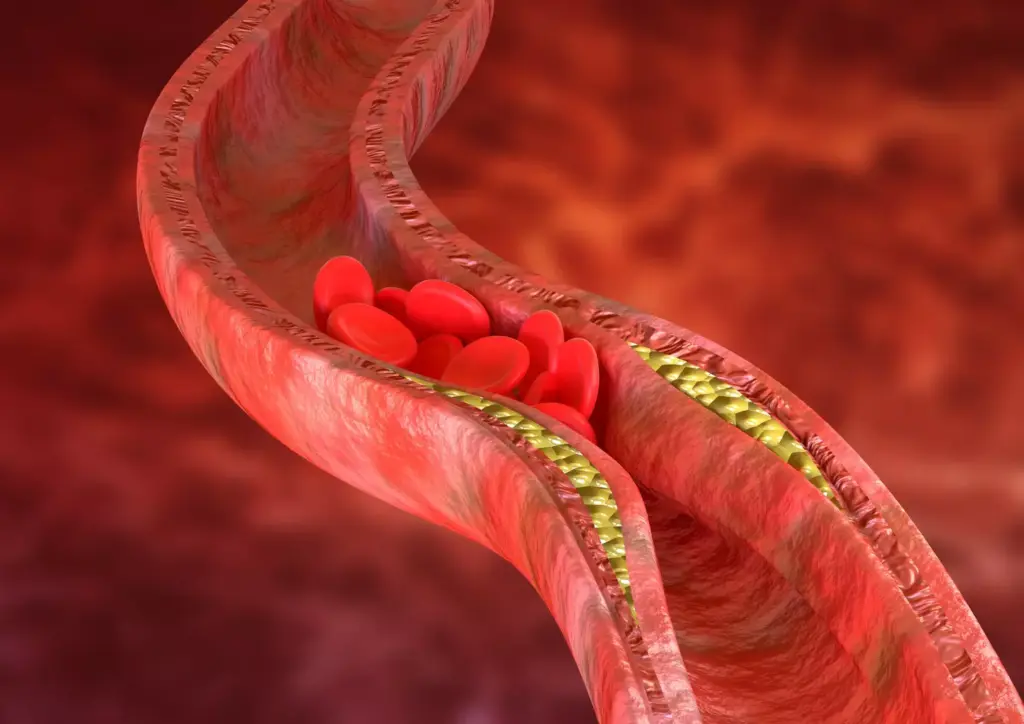
Atherosclerosis is a big problem for the aortic arch arteries. It’s when plaque builds up in the walls, causing stenosis and thrombosis. This can cut down blood flow and harm vital organs.
Consequences of Atherosclerosis:
Aneurysms and dissections are serious issues for the aortic arch. An aneurysm is when the aortic wall gets too big and might burst. Dissection is when there’s a tear in the aorta, letting blood leak into the wall.
| Condition | Description | Clinical Significance |
|---|---|---|
| Aneurysm | Dilation of the aortic wall | Risk of rupture, potentially life-threatening |
| Dissection | Tear in the intimal layer of the aorta | Potential for ischemia, organ failure, and death |
Takayasu arteritis is a rare disease that affects the aorta and its branches, including the aortic arch. It can cause stenosis, blockages, or aneurysms. Other inflammatory diseases can also affect the aortic arch, needing careful diagnosis and treatment.
We’ve talked about the different diseases of the aortic arch arteries and why they’re important. Knowing about these conditions helps doctors give the best care to their patients.
The aortic arch arteries are key to keeping our heart and blood vessels healthy. Knowing how they work is important for spotting and treating heart problems. We’ve looked at how these arteries supply blood to our head, neck, and arms.
The structure of these arteries is quite complex. They branch into three main paths: the brachiocephalic trunk, left common carotid artery, and left subclavian artery. Doctors need to know about these to give the best care to their patients.
Understanding these arteries is also vital for managing heart diseases like atherosclerosis, aneurysms, and dissections. By grasping how they function, doctors can better diagnose and treat these conditions. This leads to better care for patients.
As medical technology keeps improving, knowing about the aortic arch arteries is more important than ever. It’s essential for delivering top-notch healthcare services.
The aortic arch arteries have three main branches. These are the brachiocephalic trunk, left common carotid artery, and left subclavian artery. They carry oxygenated blood to the head, neck, and upper limbs.
The brachiocephalic trunk is the first branch of the aortic arch. It supplies blood to the right upper limb and head. This happens through its division into the right subclavian and right common carotid arteries.
Knowing the anatomy of the aortic arch arteries is key. It helps in diagnosing and treating vascular conditions. It also ensures the best care for patients in endovascular procedures.
There are common variations in the aortic arch arteries. These include different branching patterns, like the bovine arch. Such variations can affect endovascular procedures.
Blood flow in the aortic arch arteries is controlled by several factors. These include hemodynamic principles, pressure gradients, and autoregulation. These ensure a steady supply of oxygenated blood to the upper body.
To assess the aortic arch arteries, several imaging techniques are used. These include angiography, CT angiography, MR angiography, and ultrasound evaluation. Each has its own strengths and limitations.
The aortic arch arteries can be affected by several pathologies. These include atherosclerosis, aneurysms, dissections, and inflammatory conditions like Takayasu arteritis. If left untreated, these can have serious clinical implications.
The left common carotid artery supplies blood to the left side of the head and neck. It does this through its division into the internal and external carotid arteries. This is critical for maintaining blood flow to the brain and face.
The left subclavian artery supplies blood to the left upper limb. It does this through its course and major branches. This ensures proper blood flow to the arm and hand.
Subscribe to our e-newsletter to stay informed about the latest innovations in the world of health and exclusive offers!