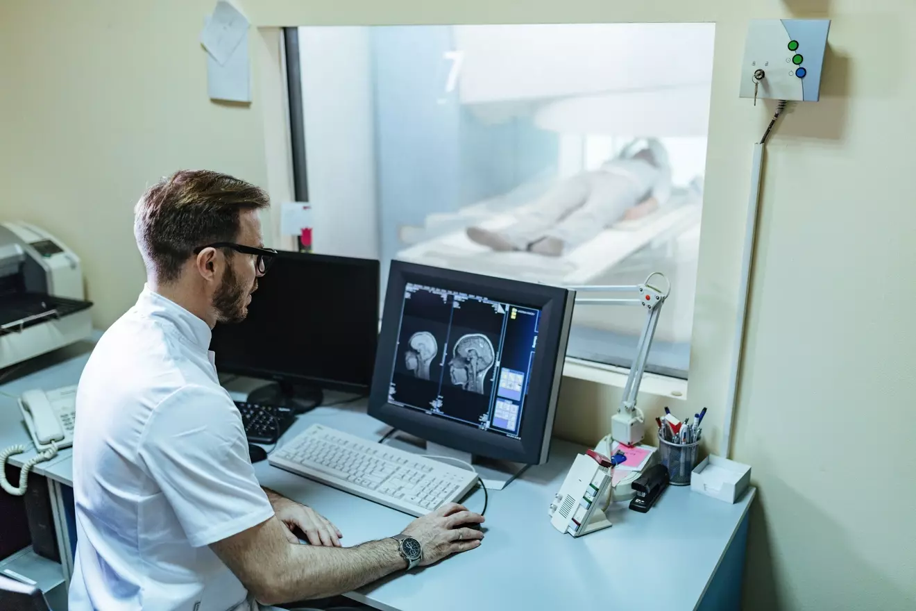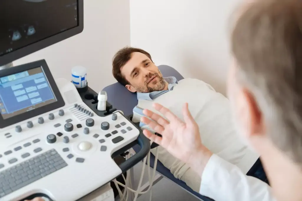
At Liv Hospital, we know how vital aortic ultrasound is for checking the abdominal aorta’s health. This method is key for spotting issues like aneurysms or dissections early. It helps doctors act fast.
Our guide will show you how to do an aorta on ultrasound. It covers the basics and what you need to know. We use info from Cureus and vascular ultrasonography eBooks to give you a full view of abdominal aorta imaging.
By using our guide, doctors can do precise and reliable checks. This leads to better health results for patients.
Key Takeaways
- Understanding the importance of aortic ultrasound in medical diagnostics.
- Learning the step-by-step process for performing an aortic ultrasound.
- Gaining insights into the technical requirements for abdominal aorta imaging.
- Recognizing the role of non-invasive imaging in detecting serious conditions.
- Appreciating the value of accurate evaluations for optimal patient care.
Understanding Aortic Ultrasound Fundamentals

Ultrasound for aorta imaging has changed vascular diagnostics. It’s non-invasive and very effective for checking aortic health. We’ll look at aortic ultrasound basics, its importance, and how ultrasound works for the aorta.
What is Aortic Ultrasound and Its Clinical Importance
Aortic ultrasound, or aortic sonogram, shows the aorta’s inside. It’s a key test for finding and tracking aorta problems. It helps doctors see the aorta’s size and spot any issues early.
Using ultrasound of aorta has many benefits:
- It’s non-invasive and painless.
- It doesn’t use radiation.
- It shows things in real-time.
- It’s cheaper than CT or MRI scans.
Basic Principles of Ultrasound Imaging for Aorta
Ultrasound for the aorta uses sound waves. High-frequency waves bounce off tissues and come back as echoes. These echoes make images of the aorta and its surroundings.
Things that affect aorta ultra sound image quality include:
- The sound wave frequency and transducer type.
- How the patient is prepared and positioned.
- The settings on the ultrasound machine.
Anatomy of the Abdominal Aorta Relevant to Sonography
The abdominal aorta starts at the diaphragm and goes down to the belly. It splits into the common iliac arteries there. Knowing the normal anatomy and any changes is key for accurate ultrasound checks.
Important spots for ultrasound aorta imaging are:
- The start of the celiac trunk.
- The superior mesenteric artery.
- The renal arteries.
- The aortic bifurcation.
Equipment and Technical Requirements

To perform an aortic ultrasound, we need specific equipment and technical skills. This ensures we get clear images of the abdominal aorta. We use the latest ultrasound technology for this purpose.
Ultrasound Machine Settings for Optimal Aortic Imaging
Getting the right settings on the ultrasound machine is key for clear aorta images. We adjust the frequency, gain, and depth to make sure the aorta shows up well. Proper machine settings help reduce errors and improve image quality.
The frequency setting is very important. It affects how deep and clear the ultrasound image is. For the abdominal aorta, we use a lower frequency (like 3-5 MHz) to get the right depth.
Transducer Selection and Frequency Considerations
Choosing the right transducer is vital for a good aortic ultrasound. We usually pick a curvilinear or convex transducer. It gives us a wider view, which is great for seeing the aorta and its surroundings.
The transducer’s frequency depends on the patient’s body and how deep the aorta is. For most adults, a 3-5 MHz transducer works well. But for obese patients, we might need a lower frequency for better penetration.
Documentation and Measurement Tools
Accurate documentation and measurement are key parts of an aortic ultrasound. We use built-in machine calipers to measure the aorta’s diameter at different points. Electronic calipers help us get precise measurements, which are important for diagnosing and tracking aortic issues.
We document everything, including images and measurements, in the patient’s file. This is important for future exams and for sharing information with other healthcare teams.
Patient Preparation for Aortic Ultrasound
Getting ready well is key for a good aortic ultrasound. A prepared patient helps the procedure succeed.
Pre-examination Instructions and Considerations
Before the aortic aneurysm sonogram, patients get clear prep instructions. They must fast to reduce bowel gas. Wearing loose, comfy clothes is also advised.
Telling patients about the procedure helps. It reduces anxiety and makes them more cooperative.
Optimal Patient Positioning Techniques
For an ultrasound of abdominal aorta, patients lie on their backs. Pillows or wedges support their back or knees for comfort.
The transducer goes in the midline, just below the xiphoid process. Slight adjustments in position can improve access and image quality.
| Positioning Technique | Benefit |
|---|---|
| Supine position | Easy access to the abdominal aorta |
| Leg elevation | Relaxation of abdominal muscles |
| Lateral tilting | Improved visualization of the aorta |
Communication and Comfort Measures During the Procedure
Keeping the patient informed is key during the abdominal aortic ultrasound. We explain each step and check on their comfort.
We use gel for the transducer and keep the room comfy. Adequate draping is also important.
By focusing on these, we ensure a smooth aortic ultrasound. This gives accurate diagnostic info.
Step-by-Step Aortic Ultrasound Procedure
When doing an aortic ultrasound, it’s important to follow a set of steps. This ensures we get clear images of the abdominal aorta. We’ll go over each step to help you understand and do the procedure well.
Initial Transducer Placement and Orientation
The first thing is to place the ultrasound transducer right. We start by putting it in the middle of the abdomen, just below the xiphoid process. The marker on the transducer should point towards the patient’s head. This is key for seeing the abdominal aorta clearly.
Transverse Scanning Technique
Next, we use the transverse scanning technique. We turn the transducer 90 degrees so it’s across the aorta. Then, we move it from the xiphoid process down to the aortic bifurcation. This lets us see the aorta in cross-section and check its size and for any problems.
Longitudinal Scanning Technique
After the transverse scan, we turn the transducer back to its original position. This is for the longitudinal scan. It’s important for seeing the aorta’s length and its walls. We move the transducer from side to side to get the whole aorta in view.
Visualization of Key Anatomical Landmarks
During the ultrasound, we look for important landmarks. These are the celiac trunk, superior mesenteric artery, and the aortic bifurcation. Seeing these helps us know where the aorta is and how big it is. We measure the aorta’s size at these points to be sure.
| Anatomical Landmark | Significance in Aortic Ultrasound |
|---|---|
| Celiac Trunk | Marks the upper limit of the abdominal aorta |
| Superior Mesenteric Artery | Helps in identifying the mid-portion of the aorta |
| Aortic Bifurcation | Defines the lower limit of the abdominal aorta |
By following these steps and looking at the key landmarks, we can do a thorough aortic ultrasound. This method is key for spotting problems and giving reliable results.
Measuring the Abdominal Aorta Correctly
To get accurate diagnoses, it’s key to follow set rules for measuring the abdominal aorta during an aortic ultrasound. Knowing the size of the abdominal aorta is vital for checking vascular health.
Standard Measurement Protocols
Using the same methods for measuring the abdominal aorta is important for getting reliable results. These methods involve checking the aorta at certain spots to get a full picture.
- Measurements are taken at the proximal, mid, and distal segments of the abdominal aorta.
- The aortic bifurcation is also evaluated as part of the standard protocol.
- Using the correct transducer frequency and machine settings is vital for accurate measurements.
Proximal Aortic Measurements
The proximal aorta is measured just below the diaphragm. Getting this measurement right is key to spotting any swelling or aneurysms early.
- Identify the celiac trunk and superior mesenteric artery as landmarks.
- Take measurements from outer wall to outer wall.
- Record the maximum diameter in both transverse and longitudinal views.
Mid-Aortic Measurements
Measurements at the mid-aorta level give more info on the aortic diameter and any problems.
- The mid-aorta is typically measured at the level of the renal arteries.
- Ensure that the measurement is perpendicular to the aortic lumen.
- Any visible plaque or thrombus should be noted.
Distal Aortic and Bifurcation Measurements
The distal aorta and bifurcation are key for checking vascular health, looking for signs of aneurysmal disease or stenosis.
When measuring the distal aorta:
- Assess the aorta just proximal to the bifurcation.
- Evaluate the bifurcation for any signs of stenosis or aneurysm.
- Document the diameter of both common iliac arteries.
By sticking to these ultrasound of abdominal aorta measurement rules, doctors can make sure their assessments are accurate. This helps in giving better care and management to patients.
Interpreting Aortic Ultrasound Images
Interpreting aortic ultrasound images means spotting normal patterns, finding common problems, and telling real findings from fake ones. We’ll show you how to do it right.
Normal Sonographic Appearance of the Aorta
A normal aorta looks like a tube with a clear center and clear walls on ultrasound. Its look can change a bit based on how you scan and the patient. In a side view, it’s round, and in a long view, it’s like a tube. We check for a smooth inside layer and a steady size along its length.
When we do an aortic scan, we check the aorta’s size, wall thickness, and look for any structural abnormalities. The normal aorta should have a uniform echo and stand out from other structures.
Recognizing Common Pathologies
On an aorta test, we can spot aneurysms, dissections, and atherosclerosis. An aortic aneurysm sonogram shows when the aorta gets too big, which is a big deal.
- Aneurysms are when the aorta gets too wide in one spot.
- Dissections show up as a flap inside the aorta.
- Atherosclerosis might look like thickening or calcification of the inside layer.
Spotting these issues on ultrasound for aorta needs a good grasp of what each looks like on ultrasound.
Differentiating Artifacts from True Findings
Artifacts can look like real problems on an aortic ultrasound. It’s key to tell real issues from fake ones to avoid mistakes. Changing the scan angle, using different transducers, or adjusting the patient’s position can help figure out what’s real.
Knowing about common artifacts and having experience are key when looking at aorta sonograms. By using our knowledge and checking with the patient’s history, we can make sure our findings are correct.
Detecting and Assessing Aortic Aneurysms
Finding and checking aortic aneurysms is vital for heart health checks. An aortic aneurysm sonogram helps spot aneurysms. It gives important info for treatment plans.
Defining Aortic Aneurysm on Ultrasound
An aortic aneurysm is when the aorta gets too big by more than 50%. On ultrasound, it looks like a bulge in the aortic wall. We look at size and any complications to confirm it.
Measurement Techniques for Aneurysm Evaluation
Getting the aneurysm’s size right is key for planning treatment. We measure it in two ways: across and lengthwise. The ultrasound abdominal aorta lets us see its size, shape, and where it is.
Risk Stratification Based on Ultrasound Findings
How risky an aneurysm is depends on its size and other details. Bigger aneurysms are more likely to burst. We look at size and other signs to decide how risky it is.
Documentation Requirements for Aneurysm Detection
Keeping detailed records is important for tracking aneurysms. We note the size, location, and any special features. The aorta ultrasound images are kept for later checks. This helps us see if the aneurysm is changing and adjust plans as needed.
Troubleshooting Common Challenges in Aortic Imaging
Getting a good aortic ultrasound can be tough. Sonographers face issues like bowel gas, body shape, and patient cooperation. These problems can make it hard to get clear images.
Overcoming Bowel Gas Interference
Bowel gas is a big problem for aortic ultrasound. Sonographers can try moving the patient or using gentle pressure to clear the gas. Asking the patient to take a deep breath can also help by moving the diaphragm and gas.
Strategies for Imaging in Obese Patients
Ultrasound in obese patients is hard because of the extra tissue. Sonographers can use a lower frequency transducer for better penetration. Adjusting the ultrasound settings and using harmonic imaging can also improve the image.
Addressing Limited Visualization Due to Patient Factors
Some patients can’t hold their breath or feel uncomfortable. Good communication and education are key. Sonographers should explain the process clearly and reassure the patient. Adjusting the patient’s position or adding support can also help.
Technical Adjustments for Improved Image Quality
Technical tweaks are vital for better ultrasound images. Changing gain, depth, and focus can make a big difference. Doppler imaging helps check blood flow. Keeping the equipment in top shape is also important.
By using these strategies, sonographers can solve common problems in aortic ultrasound. This leads to more accurate diagnoses.
Conclusion: Best Practices for Aortic Ultrasound
Following best practices for aortic ultrasound is key to making accurate diagnoses and caring for patients well. Healthcare professionals should stick to established guidelines for aortic ultrasound. This ensures top-notch abdominal aorta imaging and quality care.
Medical organizations, like the American Institute of Ultrasound in Medicine, have set guidelines for aorta sonogram and ultrasound for aorta. These guidelines highlight the need for technical skill, patient prep, and precise image measurement and interpretation during an aortic scan.
By sticking to these best practices, healthcare providers can give patients accurate diagnoses and effective treatment plans. This leads to better patient outcomes.
FAQ
What is an aortic ultrasound?
An aortic ultrasound is a test that uses sound waves to see the aorta. The aorta is the main artery that carries blood from the heart to the body.
Why is aortic ultrasound important?
It’s key for finding and watching aortic aneurysms. Aneurysms can be deadly if they burst. It also checks the aorta’s health and finds other problems.
How do I prepare for an aortic ultrasound?
You might need to fast, wear comfy clothes, and skip some meds. Our guide has all the prep steps you need.
What are the technical requirements for performing an aortic ultrasound?
You need a top-notch ultrasound machine, the right transducer, and the best settings. Our guide explains what you need in detail.
How is an aortic ultrasound performed?
A technician places a transducer on your belly. It sends sound waves to make aorta images. Our guide shows you how it’s done, step by step.
What are the standard measurement protocols for aortic ultrasound?
It measures the aorta’s diameter at certain points. Our guide tells you all about it.
How do I interpret aortic ultrasound images?
You need to know what’s normal and what’s not. Our guide helps you understand what you see.
What are the common challenges in aortic imaging, and how can they be overcome?
Challenges include gas, obesity, and limited views. Our guide offers tips for better images.
What is an abdominal aortic ultrasound?
It’s a special ultrasound for the abdominal aorta. This part of the aorta goes through the belly.
How is an aortic aneurysm diagnosed using ultrasound?
Ultrasound checks the aorta’s size and looks for bulges. Our guide explains how it works.
What are the benefits of using ultrasound for aorta imaging?
Ultrasound is safe, quick, and doesn’t use radiation. It’s also cheaper than other tests.
References
- Devin Tooma & Vi Dinh; Jessica Ahn et al. Aorta Ultrasound Made Easy: Step-By-Step Guide. POCUS 101. Available from: https://www.pocus101.com/aorta-ultrasound-made-easy-step-by-step-guide/ (POCUS 101)
- Rahman M. Abdominal Aortic Aneurysm Ultrasound Screening – Cardiologist Sherman, TX / Durant, OK. Healthy Heart Cardiology. Available from: https://www.hhcardiology.com/abdominal-aortic-aneurysm-ultrasound-screening-cardiologist-sherman-tx/ (hhcardiology.com)
- Florida Hearts. Abdominal Aortic Ultrasound — Imaging and Diagnostic Testing. Available from: https://www.floridahearts.com/contents/services/imaging-and-diagnostic-testing/abdominal-aortic-ultrasound
- American College of Emergency Physicians (ACEP). Basic Sonoguide – Abdominal Aorta. Available from: https://www.acep.org/sonoguide/basic/aorta/
- University of California San Francisco (UCSF). Abdominal Aorta Ultrasound Protocol (PDF). Available from: https://edus.ucsf.edu/sites/edus.ucsf.edu/files/wysiwyg/UCSF20ED20US20Protocol20Abdominal20Aorta_Final.pdf


