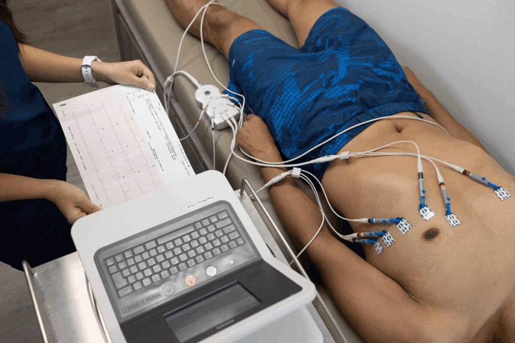
Spotting atrial tachycardia on an ECG can be tough, even for experts. Atrial tachycardia is a group of fast heart rhythms. They happen when the heart beats too quickly, usually between 100–250 beats per minute.
At Liv Hospital, we use the latest ECG tech to help our patients. We focus on you first to make sure we get your heart rhythm right. Knowing the ECG signs is key to telling atrial tachycardia apart from other heart issues.
We’re all about top-notch heart care at Liv Hospital. We use ECGs to find atrial tachycardia. This shows a fast heart rate with P waves that look different from normal heartbeats.
Key Takeaways
- Atrial tachycardia is a type of supraventricular tachycardia originating from the atria.
- ECG is key for diagnosing atrial tachycardia.
- P waves in atrial tachycardia differ from those in sinus rhythm.
- Rapid atrial rates characterize atrial tachycardia.
- Accurate diagnosis is key to effective treatment.
Understanding Atrial Tachycardia: Definition and Pathophysiology

To understand and treat atrial tachycardia well, knowing its basics is key. It’s a type of fast heart rhythm that starts in the atria.
What Defines Atrial Tachycardia
Atrial tachycardia is marked by a heart rate over 100 beats per minute. It starts outside the normal heart rhythm area. On an ECG, it shows a regular, narrow rhythm with different P waves.
Electrophysiological Mechanisms
The heart’s electrical issues cause atrial tachycardia. Abnormal automaticity means the atria fire on their own. Re-entry happens when an electrical signal keeps going in a loop.
Common Triggers and Risk Factors
Many things can start atrial tachycardia, like some medicines, caffeine, and stress. Heart problems also increase the risk.
Atrial tachycardia can show up in different ways. Focal and multifocal types have unique ECG signs.
The Clinical Significance of Atrial Tach

It’s key to grasp the importance of atrial tachycardia for good patient care. This condition can be either paroxysmal or persistent, affecting people differently. Knowing its prevalence, related conditions, and complications helps us offer better care.
Prevalence and Demographics
Supraventricular tachycardia, which includes atrial tachycardia, affects people in various ways. It’s more common in certain age groups and those with heart issues. The NCBI Bookshelf data shows its importance in medical practice.
People with heart problems are at higher risk of atrial tachycardia. We must keep this in mind when managing their condition.
Associated Conditions
Atrial tachycardia often comes with other health issues like heart disease and COPD. It’s important to spot these connections to treat atrial tachycardia right.
For example, COPD patients face a higher risk of atrial tachycardia. Electrolyte imbalances, like potassium and magnesium issues, can also trigger it.
Potential Complications
Atrial tachycardia can cause serious problems like tachycardia-induced cardiomyopathy. This happens when the heart beats too fast, making it less efficient. It can lead to heart failure.
Other issues include symptoms like palpitations, shortness of breath, and tiredness. These can really affect a person’s life quality. Treating atrial tachycardia well is key to avoiding these problems and helping patients get better.
By understanding atrial tachycardia’s clinical importance, we can give our patients better care. This knowledge helps us diagnose and treat them better, leading to better health outcomes.
ECG Feature #1: Heart Rate Characteristics in Atrial Tachycardia
Understanding heart rate characteristics is key to diagnosing atrial tachycardia. The heart rate in atrial tachycardia usually falls between 100–250 bpm. This is a vital diagnostic clue.
Typical Rate Range (100-250 bpm)
The heart rate in atrial tachycardia is usually between 100 and 250 bpm. This range is important. It helps tell atrial tachycardia apart from other tachycardias.
For example, sinus tachycardia has a rate below 100 bpm. Ventricular tachycardia, on the other hand, has a rate over 250 bpm.
Rate Variability in Different AT Types
Atrial tachycardia can be divided into different types based on its cause and location. The rate can vary a lot among these types. For instance, focal atrial tachycardia has a more steady rate.
But multifocal atrial tachycardia has a more variable rate. This is because it has multiple ectopic foci.
Rate variability is a key feature. It helps us understand the underlying cause of atrial tachycardia. Knowing this is essential for accurate diagnosis and treatment.
Rate Response to Interventions
The way atrial tachycardia responds to different treatments is very telling. For example, it may react differently to vagal maneuvers or anti-arrhythmic drugs than other tachycardias. Watching how the rate and rhythm change with these treatments can confirm the diagnosis.
Vagal maneuvers, like the Valsalva maneuver or carotid massage, can sometimes stop or slow down atrial tachycardia. This gives us clues about its cause and helps us decide how to manage it.
By grasping the heart rate characteristics, including the typical range, variability, and response to treatments, we can better diagnose and manage atrial tachycardia. This knowledge is vital for healthcare professionals dealing with tachycardia patients.
ECG Feature #2: P-Wave Morphology and Axis
Understanding P-wave morphology and axis is key for diagnosing atrial tachycardia. The P-wave on an electrocardiogram (ECG) gives clues about the arrhythmia’s origin and nature.
Normal vs. Abnormal P-Wave Appearance
In atrial tachycardia, P waves look different from those in normal heart rhythm. They might be inverted, notched, or have an unusual shape, showing they come from an ectopic source. For example, a negative P wave in lead I might point to a left atrial origin. A positive P wave in lead aVR could suggest a posterior or inferior atrial source.
Location-Specific P-Wave Changes
P-wave shapes change based on where the ectopic focus is. Focal atrial tachycardia from a specific atrial area can have unique P waves. For instance, a tachycardia from the crista terminalis might show P waves positive in inferior leads and negative in lead aVL.
P-Wave Axis Determination
Finding the P-wave axis is key to pinpointing atrial tachycardia’s source. By looking at P-wave direction in different ECG leads, we can guess the atrial activation’s direction. An abnormal P-wave axis can show an ectopic focus, helping to tell atrial tachycardia apart from other supraventricular tachycardias.
When looking at P-wave morphology and axis, remember to think about the patient’s symptoms and other tests. This full view helps make ECG diagnosis of atrial tachycardia more accurate.
ECG Feature #3: PR Interval and AV Relationship
Looking at the PR interval and AV conduction is key to telling atrial tachycardia apart from other heart rhythm issues. The PR interval shows the time from the start of the P wave to the QRS complex. In atrial tachycardia, this time can be shorter, normal, or longer.
PR Interval Variations in AT
In atrial tachycardia, the PR interval can be different. It might be shorter, normal, or longer. This depends on where the tachycardia starts and how the AV node works.
A shorter PR interval could mean the AV node is working better or there’s an extra pathway. On the other hand, a longer PR interval might show AV nodal disease or block.
AV Conduction Patterns
The way the AV node conducts signals in atrial tachycardia is very telling. We see different patterns, like 1:1 conduction and various block degrees. For example, 2:1, 3:1, or even higher blocks.
Knowing these patterns helps us diagnose and treat atrial tachycardia better.
Significance of PR Changes
Changes in the PR interval in atrial tachycardia are very important. A changing PR interval might mean the AV conduction is shifting or there are multiple atrial foci.
By studying the PR interval and AV relationship on an ECG, we can understand the heart’s rhythm better. This helps us make the right treatment choices.
ECG Feature #4: QRS Complex Characteristics
The QRS complex is key in spotting atrial tachycardia on an ECG. Atrial tachycardia usually shows a narrow QRS complex. This is unless there’s an issue with how the heart beats.
Narrow Complex Tachycardia Features
Atrial tachycardia often looks like a narrow complex tachycardia. This means the QRS duration is under 120 ms. The heart’s ventricles activate through the normal His-Purkinje system.
This narrow QRS complex is a key sign of supraventricular tachycardias. Atrial tachycardia is one of them.
Aberrant Conduction Possibilities
But sometimes, atrial tachycardia can show a wide QRS complex. This happens if there’s a bundle branch block or if the heart rate is too fast. It can make the heart act like it has a block.
When this happens, it can be hard to tell if it’s atrial tachycardia or ventricular tachycardia. It’s important to look closely at the P-wave, PR interval, and other ECG signs.
Rate-Related QRS Changes
Also, the QRS complex can change with the heart rate in atrial tachycardia. At very high rates, the QRS might change a bit. This could be a slight change in shape or a tiny difference in duration.
Knowing about these QRS changes is vital for diagnosing atrial tachycardia. By studying the ECG closely, doctors can make the right choices for their patients.
ECG Feature #5: Rhythm Regularity and Onset Patterns
Atrial tachycardia (AT) has unique ECG signs, like rhythm regularity and onset patterns. These are key for correct diagnosis. The rhythm of atrial tachycardia can be regular or irregular, based on the cause. Regular rhythms are common, but irregular ones can happen, like in multifocal atrial tachycardia.
Regular vs. Irregular AT Rhythms
Mostly, atrial tachycardia shows a regular rhythm, a key sign of this condition. But, some types, like multifocal atrial tachycardia, can have an irregular rhythm because of many ectopic foci. It’s important to know the rhythm to tell different types of atrial tachycardia apart.
Abrupt vs. Gradual Onset
The start of atrial tachycardia can be abrupt or gradual. Paroxysmal atrial tachycardia often starts and stops quickly, seen on ECG. On the other hand, some types of atrial tachycardia may slowly increase or decrease in rate. Knowing how it starts is key for diagnosing and treating atrial tachycardia.
Termination Patterns
How atrial tachycardia ends can also help diagnose it. Some episodes end abruptly, while others slow down before stopping. The way it ends can tell us about the cause and help decide treatment.
By looking closely at rhythm regularity, onset, and end patterns on an ECG, doctors can better diagnose and manage atrial tachycardia. This detailed understanding is vital for top-notch care for patients with this condition.
Focal vs. Multifocal Atrial Tach: Differential ECG Findings
ECG findings are key in telling focal from multifocal atrial tachycardia. Atrial tachycardia is a fast heart rate from the atria. Knowing the difference is important for treatment.
Uniform P-Waves in Focal AT
Focal atrial tachycardia comes from one spot in the atria. This means uniform P-waves on the ECG. The P-wave shape can hint at where the problem is.
Multiple P-Wave Morphologies in MAT
On the other hand, multifocal atrial tachycardia (MAT) has multiple P-wave morphologies. This is because of many different starting points. It makes the rhythm hard to predict and treat.
Clinical Contexts for Each Type
Focal atrial tachycardia usually happens in people with normal hearts. But, multifocal atrial tachycardia is linked to things like COPD, heart failure, or imbalances in electrolytes. Knowing the cause helps in treating it right.
When looking at ECGs, we must think about the situation and what we see. Uniform P-waves point to a single cause, while many P-wave shapes mean multiple causes. Getting it right is key to helping patients.
Distinguishing Atrial Tach from Sinus Tachycardia
It’s important to know the difference between atrial tachycardia and sinus tachycardia for better patient care. Atrial tachycardia can be told apart from sinus tachycardia by looking at the P-wave shape and how it reacts to vagal maneuvers.
P-Wave Morphology Differences
The shape of the P-wave is key in telling these two apart. In sinus tachycardia, the P-wave is usually upright in lead II and has a normal direction. But in atrial tachycardia, the P-wave shape can change a lot, depending on where the tachycardia starts.
P-wave characteristics in atrial tachycardia:
- Abnormal P-wave axis
- Variable P-wave morphology
- Often inverted or biphasic P-waves in certain leads
On the other hand, sinus tachycardia shows a consistent P-wave shape that is upright in lead II.
Onset and Termination Characteristics
The way these tachycardias start and stop is different too. Sinus tachycardia starts and ends slowly. But atrial tachycardia can start and stop quickly.
“Sinus tachycardia has a gradual onset and termination, whereas atrial tachycardia often starts and stops abruptly.” – EMCrit Project
This difference is important for making a diagnosis and deciding on treatment.
Response to Vagal Maneuvers
Vagal maneuvers also help tell these tachycardias apart. Atrial tachycardia might slow down or stop briefly with vagal maneuvers. But sinus tachycardia will slow down a bit with vagal maneuvers but won’t stop suddenly.
| Characteristics | Atrial Tachycardia | Sinus Tachycardia |
| P-wave Morphology | Variable, often abnormal | Consistent, normal |
| Onset/Termination | Abrupt | Gradual |
| Response to Vagal Maneuvers | May terminate or slow temporarily | Slows down gradually |
By looking at these differences, doctors can tell atrial tachycardia from sinus tachycardia. This helps them give the right treatment and care.
Differentiating Atrial Tach from Atrial Flutter
It’s important for healthcare workers to know how to tell apart atrial tachycardia and atrial flutter on an ECG. Both are types of supraventricular tachycardias but show different signs on an ECG.
Absence of Sawtooth Flutter Waves
A key difference is the presence of sawtooth flutter waves in atrial flutter. These waves are not seen in atrial tachycardia. Atrial flutter has a regular heart rate, usually 300 bpm, with a sawtooth pattern, as shown by the EMCrit Project. Atrial tachycardia does not have this pattern.
Rate Differences and Variability
Atrial flutter has a more consistent and faster heart rate, about 300 bpm. Atrial tachycardia has a more variable rate, between 100-250 bpm. This rate difference is key in telling these two apart.
- Atrial Flutter: Regular rate, typically around 300 bpm
- Atrial Tachycardia: Variable rate, generally between 100-250 bpm
AV Conduction Patterns
The way the heart conducts signals also varies between atrial tachycardia and atrial flutter. Atrial flutter often has a fixed AV conduction ratio, like 2:1 or 3:1. Atrial tachycardia, on the other hand, has a more variable pattern.
“The differentiation between atrial tachycardia and atrial flutter requires careful analysis of the ECG, focusing on the presence or absence of sawtooth flutter waves, atrial rate, and AV conduction patterns.”
ECG Interpretation Guidelines
By looking closely at these ECG signs, doctors can tell atrial tachycardia from atrial flutter. This helps in making the right diagnosis and treatment.
Special Variant: Atrial Tach with Block
Atrial tachycardia with block is a special type of atrial tachycardia. It needs careful ECG reading for the right diagnosis. This condition has a changing AV conduction ratio, making it hard to tell apart from other heart rhythm problems.
ECG Recognition Patterns
To spot atrial tachycardia with block on an ECG, look for a changing AV conduction ratio. This means the heart’s upper chambers beat faster than the lower chambers. The ratio of these beats isn’t always the same.
Key ECG Features:
- Variable AV conduction ratio
- Atrial rate typically between 150-250 bpm
- Presence of AV block, often 2:1 or variable
Common Mimics and Misdiagnoses
This condition can be mistaken for other heart rhythm issues because of its complex ECG pattern. It often gets confused with atrial fibrillation with a rapid ventricular response and other supraventricular tachycardias.
“The presence of AV block is a key distinguishing feature of atrial tachycardia with block, differentiating it from other supraventricular tachycardias.” – Cardiology Expert
It’s important to study the ECG closely to avoid mistakes and treat it correctly.
Clinical Significance
Atrial tachycardia with block can lead to serious symptoms and problems if not treated right. People might feel their heart racing, have trouble breathing, and feel dizzy. These symptoms can also lower the heart’s ability to pump blood well.
| Clinical Feature | Description |
| Symptoms | Palpitations, shortness of breath, dizziness |
| Complications | Decreased cardiac output, heart failure |
| Management | Rate or rhythm control strategies, addressing underlying causes |
Knowing how to read ECGs and understand the importance of atrial tachycardia with block is key. It helps doctors diagnose and treat it properly.
Conclusion: Mastering Atrial Tachycardia Interpretation
Understanding atrial tachycardia on ECG is key for correct diagnosis and treatment. This article has covered seven important ECG signs to spot atrial tachycardia.
Knowing about P-wave shape, PR interval, QRS complex, and rhythm regularity helps in diagnosing atrial tachycardia. The EMCrit Project stresses the need to grasp these ECG signs and their clinical meanings for proper management.
Atrial tachycardia is a complex issue that needs deep knowledge of its ECG signs and clinical importance. Using this knowledge in our practice can lead to better patient care and outcomes.
FAQ
What is atrial tachycardia, and how is it diagnosed?
Atrial tachycardia is a heart rhythm problem. It starts in the atria. Doctors use an electrocardiogram (ECG) to spot it. The ECG shows a fast heart rate with clear P waves.
What are the common triggers and risk factors for atrial tachycardia?
Stress, some medicines, and heart issues can trigger it. Heart disease, COPD, and imbalances in electrolytes are also risks.
How do the ECG features of atrial tachycardia differ from those of sinus tachycardia?
Atrial tachycardia has unique P-wave shapes and starts and stops differently. It also reacts differently to vagal maneuvers than sinus tachycardia.
What is the significance of P-wave morphology and axis in diagnosing atrial tachycardia?
The shape and direction of P waves can show where the arrhythmia starts. This helps find the source of the problem.
How does the PR interval and AV relationship contribute to the diagnosis of atrial tachycardia?
Changes in the PR interval and how the AV conduction works help tell it apart from other heart rhythm issues.
What are the characteristics of the QRS complex in atrial tachycardia?
It usually has a narrow QRS complex. But, it can be wide if the heart’s conduction is off. The QRS can also change with the heart rate.
What is the difference between focal and multifocal atrial tachycardia?
Focal atrial tachycardia has one P-wave shape. Multifocal atrial tachycardia has many P-wave shapes and an irregular rhythm.
How does atrial tachycardia with block present on an ECG?
It has a special ECG pattern. It’s important to diagnose it correctly to avoid mistakes.
What are the possible complications of atrial tachycardia?
It can lead to a weakened heart. This is why accurate diagnosis and treatment are key.
How does the heart rate respond to interventions in atrial tachycardia?
Knowing how the heart rate changes with treatment is important. It helps in managing the condition, as different types of atrial tachycardia react differently.
References:
- Liwanag, M., & Willoughby, C. (2023, June 26). Atrial tachycardia. In StatPearls [Internet]. Treasure Island (FL): StatPearls Publishing. https://www.ncbi.nlm.nih.gov/books/NBK542235/
- Kotadia, I. D., et al. (2020). Supraventricular tachycardia: An overview of diagnosis and management. Progress in Cardiovascular Diseases, https://www.sciencedirect.com/science/article/pii/S1470211824036510


