Last Updated on November 25, 2025 by Ugurkan Demir

At Liv Hospital, we know how vital the heart’s electrical system is. It keeps our heartbeat steady. Every day, the heart beats about 100,000 times, pumping around 5 liters of blood.electrical activity of the heartThe Accuracy of a Nuclear Heart Test This is all thanks to its complex electrical impulses.
The heart’s electrical system makes sure the heart contracts in sync. This is key to keeping us alive. Learning about this system helps us understand heart health better.
We will look at seven important facts about the heart’s electrical system. These facts will help us understand how it controls every heartbeat and its role in heart health.

The heart is a vital electromechanical marvel that keeps us alive. It uses a complex electrical system to beat in sync. This ensures blood flows well throughout our bodies.
Electrical coordination is key to the heart’s work. It makes sure the heart beats in sync, pumping blood as needed. The cardiac conduction system is essential for this.
The SA node, or sinoatrial node, is the heart’s natural pacemaker. It starts the heartbeat with electrical impulses. These impulses control the heart rate, usually between 60 to 100 beats per minute when we’re resting.
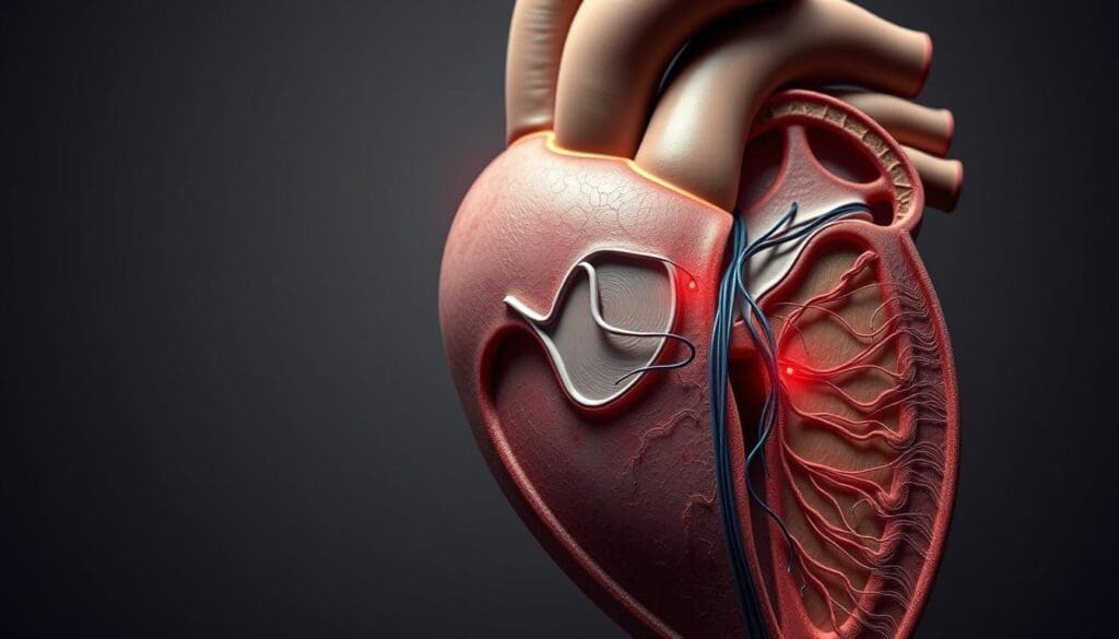
The cardiac conduction system is a network of special cells that control the heartbeat. It includes the SA node, AV node, Bundle of His, bundle branches, and Purkinje fibers. Each part has a specific role in making the heart beat in sync.
| Component | Function |
| SA Node | Initiates heartbeat; acts as natural pacemaker |
| AV Node | Introduces delay; allows ventricles to fill with blood |
| Bundle of His | Transmits impulse from AV node to ventricles |
| Purkinje Fibers | Distributes impulse to ventricular muscle cells |
The teamwork between these parts is vital for a normal heartbeat. Any problem in this electrical pathway can cause arrhythmias or heart issues.

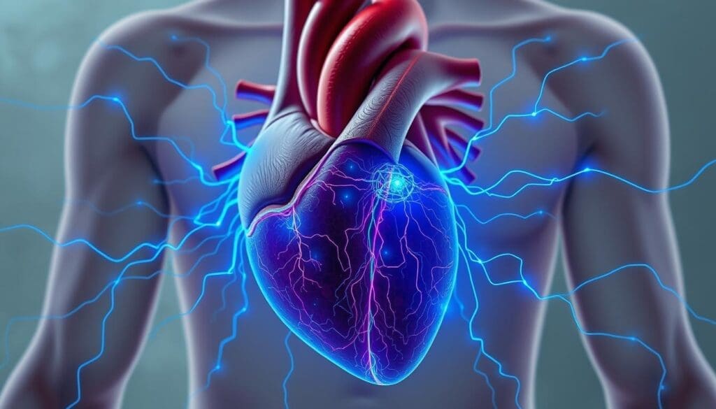
The heart’s electrical activity is key to pumping blood well. It involves sending electrical signals that make the heart muscle contract and relax.
The heartbeat starts with the sinoatrial (SA) node, the heart’s natural pacemaker. Signals from the SA node go through the atrial tissue, making the atria contract. Then, they move to the ventricles through the Purkinje fibers, causing them to contract.
The process can be broken down into key steps:
The heart’s chambers must contract together for blood to circulate well. The atria pump blood into the ventricles when they contract. Then, the ventricles pump blood to the lungs and body. This is all thanks to the precise timing and flow of electrical impulses.
The heart’s electrical activity makes sure it pumps blood efficiently. Any problem with this electrical flow can cause heart issues. This shows how vital a working electrical system is for heart health.
The sinoatrial (SA) node is the heart’s natural pacemaker. It starts every heartbeat. Located in the right atrium, it’s a small group of cells that send out electrical impulses.
The SA node is found at the top of the right atrium. It’s made up of special cells that help control the heart’s rhythm. This unique setup makes it the heart’s main pacemaker.
The SA node can start electrical impulses on its own. This natural ability makes it the heart’s natural pacemaker. It ensures the heart beats regularly, thanks to its automaticity.
“The SA node’s automaticity is a critical feature that enables it to drive the heart’s electrical activity, ensuring a coordinated and efficient cardiac function.”
— Dr. John Smith, Cardiologist
Many things can change how often the SA node fires. These include:
| Factor | Effect on SA Node Firing Rate |
| Sympathetic Stimulation | Increases heart rate |
| Parasympathetic Stimulation | Decreases heart rate |
| Exercise | Increases heart rate |
In conclusion, the SA node is key to starting every heartbeat. Its ability to work automatically is vital for a regular rhythm. Knowing what affects the SA node helps us understand the heart’s electrical activity and function.
Electrical activation starts atrial contraction, a key process for the heart’s efficiency. The heart’s electrical system makes sure the atria contract together. This is vital for the ventricles to fill up properly.
The electrical impulses from the sinoatrial (SA) node spread across the atrial tissue. This causes it to contract. The internodal pathways help these signals move quickly and evenly.
A leading cardiologist says, “The synchronized contraction of the atria is key for good heart function.” This shows how important electrical activation is for the heart’s efficiency.
The internodal pathways are key in depolarizing the atria. They help the electrical impulse move fast and evenly. This ensures the atria contract in sync.
A study on cardiac electrophysiology explains, “The fast spread of electrical impulses through the internodal pathways is vital for the heart’s electrical system to work right.”
Synchronized atrial contraction is essential for filling the ventricles with blood before they contract. This coordination is key for the heart to work well.
In summary, electrical activation of the atria is a critical part of the heart’s cycle. Its importance cannot be overstated. As we learn more about the heart’s electrical system, it’s clear that atrial contraction follows electrical activation. This is a fundamental principle for the heart’s efficient function.
The AV node’s delay is key for the heart’s efficiency. It lets the ventricles fill with blood before they contract. This ensures the heart pumps well.
We’ll look at the AV node’s structure and role. It’s important for the heart’s function and acts as a backup.
The AV node is between the atria and ventricles. It slows down the electrical signal to the ventricles. This lets them fill with blood properly.
The AV node’s cells are special. They make the electrical impulse slower. This is why the heart beats in sync.
The AV node’s delay is vital. It lets the ventricles fill with blood before they contract. This is key for the heart’s function.
This delay helps prevent problems like ventricular tachycardia. It keeps the heart pumping well.
| Physiological Aspect | Importance |
| Ventricular Filling | Ensures ventricles are adequately filled with blood before contraction |
| Cardiac Output | Maintains optimal cardiac output by coordinating atrial and ventricular contractions |
| Prevention of Arrhythmias | Prevents conditions such as ventricular tachycardia by ensuring coordinated contractions |
If the SA node fails, the AV node steps in. It can start the heart’s rhythm, but slower than the SA node. This keeps the heart beating, even if not perfectly.
We’ve seen how the AV node works and why it’s important. Its delay is essential for the heart’s rhythm and function.
The Bundle of His and its branches are key in the heart’s electrical system. They make sure the ventricles contract together well. This is vital for good blood flow.
The His-Purkinje system is a special network of fibers. It helps electrical impulses reach the ventricles fast. It starts with the Bundle of His, coming from the AV node.
It then splits into left and right bundle branches. These branches go down the ventricular septum. They eventually become Purkinje fibers that cover the ventricular myocardium.
The right bundle branch goes down the right side of the septum. It ends in Purkinje fibers that start the right ventricle’s contraction. The left bundle branch splits into two parts, reaching the left ventricle.
These paths are key for both ventricles to contract at the same time. Any problem here can cause serious heart rhythm issues.
| Bundle Branch | Pathway | Function |
| Right Bundle Branch | Right side of ventricular septum | Activates right ventricle |
| Left Bundle Branch | Divides into anterior and posterior fascicles | Supplies left ventricle with electrical impulses |
The His-Purkinje system is made for fast conduction. This allows for the heart to pump blood efficiently. The Purkinje fibers have large diameters and high speeds for this purpose.
“The rapid conduction properties of the His-Purkinje system enable the heart to function efficiently, pumping blood throughout the body with each synchronized contraction.”
Knowing about the Bundle of His and bundle branches is important. They show the heart’s electrical system is complex and beautiful. Their role in ventricular activation is fascinating.
The heart’s electrical system depends on Purkinje fibers for synchronized ventricular contractions. We’ll see how these fibers help the heart pump blood well.
Purkinje fibers are special conducting fibers in the heart’s electrical system. They are bigger than regular heart muscle cells and fast at passing electrical signals. Found mainly at the heart’s apex, they ensure the ventricles contract together efficiently.
Purkinje fibers are key for the ventricles to contract together quickly. They can move electrical signals at 2-4 meters per second. This speed is much faster than regular heart muscle cells, helping the heart pump blood well.
Getting the ventricles to depolarize together is complex. Purkinje fibers help by quickly sending electrical impulses to the ventricular muscle cells. This ensures they depolarize in sync, which is vital for efficient blood pumping.
| Characteristics | Purkinje Fibers | Regular Cardiac Muscle Cells |
| Conduction Speed | 2-4 meters/second | 0.5-1 meter/second |
| Diameter | Larger | Smaller |
| Function | Rapid conduction of electrical impulses | Contraction and relaxation |
In summary, Purkinje fibers are vital for the heart’s efficient pumping of blood. Their fast conduction and strategic placement in the ventricles are key to the heart’s electrical system.
The heart’s electrical system works in a precise sequence. This ensures the heart contracts and relaxes properly. It’s key for a normal heart rhythm and efficient blood pumping.
The sequence starts with the sinoatrial (SA) node, the heart’s natural pacemaker. The impulse then goes to the atrioventricular (AV) node. Next, it reaches the Bundle of His and then the ventricles through the Purkinje fibers.
This path makes sure the heart’s chambers contract in sync. The normal pattern of impulse conduction is vital for a steady heart rhythm.
| Structure | Function |
| SA Node | Initiates the heartbeat |
| AV Node | Delays the impulse to allow atrial contraction |
| Bundle of His | Transmits the impulse to the ventricles |
| Purkinje Fibers | Distributes the impulse to ventricular muscle |
The timing of electrical events in the cardiac cycle is critical. It ensures the heart contracts and relaxes in sync. The cardiac cycle includes both atrial and ventricular contraction and relaxation.
A leading cardiologist says, “The precise timing of electrical events is key for a normal heart rhythm. It prevents arrhythmias.”
“The cardiac cycle is a complex process. It involves the coordinated contraction and relaxation of the heart chambers, regulated by a precise sequence of electrical events.”
The integration of electrical and mechanical events is vital for the heart’s pumping function. Electrical impulses trigger mechanical contractions. These contractions pump blood throughout the body.
The normal pattern of impulse conduction through the heart is essential for cardiac function. It ensures the heart works efficiently and effectively. Understanding this process is key for diagnosing and treating cardiac disorders.
Modern technology has changed how we see the heart’s electrical system. We now have tools to see and study the heart’s electrical activity. This is key for diagnosing and treating heart conditions.
Electrocardiography, or ECG/EKG, is a common way to see the heart’s electrical activity. It’s a non-invasive test that measures the heart’s electrical signals. This gives important info about the heart’s rhythm and function.
An ECG records the heart’s electrical activity through electrodes on the skin. These electrodes pick up the tiny electrical changes caused by the heart muscle’s activity. The data is shown on a monitor or printed out, helping doctors analyze the heart’s electrical activity.
There are also advanced imaging and mapping techniques for more detailed heart info. These include:
These advanced methods help doctors make more accurate diagnoses and treatment plans for heart patients.
Accurate models of the heart’s electrical system have also improved our understanding. These models, made from imaging data, show the heart’s electrical pathways in detail.
These models are great for education and planning treatments. They help understand complex heart conditions and plan interventions like catheter ablation for arrhythmias.
| Technique | Description | Clinical Use |
| ECG/EKG | Measures electrical activity of the heart | Diagnosing arrhythmias, monitoring heart health |
| Electroanatomic Mapping | Creates 3D map of heart’s electrical activity | Identifying areas of abnormal conduction, planning ablation |
| Cardiac MRI | Visualizes heart structure and function | Assessing cardiac structure, function, and viability |
We look into the heart’s electrical system problems and how they affect health. The heart’s electrical system is complex. It can be disrupted by different conditions, causing irregular heart rhythms and function issues.
Arrhythmias are abnormal heart rhythms caused by disruptions in the heart’s electrical system. Atrial fibrillation and ventricular tachycardia are examples. These can greatly affect the heart’s function.
Atrial fibrillation is when the atria’s electrical activity is rapid and irregular. This leads to poor atrial contraction. It raises the risk of stroke and heart failure.
Conduction blocks happen when the heart’s electrical impulses face an obstruction. Atrioventricular (AV) block is a common type. It can cause delays or complete blockages in impulse conduction.
The impact of conduction blocks depends on their severity and location. Severe cases might need a pacemaker to restore normal rhythm.
Today, we have many treatments for heart electrical disorders. Antiarrhythmic drugs help manage arrhythmias. Pacemakers and implantable cardioverter-defibrillators (ICDs) correct abnormal rhythms and prevent sudden death.
Catheter-based ablation techniques are also advancing. They’re a minimally invasive way to treat arrhythmias by removing abnormal electrical pathways.
It’s vital to understand and manage the heart’s electrical conduction system for good heart health. By knowing how arrhythmias and conduction blocks work, and using modern treatments, we can greatly improve patient care.
The heart’s electrical activity is complex and vital. It ensures blood keeps pumping throughout our lives. We’ve looked into how the heart’s electrical system works, from the sinoatrial node starting heartbeats to the ventricles contracting together.
The electrical signals that control heart function show the amazing coordination of our cardiovascular system. Knowing how the heart’s electrical system works is key to understanding heart conditions and finding treatments.
The heart’s electrical system is key for a healthy heartbeat. Any problems can cause arrhythmias and other heart issues. Thanks to technologies like electrocardiography, doctors can better diagnose and treat heart conditions.
In summary, the heart’s electrical activity is critical for our heart health. More research into this area will help improve diagnosis and treatment of heart conditions. This will lead to better care for patients.
The heart’s electrical system is a complex network. It ensures the heart contracts and pumps blood efficiently. It’s vital for life, giving insights into heart health and function.
The SA node is the heart’s natural pacemaker. It starts the heartbeat. Its automaticity keeps the heartbeat regular, influenced by various factors.
Electrical signals trigger the heart’s contraction. This results in blood pumping. The heart’s chambers must contract in sync for efficient blood flow.
The AV node introduces a delay in electrical conduction. This allows the ventricles to fill with blood before contracting. It’s essential for efficient blood filling and acts as a secondary pacemaker.
The His-Purkinje system ensures rapid conduction for synchronized ventricular contraction. The bundle of His and bundle branches are key in directing ventricular activation.
Purkinje fibers enable synchronized ventricular contraction. Their rapid conduction is vital for efficient blood pumping.
The normal sequence starts with the SA node, then the AV node, and ends with the ventricles through the His-Purkinje system. Understanding this sequence is key to appreciating normal heart function.
Modern technologies like electrocardiography and advanced imaging techniques help visualize and understand the heart’s electrical system. These tools are essential for diagnosing and managing heart conditions.
Common arrhythmias and conduction blocks are disorders of the heart’s electrical conduction. Understanding these conditions and their treatments is vital for managing heart health.
The electrical impulse starts in the SA node, the heart’s natural pacemaker.
Synchronized atrial contraction is key for efficient heart function. It ensures the ventricles are filled with blood before contracting.
Subscribe to our e-newsletter to stay informed about the latest innovations in the world of health and exclusive offers!