Last Updated on November 25, 2025 by Ugurkan Demir

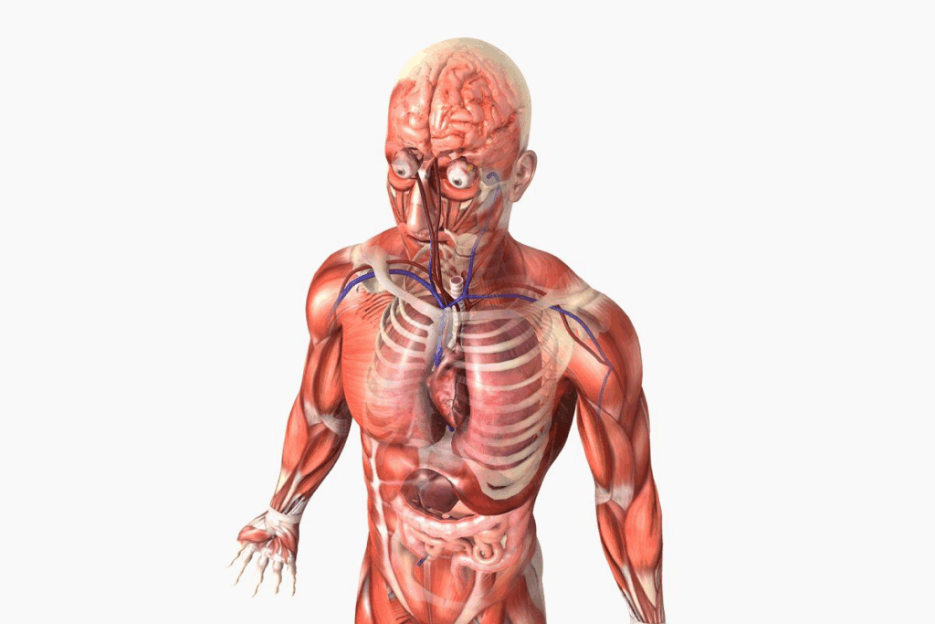
The human heart is a vital organ that plays a critical role in our overall health. It is located in the mediastinum, often called the ‘cardia.’ We will explore seven essential facts about its size, shape, and anatomy.Discover real facts about the heart human structure, size, and how it functions.
The heart’s size is often compared to a fist, weighing between 250-350 grams. Understanding its anatomy is key to appreciating its complexity and resilience. At Liv Hospital, we use cutting-edge medical standards to provide top-notch care for our patients.

The human heart is at the heart of our circulatory system. It’s a vital organ that pumps blood all over the body. This muscle works hard, bringing oxygen and nutrients to our tissues and organs, and taking away waste.
The heart’s role in keeping us alive is huge. Without it, we would quickly run out of oxygen and nutrients. Knowing how the heart works helps us see its importance for our health.
The human heart is built for endurance. It beats about 100,000 times every day. Its design and function work together to pump blood well across the body.
| Heart Function | Description |
| Pumping Blood | The heart pumps blood throughout the body, supplying oxygen and nutrients. |
| Regulating Blood Pressure | The heart helps regulate blood pressure through its pumping action. |
| Maintaining Circulation | The heart ensures continuous blood circulation, essential for overall health. |
Knowing about heart anatomy is key for diagnosing and treating heart problems. Doctors use this knowledge to spot issues and create good treatment plans.
By understanding the heart’s role in our survival and its anatomy, we can see why keeping our heart healthy is so important.

To fully understand the heart, we must look at its scientific name, location, and protective layers. The heart is a key organ in our body’s system. It is called the ‘cardia’ or ‘cardiac’ in scientific terms. Knowing where it is and what protects it helps us understand our body better.
The word ‘cardia’ comes from the Greek ‘kardia,’ meaning heart. In medicine, ‘cardiac’ means anything heart-related. This term is key for doctors to talk about heart issues worldwide.
The heart sits in the mediastinum, a part of the chest. It’s between the sternum and the spine. The heart is mostly on the left side, with two-thirds of it there.
The pericardium is a double-layered sac around the heart. It protects the heart and helps it move. The pericardium has an outer and inner layer, with the inner layer divided further. It makes sure the heart moves smoothly.
| Layer | Description | Function |
| Fibrous Pericardium | Outer layer of the pericardium | Provides structural support |
| Serous Pericardium | Inner layer, divided into parietal and visceral layers | Reduces friction and facilitates heart movement |
| Parietal Layer | Outer part of the serous pericardium | Lining the fibrous pericardium |
| Visceral Layer (Epicardium) | Inner part of the serous pericardium, attached to the heart | Directly adheres to the heart, reducing friction |
The size of an adult human heart is truly amazing. It’s a key part of our survival, yet often misunderstood.
An adult heart is about 12 cm long, 8-8.5 cm wide, and 6 cm thick. These sizes can vary, but they give us a basic idea of the heart’s size.
Many people compare the heart to a closed fist. This is pretty accurate. The heart is roughly the same size as a clenched fist. This makes sense, as the heart is a muscle that pumps blood, and its size matches the body’s size.
Think of the heart as the size of a large grapefruit or a small melon. These comparisons help us understand the heart’s size better. They make it easier to see its role in our body.
Knowing the true size of the human heart helps us appreciate its anatomy and importance. It shows how efficient and vital it is for our health.
The human heart’s shape is truly remarkable. It’s often seen as an inverted cone. This shape is both simple and very effective for its job.
The heart has a wide base and a sharp tip. This shape helps the heart pump blood well around the body. The base goes up, and the tip points down and left.
The heart’s base is where big blood vessels connect. It gives the heart a solid base for pumping. The tip, or apex, is the lowest point and points left.
The heart points down and left because of its chest position and blood vessel attachments. This helps blood flow efficiently. It’s a result of evolution to make the heart work better.
Key aspects of the heart’s shape include:
Knowing the heart’s shape helps us understand its role in our body’s circulatory system.
When we think about the heart’s color, we see a mix of tissue and blood flow. The heart, a muscular organ, pumps blood all over the body. Its color comes from its makeup and how it works.
The heart’s real color is dark red. This is because of its blood and muscle tissue. The heart’s myoglobin and deoxygenated blood make it look dark red.
The Role of Myoglobin: Myoglobin is a protein in muscle that stores oxygen. The heart has lots of myoglobin, making it dark red.
Blood flow changes how we see the heart’s color. The heart pumps blood, which affects its color. Both oxygenated and deoxygenated blood make it dark red.
| Factor | Influence on Heart Color |
| Myoglobin Content | Contributes to dark red color |
| Blood Flow | Affects overall coloration, maintaining dark red hue |
| Oxygenation Level | Influences shades of red, with deoxygenated blood contributing to darker tones |
Knowing what makes the heart’s color special helps us understand its role. The heart’s dark red color shows its key role in keeping us alive by circulating blood.
Knowing the average weight of the human heart is key to understanding its health. The heart’s weight can change based on gender and overall health.
An adult human heart weighs about 250-300 grams for women and 300-350 grams for men. These numbers help us understand heart health better.
Studies show that male hearts are usually heavier than female hearts. This is mainly because of differences in body size and muscle mass between genders.
Age plays a big role, as the heart changes with time. People who are more physically fit may have bigger hearts due to more work for the heart. Health issues like high blood pressure can also affect heart weight.
| Category | Average Heart Weight (grams) |
| Adult Female | 250-300 |
| Adult Male | 300-350 |
“The weight of the heart is a critical indicator of its overall health and function. Variations from the average weight can signal possible issues that need medical attention.”
– Cardiovascular Expert
By knowing the average heart weight and what affects it, we can better understand heart health.
The human heart is often seen as separate parts, but it’s actually one continuous muscle.
Many think the heart has different chambers and valves, making it seem like it’s not one. But, the heart is a single organ with complex structures. These work together to pump blood efficiently.
The heart is a single unit made of muscle. Its walls are made of cardiac muscle cells that work together. This allows for consistent blood flow.
The idea of separate parts comes from its internal divisions. But, these are just chambers within one organ, not separate organs.
The heart has four chambers: the right and left atria, and the right and left ventricles. These chambers work together to pump and receive blood.
| Chamber | Function |
| Right Atrium | Receives deoxygenated blood from the body |
| Right Ventricle | Pumps deoxygenated blood to the lungs |
| Left Atrium | Receives oxygenated blood from the lungs |
| Left Ventricle | Pumps oxygenated blood to the body |
Heart valves are key for blood flow in one direction. There are four valves: the tricuspid, pulmonary, mitral, and aortic. Each valve is vital for blood circulation.
“The heart’s valves are like the doors of a house, opening and closing to allow or block the flow of people. In the heart, they ensure that blood flows forward, not backward.”
The heart’s structure, with its single muscle, chambers, and valves, shows its complexity and importance. Knowing this helps us understand how it works and how it can be affected by conditions.
We often think the human heart is always the same size and shape. But, there are natural changes that are key to know. These changes come from age, gender, and how fit we are, which all affect heart size.
As we get older, our heart changes in size and how it works. Studies show that older adults’ hearts get bigger. This is because of aging and health issues like high blood pressure.
Men and women’s hearts are different. Men usually have bigger hearts, even when you compare them to body size. These differences are important for doctors to know when treating heart problems.
People who exercise a lot see changes in their heart. It gets better at pumping blood and might grow. This is called the “athletic heart syndrome.” It’s safe and helps the heart work better during exercise.
To show how different hearts can be, here’s a table with some key differences:
| Population | Average Heart Weight (grams) | Average Heart Size (cm) |
| Young Adult Males | 280-340 | 12-14 |
| Young Adult Females | 230-280 | 10-12 |
| Athletes | 300-400 | 13-15 |
| Older Adults | 250-350 | 12-14 |
Knowing about these heart size changes is vital for doctors and researchers. It helps us understand and treat heart issues better. This leads to better health outcomes for everyone.
The human heart has four chambers. These chambers work together to pump blood all over the body. This setup is key for the heart to do its job well.
The upper chambers are called atria. There are two: the right and left atrium. The right atrium gets blood that’s low in oxygen from the body. The left atrium gets blood full of oxygen from the lungs.
The atria hold blood until it’s time for the ventricles to pump it out. They make sure the ventricles are full before they contract.
The atria are separated by a thin wall. This wall keeps oxygen-rich and oxygen-poor blood from mixing. The atria’s walls are thinner than the ventricles because they don’t need to push blood as hard.
The lower chambers are called ventricles. There are two: the right and left ventricle. The right ventricle sends blood to the lungs to get oxygen. The left ventricle sends oxygen-rich blood to the rest of the body.
The ventricles have thicker walls than the atria. They need to push blood further, so they need more strength. The left ventricle is thicker because it has to pump blood all over the body.
The four chambers work together to keep blood flowing right. The heart’s cycle makes sure blood is pumped well. Knowing how the heart’s chambers work is important for treating heart problems.
| Chamber | Function | Blood Type | Destination |
| Right Atrium | Receives blood from the body | Oxygen-depleted | Right Ventricle |
| Left Atrium | Receives blood from the lungs | Oxygen-rich | Left Ventricle |
| Right Ventricle | Pumps blood to the lungs | Oxygen-depleted | Lungs |
| Left Ventricle | Pumps blood to the body | Oxygen-rich | Body |
The heart’s blood vessels are key to the body’s circulation. They make sure blood flows well everywhere. The system includes big arteries, important veins, and the coronary system. All work together to keep the heart healthy.
The heart links to major arteries for blood flow. The aorta is the biggest, starting from the left ventricle. It sends oxygen-rich blood all over. The pulmonary arteries carry blood to the lungs.
Veins are vital for bringing blood back to the heart. The superior and inferior vena cava bring deoxygenated blood to the right atrium. The pulmonary veins carry oxygen-rich blood to the left atrium.
The coronary system gives blood to the heart muscle. The coronary arteries branch from the aorta. This system is essential for the heart’s health.
In summary, the heart’s vessels and system ensure blood flows well. Knowing about major arteries, veins, and the coronary system helps us understand heart health.
Many health problems can change the heart’s size and shape. Knowing about these issues helps doctors diagnose and treat heart problems.
Cardiomegaly means the heart gets bigger. It can happen for many reasons like high blood pressure or heart valve issues. A big heart might not work as well and could need medical help.
Key factors contributing to cardiomegaly include:
Heart disease can change the heart’s structure and how it works. For example, coronary artery disease can cause heart attacks. These attacks can scar the heart and change its shape.
| Heart Disease Type | Effect on Heart Anatomy |
| Coronary Artery Disease | Reduced blood flow, possible scarring |
| Heart Valve Disease | Changes in valve structure, affecting blood flow |
Congenital heart defects are problems in the heart that babies are born with. These defects can affect the heart’s walls, valves, or blood vessels. They can lead to serious heart problems.
It’s important to understand these conditions to give the right care. New medical technologies and surgeries have greatly helped people with congenital heart defects.
We’ve looked into the human heart’s details, showing its unique traits and key roles. The heart is a complex organ with amazing features. These include its size, shape, and detailed anatomy.
Knowing about heart anatomy helps us see its role in keeping us healthy. Learning about the seven key facts about the human heart shows its complexity and importance in our body’s system.
The heart’s complexity is seen in its structure, function, and how it reacts to different situations. We’ve seen how age, gender, and fitness level can affect the heart. This shows the need for care tailored to each person.
Understanding the heart’s complexity helps us see why keeping it healthy is so important. It also encourages us to seek medical help when needed. This knowledge lets us take charge of our heart health and make smart choices for our well-being.
The human heart is called “cardia.” It comes from the Greek word “kardia.” This word is the base for many heart-related medical terms.
The heart is in the mediastinum. This is the middle part of the chest. It’s surrounded by the pericardium, which protects it.
A human heart is about the size of a closed fist. It’s usually 12 cm long, 8 cm wide, and 6 cm thick.
The heart looks like an inverted cone. Its base is up, and the tip points down and left.
The heart is dark red. This is because of its blood and the tissue inside.
A heart weighs differently for men and women. Men’s hearts are usually 250-300 grams. Women’s hearts are 200-250 grams.
No, the heart is one muscle. It has four chambers: the right and left atria, and the right and left ventricles.
The heart’s size varies. But on average, it’s 12 cm long, 8 cm wide, and 6 cm thick.
Exercise can change the heart’s size and shape. This is known as “athlete’s heart.” It happens when the heart adapts to more activity.
The heart has four chambers. These are the right atrium, left atrium, right ventricle, and left ventricle. Each plays a key role in how the heart works.
The coronary circulation system is a network of blood vessels. They supply oxygen and nutrients to the heart muscle. This is essential for the heart to function well.
Subscribe to our e-newsletter to stay informed about the latest innovations in the world of health and exclusive offers!