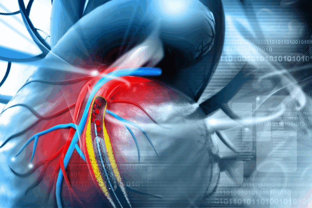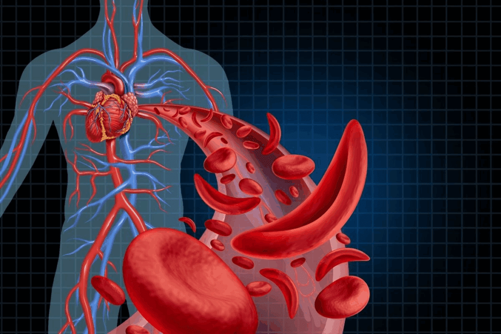
The human heart is a vital organ that needs a complex network of blood vessels to work right. At the heart of this network are the coronary arteries. They are key in bringing oxygen-rich blood to the heart’s different parts.Discover how many arteries go to the heart with 8 key facts about coronary arteries.
The coronary arteries split off first from the aorta. They give the heart’s layers the nutrients and oxygen they need. Knowing how the coronary circulation works in the heart is key to keeping it healthy.
At Liv Hospital, we know how important the coronary arteries are for heart health. Our team of experts is ready to give top-notch care and support to patients. We help those looking for the latest in medical treatments.

The heart needs oxygen and nutrients to work well. This is thanks to the coronary circulation. It’s a network of arteries that keeps the heart pumping blood efficiently. The heart’s blood supply is key to its performance.
The heart muscle, or myocardium, can’t get enough oxygen and nutrients from the blood it pumps. The coronary arteries give it what it needs directly. About 5% of the heart’s output goes to the myocardium through these arteries.
“The coronary circulation is the heart’s lifeline,” say heart experts. It gives the heart the oxygen and nutrients it needs to keep working. This blood supply is vital for the heart to pump blood effectively, no matter the situation.
Oxygen demand and coronary blood flow are closely linked. When the heart works harder, it needs more oxygen. This leads to more blood flow to meet that need. The heart’s circulation can change to meet different conditions, like exercise or stress.
In short, the coronary circulation is essential for the heart’s function. It ensures the heart muscle gets the oxygen and nutrients it needs. Knowing this helps us understand heart health and why keeping the coronary circulation healthy is important.

To find out how many arteries go to the heart, we need to look at the coronary artery system. The heart gets blood from two main coronary arteries. These arteries split into smaller ones to cover the whole heart. Knowing this is key to understanding how the heart works and staying healthy.
The heart’s blood flow comes from the left coronary artery and the right coronary artery. They start at the aortic root, just above the aortic valve. The left artery is bigger and feeds more of the heart muscle.
The right artery is smaller but just as important. It supplies the right atrium and parts of the ventricles, and the heart’s back side.
There’s a big network of smaller branches from the two main arteries. The left artery splits into the Left Anterior Descending (LAD) artery and the Left Circumflex (LCx) artery. The LAD goes down to the heart’s tip, covering the front and middle parts.
The LCx wraps around the heart, reaching the sides and sometimes the back. The right artery has branches like the Posterior Descending Artery (PDA), which goes to the heart’s back. It also has branches for the right ventricle.
“The coronary circulation is a complex network that is essential for maintaining the viability and function of the heart muscle.” – Medical Expert
| Artery | Primary Supply Area |
| Left Coronary Artery | Anterior wall, lateral wall, and part of the posterior wall |
| Right Coronary Artery | Right atrium, parts of right and left ventricles, posterior aspect |
| Left Anterior Descending (LAD) | Anterior wall and interventricular septum |
| Left Circumflex (LCx) | Lateral and sometimes posterior walls |
Knowing about the heart’s arteries is key for diagnosing and treating heart disease. The complex network of coronary arteries shows how vital heart circulation is.
The left coronary artery is key for the heart’s blood supply. It starts from the left aortic sinus of the aorta, just above the aortic valve. This artery is critical for supplying blood to a significant portion of the heart muscle.
It covers the anterior wall, lateral wall, and part of the posterior wall of the left ventricle. It also supplies the anterior two-thirds of the interventricular septum.
The left main coronary artery, also known as the left main stem or LM heart artery, is the start of the left coronary artery system. It is usually short, between 5 to 10 mm long, but can vary. The LM heart artery originates from the left aortic sinus and runs between the pulmonary trunk and the left atrial appendage.
One of the key characteristics of the LM heart artery is its variability in length and branching pattern. In some individuals, it may be quite long, while in others, it may be very short or even absent. This variability is important for clinicians to consider during diagnostic and interventional procedures.
The left anterior descending (LAD) artery is one of the two major branches of the left main coronary artery. It is often referred to as the “widow maker” due to the high mortality associated with occlusions in this artery. The LAD supplies blood to the anterior wall of the left ventricle and the anterior two-thirds of the interventricular septum.
The LAD runs down the anterior interventricular groove towards the apex of the heart. Along its path, it gives off several diagonal branches that supply the lateral wall of the left ventricle and septal perforators that supply the interventricular septum.
The left circumflex (LCx) artery is the other major branch of the left main coronary artery. It supplies blood to the lateral and posterior walls of the left ventricle. The LCx artery runs in the left atrioventricular groove, encircling the mitral valve.
The territory supplied by the LCx artery can vary significantly among individuals. In some cases, it may give off large obtuse marginal branches that supply a significant portion of the lateral wall. The LCx artery may also give off branches to the left atrium.
The right coronary artery is a key part of the heart’s blood flow. It starts from the anterior aortic sinus. It helps supply blood to the right atrium, right ventricle, and parts of the left ventricle.
The right coronary artery begins from the anterior aortic sinus of the aorta, just above the aortic valve. It then moves through the atrioventricular groove towards the heart’s crux. This is where the atrioventricular groove meets the posterior interventricular groove.
The right coronary artery has a significant branch called the posterior descending artery (PDA). Also known as the coronary artery posterior, it runs along the posterior interventricular groove. It supplies the posterior third of the interventricular septum and parts of the left and right ventricles.
The PDA is key because it often supplies the inferior wall of the heart.
“The posterior descending artery is a critical branch that supplies blood to the posterior aspect of the heart, including the interventricular septum.” – Cardiology Expert
The right coronary artery also has marginal branches. These supply the right margin of the heart. The most important is the right marginal artery, which runs along the acute margin of the heart. It supplies the right ventricle.
These branches are vital for ensuring the right ventricle gets enough blood.
| Artery | Origin | Course | Supply |
| Right Coronary Artery | Anterior aortic sinus | Atrioventricular groove | Right atrium, right ventricle, parts of left ventricle |
| Posterior Descending Artery (PDA) | Right Coronary Artery | Posterior interventricular groove | Posterior third of interventricular septum, parts of left and right ventricles |
| Right Marginal Artery | Right Coronary Artery | Acute margin of the heart | Right ventricle |
A network of secondary branches and smaller vessels is key to the heart’s blood supply. These vessels ensure the heart gets the oxygen and nutrients it needs to work well.
The left circumflex artery has obtuse marginal (OM) branches. These are important for the left ventricle’s lateral wall. OM arteries, or OM heart vessels, differ in number and size among people. They are vital for heart muscle health.
The left anterior descending (LAD) artery has diagonal branches for the left ventricle’s anterolateral wall. These are key for heart perfusion. The LAD also has septal perforators for the interventricular septum. The right coronary artery (RCA) has acute marginal branches for the right ventricle.
| Vessel | Origin | Area Supplied |
| Obtuse Marginal (OM) Arteries | Left Circumflex Artery | Lateral wall of the left ventricle |
| Diagonal Branches | Left Anterior Descending (LAD) Artery | Anterolateral wall of the left ventricle |
| Septal Perforators | Left Anterior Descending (LAD) Artery | Interventricular septum |
| Acute Marginals | Right Coronary Artery (RCA) | Right ventricle |
It’s important to understand the secondary branches and smaller vessels for heart health. Their role in maintaining heart function is huge. Seeing these vessels through imaging is key for diagnosing and treating heart disease.
It’s important to know how the coronary arteries deliver blood to the heart. They send oxygen-rich blood to all parts of the heart. This includes the left and right atria, ventricles, and the interventricular septum.
The ventricles are the heart’s main pumping chambers. They need a lot of blood. The left ventricle, with its thick walls, pumps blood all over the body. It gets a lot of its blood from the left anterior descending (LAD) artery.
The right coronary artery (RCA) mainly supplies the right ventricle. Sometimes, the left circumflex artery (LCx) also helps with this.
The atria get their blood from both the right and left coronary arteries. The right atrium mainly gets its blood from the RCA. The left atrium gets its blood from the LCx.
The interventricular septum, which separates the ventricles, gets blood from both the LAD and the PDA. The PDA usually comes from the RCA.
The heart’s blood supply has overlap and redundancy. This ensures the heart muscle stays perfused even if one artery is blocked. For example, the LAD and LCx can overlap, as can the RCA and LCx.
This redundancy is key for keeping the heart working even with coronary artery disease.
To show how blood is distributed in the heart, here’s a table:
| Heart Region | Primary Arterial Supply | Secondary Supply |
| Left Ventricle | LAD | LCx |
| Right Ventricle | RCA | LCx (in some cases) |
| Left Atrium | LCx | RCA (small branches) |
| Right Atrium | RCA | – |
| Interventricular Septum | LAD, PDA | – |
The heart’s complex network of coronary arteries makes sure all parts get the blood they need. Knowing this network is vital for diagnosing and treating heart disease.
Understanding coronary artery dominance patterns is key to knowing how the heart gets its blood. The way blood flows to the heart muscle varies from person to person. This is because of differences in how the coronary arteries distribute blood.
The right-dominant circulation is the most common. In this pattern, the right coronary artery (RCA) supplies the posterior descending artery (PDA). This artery runs along the heart’s posterior interventricular groove. About 70% to 80% of people have the PDA coming from the RCA, showing a right-dominant circulation.
This pattern is important because it affects how blood reaches different parts of the heart.
Characteristics of Right-Dominant Circulation:
While right-dominant circulation is common, there are other patterns. In left-dominant circulation, the left coronary artery (LCA) supplies the PDA, found in about 10% of people. Co-dominant circulation is when both the RCA and LCA supply the posterior heart.
| Dominance Pattern | Frequency | PDA Origin |
| Right-Dominant | 70-80% | RCA |
| Left-Dominant | ~10% | LCA |
| Co-Dominant | Variable | Both RCA and LCA |
The dominance pattern is very important in treating coronary artery disease (CAD). Knowing the dominance pattern helps doctors diagnose and treat CAD. This is because blockages in different arteries have different effects in different patterns.
Coronary artery dominance affects how the heart gets oxygen. The different patterns show how complex the heart’s blood flow is and how it adapts to the heart’s needs.
Understanding the coronary circulation is key for diagnosing and treating heart issues. Seeing how blood flows to the heart muscle is vital for heart health.
Coronary circulation diagrams are vital for grasping the complex network of arteries that feed the heart. These diagrams show the blood supply heart anatomy. They reveal how coronary arteries branch out and spread oxygenated blood to the heart muscle.
Healthcare experts use these diagrams to understand the link between coronary artery anatomy and heart function. This knowledge is essential for spotting coronary artery disease and planning treatments.
Medical imaging has improved, letting us see coronary arteries in great detail. Coronary artery photos from angiography and other methods give us important insights into the heart’s blood flow.
Today’s imaging, like coronary angiography, cardiac CT, and MRI, helps us see the arteries and spot problems. These images are key for making treatment choices, like angioplasty or bypass surgery.
By mixing coronary artery photos with patient data, doctors can create treatment plans that meet each person’s needs.
It’s key to know about the different ways coronary arteries can be structured. These arteries are vital for the heart’s blood supply. They can start from different places, spread out in various ways, and follow different paths.
One common difference is how the coronary arteries start. Usually, the left and right ones come from the left and right aortic sinuses. But sometimes, they share a common trunk. This is something doctors need to know, as it changes how they treat heart problems.
The way these arteries spread out can also differ. For example, the heart’s blood supply can be mostly from the right, left, or both sides. Knowing this helps doctors read heart scans and plan treatments.
Some rare coronary artery issues, like an anomalous origin, can be serious. They might cause heart problems or even sudden death, mainly when exercising. Spotting these issues needs careful attention and advanced scans.
These rare problems need special care. Doctors might need to operate or change what activities patients can do. Spotting these issues is vital for the best care for heart disease patients.
Doctors can better diagnose and treat heart disease by understanding both common and rare coronary artery variations. This knowledge is essential for top-notch care for heart patients.
Coronary anatomy is key in diagnosing and treating coronary artery disease. It’s vital for managing this disease, a major cause of death globally. We’ll look at how coronary artery disease and blockages affect us. We’ll also explore interventional procedures and surgical options.
Coronary artery disease happens when arteries narrow or block due to plaque buildup. This reduces blood flow to the heart, leading to angina, heart attacks, or other issues. Knowing the coronary artery anatomy is critical for treating this disease well.
Interventional methods like angioplasty and stenting open blocked arteries. Surgery, like CABG, might be needed for severe or complex blockages. The choice between these options depends on the blockage’s location, severity, and the patient’s health.
Knowing about coronary arteries is key to keeping your heart healthy. These arteries are vital for bringing blood to the heart. They play a big role in keeping your heart working well.
We’ve looked at the complex system of coronary arteries. This includes the left and right coronary arteries and their branches. It’s important to understand this to see why a healthy heart is so critical.
Learning about coronary arteries helps us see why we need to take care of our heart’s arteries. This knowledge lets us take steps to keep our heart healthy. It also helps us lower the risk of heart diseases.
In wrapping up our talk on coronary arteries, it’s clear that knowing about them is essential for heart health. By focusing on heart health, we can aim for a better future.
The heart gets blood from two main arteries, the left and right coronary arteries. These arteries split into many smaller ones.
The coronary arteries bring oxygen-rich blood to the heart muscle. This lets the heart work right and keep pumping.
Coronary circulation is the system of blood vessels that feeds the heart muscle. It’s key because the heart needs oxygen to work well.
The left coronary artery mainly feeds the left ventricle and atrium. The right coronary artery supplies the right ventricle and atrium, plus the heart’s back side.
The left coronary artery splits into the Left Anterior Descending (LAD) and Left Circumflex (LCx) arteries. These supply different heart areas.
Coronary artery dominance patterns, like right-dominant and left-dominant, help doctors understand heart disease. They also guide treatment plans.
Coronary circulation is shown through diagrams, photos, and imaging like angiography and cardiac CT scans.
Variations in coronary arteries can raise the risk of heart disease. They also complicate treatments.
Coronary artery disease can block arteries, cutting off blood to the heart. This can lead to heart attacks or other serious issues.
Treatments include angioplasty and stenting, and sometimes surgery like coronary artery bypass grafting.
Knowing coronary anatomy is key for diagnosing and treating heart disease. It helps plan treatments and surgeries.
Subscribe to our e-newsletter to stay informed about the latest innovations in the world of health and exclusive offers!
WhatsApp us