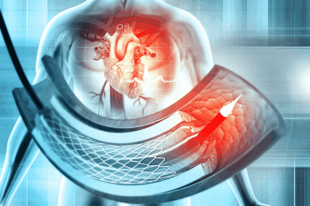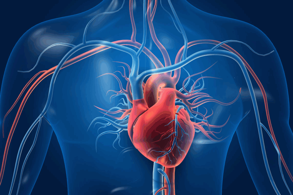
At Liv Hospital, we know how vital coronary vessels are for heart health. The heart needs a steady blood supply to work right. The coronary arteries are key in bringing oxygen-rich blood to the heart muscle.
The coronary circulation is a network of vessels that feed and drain the heart. These arteries circle the heart like a crown, keeping it healthy. Blockages in these arteries lead to heart disease worldwide.
We’ll dive into important facts about coronary arteries, heart areas, and the paths that keep your heart pumping. Knowing the details of blood supply heart anatomy helps us understand heart health better.

Coronary vessels are key for bringing oxygen and nutrients to the heart. The heart, being a muscle, needs these to keep working. This is done through the coronary circulation, a network of vessels that feed the heart muscle.
The main arteries for this are the left and right coronary arteries. They start from the aortic sinuses in the aorta, the biggest artery. The left main (LM) heart artery, a part of the left coronary artery, splits into smaller branches. These supply different parts of the heart.
The heart’s blood supply comes from coronary arteries. These arteries split into smaller ones, making a detailed network. This network is vital for the heart’s health and function.
| Coronary Artery | Region Supplied | Function |
| Left Main (LM) Heart Artery | Left ventricle, left atrium | Supplies blood to the left side of the heart |
| Right Coronary Artery | Right ventricle, right atrium, parts of the left ventricle | Supplies blood to the right side of the heart and parts of the left ventricle |
Getting oxygen to the heart muscle is key. The heart muscle, or myocardium, needs oxygen to keep pumping. The coronary arteries make sure the myocardium gets enough oxygen and nutrients by bringing blood rich in oxygen.
The coronary circulation diagram shows the heart’s blood vessel network. Knowing this diagram helps us understand how oxygen and nutrients reach the heart muscle.
In short, the heart’s blood supply comes from the left and right coronary arteries. These arteries branch out to give the heart muscle the oxygen and nutrients it needs. The coronary circulation diagram is a great tool for seeing this complex network.

The heart’s blood supply comes from two main arteries: the left and right coronary arteries. Knowing how these arteries work is key to treating heart disease.
The left main (LM) artery starts from the left aortic sinus. It’s vital for the heart’s blood flow. It splits into two main branches: the left anterior descending (LAD) artery and the left circumflex (LCx) artery.
The LAD artery goes down the front of the heart, supplying the front wall. The LCx artery runs along the left side, giving blood to the sides and back of the heart.
The right coronary artery (RCA) starts from the right aortic sinus. It runs along the right side of the heart, supplying blood to the right atrium, parts of the left atrium, and the right ventricle. It has a branch called the right marginal artery for the right ventricle’s side.
In most people, the RCA also has a branch called the posterior descending artery (PDA). This artery goes to the back of the heart, supplying the back third of the heart’s wall.
Doctors and surgeons need to know about the left and right coronary arteries. This knowledge helps them with heart surgeries and treatments.
The coronary circulation is a complex network with several key branches. These branches are vital for supplying blood to the heart muscle. This ensures the heart works properly. We will look at two important branches: the obtuse marginal artery and the posterior descending artery.
The obtuse marginal (OM) artery comes from the left circumflex artery. It supplies the lateral wall of the left ventricle, a key area for heart function. The OM artery’s function is to provide oxygenated blood to this area, keeping the heart muscle healthy and functional.
A leading cardiologist notes, “The obtuse marginal artery is a vital vessel that can significantly impact the heart’s overall performance.” This shows how important this artery is for heart health.
The posterior descending artery (PDA) is another key branch in the coronary circulation. It usually comes from the right coronary artery and supplies the posterior third of the interventricular septum. This region is vital for the heart’s electrical conduction system, and the PDA plays a key role in maintaining its function.
The posterior circulation, which includes the PDA, is critical for supplying blood to the heart’s posterior regions. This circulation ensures that the heart muscle receives the necessary oxygen and nutrients for optimal performance.
As we continue to explore the coronary circulation, it becomes clear that understanding these branches is vital for diagnosing and treating coronary artery disease. By recognizing the importance of these vessels, we can better appreciate the complexities of heart anatomy and function.
Knowing how blood flows to the heart is key for treating heart disease. The heart’s blood supply is a detailed network. It makes sure the heart muscle gets enough oxygen and nutrients.
The Left Anterior Descending (LAD) artery mainly feeds the anterior heart region. It’s a big branch of the left coronary artery. It’s vital for the blood supply to the front part of the left ventricle and the interventricular septum.
The LAD has branches like the diagonal arteries. These supply the lateral wall of the left ventricle. This ensures the heart’s front gets plenty of oxygen-rich blood.
The Left Circumflex (LCx) artery and its branches, like the obtuse marginal (OM) branches, feed the lateral wall. The LCx artery goes around the heart’s side. It provides blood to the left ventricle’s lateral and posterior walls.
The posterior heart region gets its blood mainly from the Right Coronary Artery (RCA) or the Left Circumflex artery. In right-dominant circulation, the RCA has the posterior descending artery (PDA). This artery supplies the posterior third of the interventricular septum.
The posterior descending artery is key for the heart’s back. It makes sure this area gets enough blood.
In summary, the heart’s blood supply is complex and essential. It ensures every part of the heart gets the oxygen and nutrients it needs. Knowing this is vital for treating heart disease.
The heart’s blood supply is kept up by a complex system called coronary circulation. This network is key for delivering oxygen and nutrients to the heart muscle. It lets the heart work right. The way coronary circulation spreads out can differ a lot between people, affecting heart health.
About 85% of people have a right dominant circulation. This means the right coronary artery leads to the posterior descending artery. On the other hand, 10% have a left dominant circulation. Here, the left circumflex artery leads to the posterior descending artery.
Knowing if someone has a right or left dominant circulation is key for heart disease treatment. It helps us understand the risk for heart problems. We need to look at these differences when checking on heart health.
| Dominance Pattern | Prevalence | Characteristics |
| Right Dominant | 85% | Right coronary artery gives rise to the posterior descending artery |
| Left Dominant | 10% | Left circumflex artery gives rise to the posterior descending artery |
Coronary anatomy can vary a lot, not just in dominance. The start, path, and branches of coronary arteries can differ. These differences can affect how we diagnose and treat heart disease.
“Understanding the variations in coronary anatomy is essential for accurate diagnosis and effective treatment of coronary artery disease.”
It’s important to know about these differences when looking at images and planning treatments. New imaging methods help spot these variations. They guide us in making the best treatment plans.
In summary, the patterns of coronary circulation, including right vs. left dominance and other anatomy variations, are very important for heart health. Knowing these patterns helps us give the best care to those with heart disease.
The coronary arteries are key in getting blood to the heart’s chambers and ventricles. The heart needs oxygen and nutrients to work well. The coronary circulation makes sure the heart muscle gets what it needs.
The ventricles pump blood and need a lot of it. The left anterior descending artery (LAD) and the posterior descending artery (PDA) are important for this. The LAD goes to the front and part of the middle wall. The PDA, from the right coronary artery (RCA), goes to the back and bottom of the left ventricle.
“The way the ventricle arteries spread out is key for the heart’s pumping,” say heart experts. People can have different patterns, like right-dominant or left-dominant circulation.
The atria need blood too, even though they’re thinner. The sinoatrial (SA) nodal branch, from the right coronary artery (RCA), feeds the SA node. This is a big part of the heart’s electrical system. The atrial branches from the RCA and the left circumflex artery (LCx) make sure the atria get enough oxygen.
The heart’s electrical system, including the atrioventricular (AV) node and the bundle of His, gets its blood from the AV nodal branch. This branch often comes from the RCA. This network helps the heart’s electrical signals move well.
In summary, getting blood to the heart’s chambers and ventricles is complex but vital. Knowing how the ventricle arteries and atrial circulation work, and how they supply the heart’s electrical system, helps us understand the heart’s function. It also helps us find and fix any problems.
Coronary artery disease is complex. It involves atherosclerosis and plaque buildup. These factors harm the coronary arteries, which are key to the heart’s blood supply.
Atherosclerosis is a chronic inflammation that leads to coronary artery disease. It causes plaques to form in the arteries. These plaques can rupture, leading to heart attacks.
Atherosclerosis is when lipids, inflammatory cells, and fibrous elements build up in arteries. It starts with fatty streaks and grows into more serious lesions.
Many factors contribute to plaque formation, like lipid metabolism disorders, high blood pressure, and smoking. As plaques grow, they block blood flow, causing heart problems.
Blocked coronary arteries can cause angina pectoris and myocardial infarction. Angina happens when the heart doesn’t get enough oxygen, usually during stress or exercise.
Myocardial infarction, or heart attack, occurs when a blocked artery damages heart muscle. This is often due to a plaque rupture causing blood clots.
Knowing the effects of blocked arteries is key to treating coronary artery disease.
Coronary vessel imaging has changed cardiology a lot. It helps doctors diagnose and treat heart issues better. This imaging is key to understanding heart vessel structure and function. It’s vital for managing heart disease.
Coronary angiography lets doctors see the heart’s arteries. It uses a contrast agent to spot blockages. Coronary artery photos from this help plan treatments.
A top cardiologist says, “Coronary angiography is the best way to find heart disease. It gives clear images for treatment.”
“Seeing heart arteries live has changed how we treat heart disease,” said Medical Expert, a famous cardiologist.
| Imaging Technique | Description | Advantages |
| Coronary Angiography | Involves injecting contrast agent into coronary arteries | Provides detailed images of coronary arteries, guides interventions |
| CT Coronary Angiography | Non-invasive imaging using CT scans | Less invasive, useful for assessing plaque burden |
Coronary circulation diagrams are key for understanding heart vessel anatomy. They come from advanced scans like CT or MRI. These diagrams help doctors plan treatments.
Advanced imaging, like coronary circulation diagrams, has made diagnosis better. These tools help doctors create treatment plans that fit each patient’s needs.
We use these advanced imaging methods for full patient care. By combining coronary artery photos and angiography with detailed diagrams, we make accurate diagnoses and effective treatments.
Research on coronary vessels has greatly improved our understanding of heart disease. At Liv Hospital, we aim to offer top-notch healthcare. We also support international patients fully.
Treatment for heart disease has seen big changes. New methods are making care better for patients. Our work in prevention and treatment shows the need for ongoing research.
New paths in coronary vessel research are showing promise. The future of heart health will blend new tech and care tailored to each patient. We’re committed to giving the best care for heart disease patients. We use the latest research to guide our treatments.
Coronary vessels carry oxygen-rich blood to the heart muscle. This is key to keeping the heart healthy.
The left coronary artery splits into the LAD and LCx. The right coronary artery leads to the right marginal artery and often the posterior descending artery.
Knowing about coronary circulation helps in diagnosing and treating heart disease. It’s vital for blood flow to the heart.
The dominance pattern affects heart health risks. It’s important for understanding heart anatomy.
Coronary arteries are key in supplying blood to the heart muscle. They also reach the ventricles and atria.
Coronary artery disease is caused by plaque buildup. It can lead to heart blockages, chest pain, and heart attacks.
Techniques like angiography and advanced imaging help diagnose and treat heart disease. They show the state of coronary vessels.
Knowing the blood supply is key for treating heart disease. It ensures the heart gets enough oxygen and nutrients.
These arteries are vital in coronary circulation. They supply blood to specific heart areas.
Different coronary anatomy can impact heart health risks. It’s important for understanding heart circulation.
National Center for Biotechnology Information. (2025). Coronary Vessels 9 Key Facts on Heart Arteries. Retrieved from https://pubmed.ncbi.nlm.nih.gov/12557422/
Subscribe to our e-newsletter to stay informed about the latest innovations in the world of health and exclusive offers!
WhatsApp us