Last Updated on November 25, 2025 by Ugurkan Demir

Early detection is key in treating bladder cancer. Our hospital uses the latest diagnostic tools to spot the disease early. This means we can start treatment quickly, leading to better results.
We put our patients first, creating treatment plans that fit their needs. Our team works closely with each person to ensure they get the care they deserve.
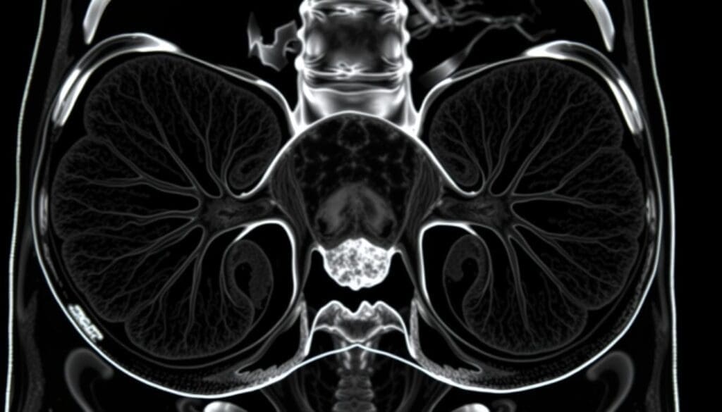
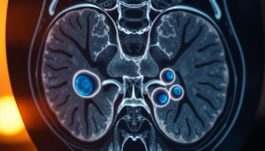
Explore 10 visualization tools for bladder cancer, including key bladder cancer photos pictures and images used in diagnosis.
Managing bladder cancer well needs good visualization. Tools like white light cystoscopy and narrow-band imaging have changed urology a lot.
These methods help doctors spot tumors early. This means they can start treatment quickly.
White light cystoscopy is a key diagnostic tool. It lets doctors see inside the bladder. This is vital for spotting bladder cancer and other issues.
Knowing the good and bad sides of this method helps patients. It aids in making choices about their health care.

Blue light cystoscopy is a tool that helps find bladder cancer better. It uses a special light to spot tumors that white light can’t see.
Research shows it can lower the chance of cancer coming back. It also helps in treating bladder cancer more effectively. This is key for those with serious tumors or past cancer.
Adding blue light cystoscopy to the tools for diagnosing bladder cancer is a big plus. It makes it easier for doctors to see tumors, helping them treat patients better.
Narrow-band imaging (NBI) is a key tool in finding and diagnosing bladder cancer. It shows the blood vessels and lesions on the bladder wall. This makes tumors easier to spot, leading to better diagnosis and treatment.
NBI shines specific light wavelengths on the bladder. This makes it easier to see abnormalities. It’s great for spotting flat lesions and tumors that white light can’t show.
NBI has many advantages in bladder cancer detection. It gives a clear view of the bladder wall. This helps doctors find tumors early, which improves treatment results and patient chances of recovery.
In clinics, NBI helps in diagnosing and treating bladder cancer. It’s great for finding small tumors or lesions that other methods can’t see. With NBI, doctors can make better treatment plans and care for patients better.
NBI is a powerful tool in bladder cancer detection. It highlights blood vessels and lesions, making it vital for doctors. Using NBI, doctors can improve patient care and treatment plans.

We use ultrasound imaging to check for bladder cancer in different ways. This method is non-invasive and helps us see the bladder wall and find tumors.
Ultrasound lets us check the bladder wall’s thickness and look for any issues. Transabdominal ultrasound is often used. It gives a full view of the bladder and nearby areas.
The process includes:
Ultrasound helps spot bladder cancer tumors by finding oddities in the bladder wall. Transurethral ultrasound gives a closer look at the tumor and its location.
Signs of bladder cancer tumors on ultrasound are:
Ultrasound has many uses in bladder cancer care. It’s good for:
Using ultrasound, doctors can make better choices for patient care and better results.

CT urography is a vital tool for diagnosing and staging bladder cancer. It uses multi-phase imaging to give a detailed view of the urinary tract. This helps doctors detect and manage bladder cancer more effectively.
CT urography uses a multi-phase imaging protocol. It includes non-contrast, corticomedullary, nephrographic, and excretory phases. This approach ensures all parts of the urinary tract are seen, helping spot even small lesions.
The multi-phase protocol is key for finding bladder cancer at different stages. It helps measure the tumor’s size, location, and if it has spread to nearby tissues.
CT urography is great for spotting upper tract urothelial carcinoma (UTUC). UTUC is a rare but aggressive cancer in the upper urinary tract.
CT urography shows the whole urinary tract, from kidneys to bladder. It’s a key tool for finding UTUC, even small tumors that other methods miss.
CT urography is not just for finding bladder cancer and UTUC. It’s also key for tumor staging and finding metastasis. The detailed images help doctors see how far the tumor has spread, which is vital for treatment planning.
Being able to accurately stage tumors and find metastasis greatly impacts patient care. It leads to more tailored treatment plans and better treatment chances.

Advanced MRI techniques are key in seeing and staging bladder cancer. MRI is great for its clear view of soft tissues. This is vital for accurate tumor staging and planning treatments. MRI’s ability to use many parameters helps fully assess bladder cancer.
Multiparametric MRI uses T2-weighted imaging, diffusion-weighted imaging (DWI), and dynamic contrast-enhanced (DCE) MRI. This mix gives a detailed look at bladder tumors. DWI is great for spotting cancer because it shows where water moves slowly.
MRI’s clear view of soft tissues is key for staging bladder cancer. It helps see how far the tumor has spread. This is important for choosing the right treatment.
DCE-MRI uses contrast agents to look at tumor blood flow. It helps find aggressive tumors and check how well treatments work.
The table below shows the main MRI methods for bladder cancer and what they do:
| MRI Technique | Application |
| Multiparametric MRI | Comprehensive tumor characterization |
| Diffusion-Weighted Imaging | Detection of malignant lesions |
| Dynamic Contrast-Enhanced MRI | Assessment of tumor vascularity and perfusion |
In summary, MRI methods are a strong tool for seeing bladder cancer. They give vital info for staging and planning treatments. By using many MRI techniques, doctors can make better choices.
PET-CT imaging has changed how we find metastatic bladder cancer. It gives a full view of the disease. This is because it combines PET’s functional info with CT’s body details. This helps doctors plan better treatments.
The benefits of using PET-CT for finding metastatic disease are:
Even though PET-CT is very useful, it has some downsides:
In summary, PET-CT imaging is a key tool for managing metastatic bladder cancer. Its ability to show both the disease’s function and structure makes it vital for patient care.
Photoacoustic imaging is a new field that mixes optical and ultrasound imaging. It creates detailed pictures of tissues. This method is promising for diagnosing and treating many health issues, like cancer.
This technology works by using light to heat tissues, which then makes sound waves. This way, it lets doctors see inside the body without surgery.
Photoacoustic imaging is great for finding bladder cancer early. It helps doctors spot tumors before they grow big. This could lead to better treatment results.
Its main advantages are its high accuracy and detailed images. These features make it a key tool for doctors. It helps them diagnose and treat diseases more effectively.
In short, photoacoustic imaging is a game-changer in medicine. It offers clear images of tissues and is non-invasive. This makes it perfect for diagnosing many health problems, including bladder cancer.
Artificial intelligence is changing how we diagnose bladder cancer. It makes image interpretation more accurate and faster. AI algorithms work with medical images to spot bladder cancer early.
This helps doctors catch tumors before they grow. It also cuts down on mistakes in diagnosis. Thanks to AI, patients get better care and have a higher chance of survival.
Our journey through bladder cancer care shows how big a leap visualization techniques have made. These new imaging tools help us diagnose and treat better. At Liv Hospital, we’re dedicated to top-notch care for our patients.
We’re excited to keep exploring new ways to fight bladder cancer. This will help us achieve even better results for our patients.
Imaging is key in finding and managing bladder cancer. It lets doctors see tumors and how far the disease has spread.
Many imaging methods are used, like white light cystoscopy, narrow-band imaging, and ultrasound. Also, CT scans, MRI, and PET-CT scans are used.
White light cystoscopy uses a scope to look inside the bladder. It helps doctors spot tumors and other issues.
Narrow-band imaging makes blood vessels and tumors stand out. This helps find bladder cancer early, when it’s easier to treat.
Ultrasound uses sound waves to create images of the bladder. It helps find tumors and see how big they are.
CT scans give detailed pictures of the bladder and nearby areas. They help find tumors and see how big they are.
MRI uses a magnetic field and radio waves to make detailed images. It helps find tumors and see how big they are.
PET-CT scans combine PET and CT scans. They help find and stage bladder cancer by showing how active the cancer is.
Artificial intelligence helps analyze images from CT and MRI scans. It can spot bladder cancer more accurately than humans.
The future of bladder cancer care will see better imaging like MRI and CT scans. Artificial intelligence will also play a big role in making diagnoses and treatment plans better.
National Center for Biotechnology Information. (2025). Visualization Tools for Bladder Cancer Early detection is. Retrieved from https://pubmed.ncbi.nlm.nih.gov/11196988/
Subscribe to our e-newsletter to stay informed about the latest innovations in the world of health and exclusive offers!