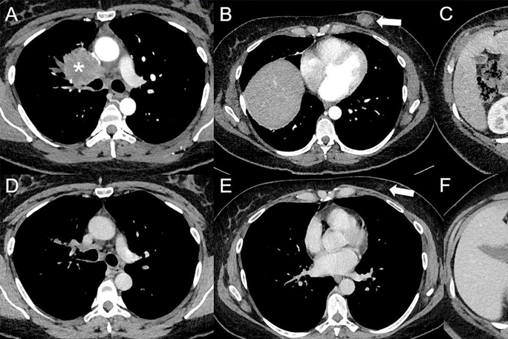
A PET scan is a diagnostic examination that involves getting images of the body based on the detection of radiation from the emission of positrons. This medical imaging technique plays a crucial role in cancer detection and diagnostic imaging. However, there are certain cancers not visible PET scan, including some slow-growing tumors, low-grade lymphomas, and prostate cancer, since these may not absorb enough of the radioactive tracer to appear clearly. To ensure accurate results, proper preparation is essential, and in some cases, additional imaging methods like MRI or CT scans may be recommended alongside PET for comprehensive evaluation.
Understanding the preparation required for a PET scan can help reduce anxiety and ensure a smooth experience.

Positron Emission Tomography (PET) scans represent a significant advancement in medical imaging, offering a unique window into the body’s functions. This technology has revolutionized the field of diagnostic imaging by providing detailed insights into the body’s cellular activity.
PET scans use a radioactive material, known as a radiopharmaceutical, which is made up of a radioactive isotope attached to a substance used in the body, typically glucose. This radiopharmaceutical is injected into the body, where it accumulates in areas with high chemical activity, such as growing cancer cells. The PET scan detects the radiation emitted by the radiopharmaceutical, creating detailed images of the body’s internal structures and functions.
Unlike other imaging tests that primarily provide anatomical information, PET scans offer functional information about the body’s tissues and organs. For instance, while CT scans and MRI provide detailed images of internal structures, PET scans reveal how these structures are functioning at a cellular level. This unique capability makes PET scans particularly valuable for diagnosing and monitoring conditions like cancer, neurological disorders, and cardiovascular diseases.
| Imaging Test | Primary Use | Information Provided |
| PET Scan | Cancer diagnosis, neurological disorders, cardiovascular diseases | Functional information about tissues and organs |
| CT Scan | Anatomical imaging, injury diagnosis | Detailed anatomical images |
| MRI | Soft tissue imaging, neurological disorders | High-resolution images of soft tissues |
The accuracy of PET scans in detecting various medical conditions, including cancer, is well-documented. However, it’s essential to understand the limits of PET scan technology, particularly in detecting certain types of cancer or in cases where the radiopharmaceutical uptake is low. Understanding these aspects is crucial for interpreting PET scan results accurately and making informed decisions about patient care.

PET scans have become a crucial diagnostic tool for various medical conditions. Their ability to provide detailed images of the body’s metabolic activity makes them invaluable in diagnosing and managing a range of health issues.
PET scans are widely used in oncology for cancer diagnosis, staging, and monitoring treatment response. They help identify the extent of cancer spread, which is critical for determining the appropriate treatment plan. The sensitivity of PET scans in detecting cancerous tissues makes them a preferred choice for assessing cancer progression.
In neurology, PET scans are used to diagnose and monitor conditions such as Alzheimer’s disease, Parkinson’s disease, and epilepsy. They provide insights into the brain’s metabolic activity, helping clinicians diagnose neurological disorders more accurately. The detailed imaging offered by PET scans enables healthcare providers to understand the progression of these conditions.
PET scans are also applied in cardiology to assess myocardial viability and blood flow. They help diagnose coronary artery disease and evaluate the heart’s function. The information obtained from PET scans is crucial for determining the best course of treatment for cardiovascular conditions.
| Medical Condition | PET Scan Application | Benefits |
| Cancer | Diagnosis, Staging, Treatment Monitoring | Accurate assessment of cancer spread and treatment response |
| Neurological Disorders | Diagnosis, Monitoring Disease Progression | Detailed insights into brain metabolic activity |
| Cardiovascular Diseases | Myocardial Viability, Blood Flow Assessment | Evaluation of heart function and diagnosis of coronary artery disease |
The table above illustrates the diverse applications of PET scans across different medical conditions, highlighting their diagnostic value and the benefits they offer in patient care.
Different types of PET scans are utilized in medical diagnostics, each with its unique applications. The variety in PET scans allows healthcare professionals to choose the most appropriate diagnostic tool based on the patient’s condition and the specific information required.
FDG-PET (Fluorodeoxyglucose-PET) scans are among the most commonly used PET scans. They involve the use of a radioactive glucose molecule, FDG, which is taken up by cells in the body. Since cancer cells metabolize glucose at a higher rate than normal cells, FDG-PET scans are particularly useful in oncology for detecting tumors, assessing cancer spread, and monitoring treatment response.
Amyloid PET scans are specifically designed to detect amyloid plaques in the brain, a hallmark of Alzheimer’s disease. These scans use a different tracer that binds to amyloid, helping in the diagnosis of Alzheimer’s and differentiation from other causes of dementia. Amyloid PET scans are valuable in neurology for assessing patients with cognitive decline.
Combining PET with other imaging modalities like CT or MRI enhances diagnostic accuracy. Combined PET/CT scans provide both metabolic information from PET and anatomical details from CT, aiding in precise localization of abnormalities. Similarly, PET/MRI scans offer a powerful diagnostic tool by combining the metabolic data from PET with the excellent soft tissue contrast of MRI, particularly useful in complex cases or when detailed soft tissue imaging is required.
The choice of PET scan type depends on the clinical question being addressed, the part of the body being examined, and the patient’s specific condition. Understanding the different types of PET scans and their applications is crucial for effective diagnosis and treatment planning.
To get the most out of your PET scan, it’s crucial to follow the pre-scan instructions carefully. Proper preparation is essential for obtaining accurate and reliable diagnostic results.
Preparation for a PET scan begins 24-48 hours before the actual procedure. During this time, patients are advised to follow a limited carbohydrate diet to help ensure that the PET scan images are clear and accurate. It’s also recommended to avoid eating or drinking anything except water for 6 hours before the exam.
Additionally, patients should inform their healthcare provider about any medications they are taking, as some may interfere with the PET scan results. It’s also a good idea to stay hydrated by drinking plenty of water during this period.
On the day of your PET scan, it’s essential to bring certain items to your appointment. These include:
Having these items readily available will help ensure a smooth and efficient process.
When preparing for your PET scan, wear comfortable, loose-fitting clothing that is easy to remove if needed. Avoid wearing clothing with metal zippers, buckles, or jewelry, as these can interfere with the imaging process. You may be asked to change into a hospital gown during the procedure.
It’s also a good idea to leave any valuable accessories at home to avoid loss or damage during the scan.
A crucial aspect of PET scan preparation is adhering to specific dietary guidelines to ensure accurate results. The dietary restrictions are designed to optimize the uptake of the radiotracer used in PET scans, which is crucial for obtaining clear and useful images.
Patients are typically advised to avoid consuming foods and beverages that could interfere with the PET scan results. This includes avoiding sugary foods and drinks, as well as those high in carbohydrates, for a certain period before the scan. Caffeine and alcohol should also be avoided as they can affect the body’s metabolism and potentially alter the distribution of the radiotracer.
It’s also recommended to limit or avoid foods that are high in sugar or simple carbohydrates for at least 24 hours before the scan. This helps in maintaining stable blood glucose levels, which is particularly important for FDG-PET scans used in cancer detection.
Patients are usually required to fast for a certain period before undergoing a PET scan. The fasting requirements can vary, but typically, patients are advised not to eat or drink anything except water for 4 to 6 hours before the scan. This fasting period helps ensure that the body is in a fasting state, which can improve the uptake of the radiotracer.
| Fasting Duration | Allowed Intake | Prohibited Intake |
| 4-6 hours | Water | Food, sugary drinks, caffeine |
| 24 hours | Low-carb, low-sugar diet | High-carb, high-sugar foods |
Adhering to these dietary restrictions and fasting requirements is crucial for the success of the PET scan. By following these guidelines, patients can help ensure that their PET scan results are accurate and reliable, which is vital for diagnosis and treatment planning.
Preparing for a PET scan involves more than just showing up at the appointment; it requires careful consideration of your medications. Certain medications can affect the accuracy of PET scan results, making it crucial for patients to inform their healthcare providers about all the medications they are taking.
Some medications can alter the distribution of the radiotracer used in PET scans, potentially leading to inaccurate results. For instance, certain diabetes medications can affect blood sugar levels, which in turn may impact the uptake of the radiotracer. It’s essential to discuss your medication regimen with your doctor to determine the best course of action before your PET scan.
For diabetic patients, managing medication before a PET scan is critical. The goal is to ensure that blood sugar levels are within a range that won’t interfere with the PET scan results. This may involve adjusting the dosage or timing of diabetes medications.
By carefully managing your medications and following your doctor’s instructions, you can help ensure that your PET scan results are accurate and reliable.
The preparation for a PET scan is not one-size-fits-all; it differs significantly depending on whether you’re undergoing a brain, cardiac, or whole-body scan. Each type of scan has its unique requirements to ensure that the images obtained are clear and useful for diagnosis.
For a brain PET scan, patients may be required to arrive with a companion, as the scan can be lengthy and may cause disorientation in some cases. It’s essential to follow the specific dietary instructions provided by your healthcare provider, as certain foods or beverages may interfere with the scan results.
Preparing for a cardiac PET scan involves avoiding caffeine and certain medications that can affect heart rate, as directed by your doctor. Patients are also advised to wear comfortable clothing and avoid heavy meals before the scan.
For a whole-body PET scan, patients are typically required to fast for a certain period before the scan and may need to avoid strenuous exercise. It’s also recommended to stay hydrated and follow any specific instructions given by your healthcare provider regarding medication and diet.
Understanding these special preparations can help ensure that your PET scan is conducted smoothly and that the results are as accurate as possible. Always consult with your healthcare provider for personalized instructions.
The day of your PET scan is a crucial step in your diagnostic journey, involving a few key procedures before the actual scan takes place. Understanding what to expect can help reduce anxiety and ensure that you’re prepared for the process.
Upon arrival, you’ll check in at the reception desk, where you’ll be asked to provide identification and any necessary paperwork. After check-in, you’ll be directed to a waiting area until you’re called for preparation. It’s a good idea to arrive a little early to complete any last-minute paperwork.
Once you’re called, you’ll be taken to a room where a healthcare professional will administer a radiotracer injection into a vein in your arm. This radiotracer is a crucial component of the PET scan procedure, as it helps highlight areas of the body that are being examined.
After the radiotracer injection, you’ll be required to wait for about an hour before the scan. This waiting period allows the radiotracer to be absorbed by the part of the body being studied. During this time, you may be asked to remain quiet and still to ensure the radiotracer distributes evenly.
The patient experience during this waiting period can vary, but most facilities are designed to be comfortable and relaxing. Once the waiting period is over, you’ll be taken to the PET scanner for the imaging procedure, which is a critical part of diagnostic imaging.
By understanding the steps involved in the PET scan process, you can better prepare yourself for what to expect, making the experience as smooth as possible.
When preparing for a PET scan, understanding the procedure can help alleviate concerns and anxiety. The PET scan is a sophisticated diagnostic tool that requires patients to be still and cooperative during the scanning process.
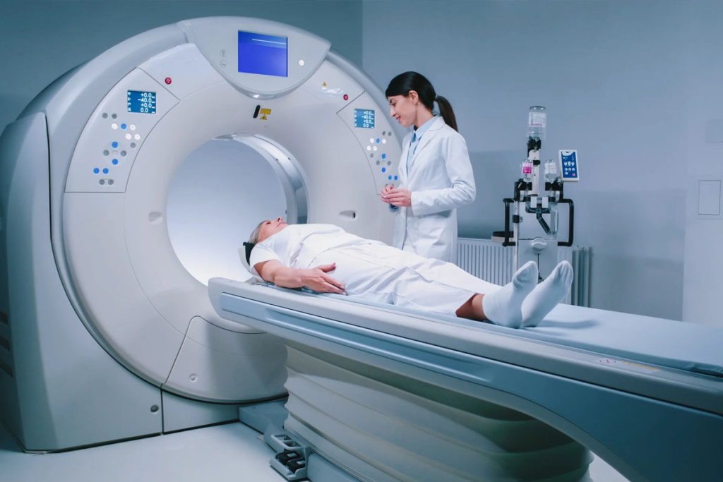
Patients are required to lie still on an exam table, which moves through the PET scanner. The duration of the scan can vary but typically lasts between 30 to 60 minutes. It is crucial to remain as still as possible to ensure that the images produced are clear and useful for diagnostic purposes.
The table below outlines the typical steps and timeline for a PET scan procedure:
| Step | Duration | Description |
| Preparation | 15-30 minutes | Getting ready for the scan, including radiotracer injection. |
| Scanning | 30-60 minutes | The actual PET scan procedure. |
| Total Time | 45-90 minutes | Overall time spent at the scanning facility. |
For patients who experience claustrophobia or anxiety, there are several strategies that can help make the PET scan procedure more comfortable. Open PET scanners are available for those who feel anxious about enclosed spaces. Additionally, patients can discuss their concerns with their healthcare provider, who may prescribe medication to help manage anxiety.
“The key to a successful PET scan is preparation and understanding of the process. Patients who are well-informed tend to experience less anxiety.” – Dr. Jane Smith, Radiologist
By understanding what to expect during the PET scan procedure, patients can better prepare themselves, reducing anxiety and ensuring a smoother experience.
Once your PET scan is complete, you’ll need to follow certain guidelines to help your body eliminate the radiotracer and minimize any potential risks. Proper post-scan care is essential for ensuring your safety and the accuracy of the scan results.
Drinking plenty of fluids is crucial after a PET scan to help flush out the radiotracer from your body. Hydration plays a key role in eliminating the tracer, reducing the risk of any adverse effects. It’s recommended to drink water or other non-caffeinated beverages throughout the day following your scan.
While most patients can resume their normal activities immediately after a PET scan, there are some precautions to consider. You may be advised to avoid close contact with pregnant women, young children, or individuals with compromised immune systems for a few hours after the scan. Additionally, it’s a good idea to follow any specific instructions provided by your healthcare provider regarding activity restrictions.
By following these guidelines, you can ensure a safe and effective recovery after your PET scan. If you have any concerns or questions, don’t hesitate to reach out to your healthcare team for guidance.
PET scans have revolutionized cancer diagnosis, but understanding their limitations is crucial for comprehensive patient care. While PET scans are highly effective for many types of cancer, there are instances where they may not provide a clear diagnosis.
One of the primary limitations of PET scans is their reliance on the uptake of Fluorodeoxyglucose (FDG), a glucose molecule tagged with a radioactive tracer. Cancers with low FDG uptake can be particularly challenging to detect using PET scans.
Certain types of cancers are known to have low metabolic activity, making them less visible on PET scans. These include:
For cancers that are not readily visible on PET scans, alternative imaging methods can provide valuable diagnostic information. These include:
In some cases, a combination of imaging modalities may be used to ensure an accurate diagnosis. Understanding the strengths and limitations of each imaging technique is crucial for selecting the most appropriate diagnostic pathway.
By recognizing the limitations of PET scans and utilizing alternative imaging methods when necessary, healthcare providers can ensure that patients receive the most accurate diagnosis and effective treatment plan.
PET scans, while valuable for diagnostic purposes, come with certain risks that patients should be aware of. The primary concerns include radiation exposure and the potential for allergic reactions to the radiotracer used during the scan.
The dose of radioactive material given during a PET scan is small, but there is still a risk of radiation exposure. This exposure is a concern because it has the potential to increase the risk of developing cancer later in life. However, it’s worth noting that the benefits of the PET scan often outweigh the risks, especially when used for diagnosing and staging cancer or monitoring treatment response.
Some patients may experience an allergic reaction to the radiotracer used in PET scans. Symptoms can range from mild to severe and include itching, hives, and difficulty breathing in severe cases. Other complications can include:
It’s essential for patients to discuss their medical history and any concerns with their healthcare provider before undergoing a PET scan. This includes informing them about any allergies, previous reactions to contrast agents, or other medical conditions that could affect the safety of the procedure.
Understanding insurance coverage for PET scans is crucial for patients to manage their diagnostic expenses effectively. PET scans are a valuable diagnostic tool, but their cost can be significant, making insurance coverage a critical factor in accessing this technology.
Insurance coverage for PET scans varies among different providers and policies. Medicare typically covers PET scans for certain medical conditions, such as cancer diagnosis and staging, neurological disorders, and cardiovascular diseases, when deemed medically necessary. Private insurance companies also cover PET scans, but the extent of coverage depends on the specific policy and the patient’s condition.
It’s essential for patients to verify their insurance coverage before undergoing a PET scan. This involves checking the policy details, understanding any out-of-pocket expenses, and confirming that the PET scan is covered for their specific medical condition.
Even with insurance coverage, patients may incur out-of-pocket expenses for PET scans, including deductibles, copays, and coinsurance. For those facing financial hardship, various forms of financial assistance may be available, such as patient assistance programs offered by hospitals, non-profit organizations, or the manufacturers of PET scan equipment and radiopharmaceuticals.
Patients should discuss their financial options with their healthcare provider or a financial counselor to understand the available assistance programs and how to apply for them.
When it comes to PET scans, certain patient groups necessitate distinct considerations and preparations. This is crucial for ensuring both the safety and the diagnostic efficacy of the procedure.
Pediatric patients undergoing PET scans require special care and preparation. The dosage of the radiotracer must be adjusted according to the child’s age and weight to minimize radiation exposure. Additionally, efforts are made to make the experience as non-threatening as possible, often involving sedation or distraction techniques to keep the child calm during the procedure.
For pregnant women, the decision to undergo a PET scan is made with caution, weighing the benefits against the potential risks to the fetus. Alternative imaging methods are considered whenever possible. When a PET scan is necessary, the radiotracer dose is minimized, and the woman is informed about the potential risks. Breastfeeding women are advised on how to safely resume breastfeeding after the procedure, often with guidance on expressing and storing milk beforehand.
Elderly patients, especially those with mobility issues, require additional support and accommodations. This may include assistance with positioning on the scanning table, provision of extra blankets for comfort, or allowing extra time for the procedure. Caregivers or family members are often encouraged to be present to provide support and help with any needs during the scan.
By addressing the unique needs of these patient groups, healthcare providers can ensure that PET scans are conducted safely and effectively, providing valuable diagnostic information while minimizing risks and discomfort.
Proper preparation is key to ensuring the best results from a PET scan. As discussed, a PET scan is a sophisticated diagnostic tool that requires careful preparation to achieve diagnostic accuracy.
Patient preparation plays a crucial role in the diagnostic effectiveness of a PET scan. By following the guidelines outlined, individuals can help ensure that their PET scan results are reliable and accurate.
Diagnostic accuracy is paramount in determining the best course of treatment. A well-prepared PET scan can provide valuable insights into various medical conditions, enabling healthcare professionals to make informed decisions.
In conclusion, to achieve the best results from a PET scan, it is essential to adhere to the recommended preparation guidelines. This will not only enhance diagnostic accuracy but also contribute to effective patient care.
A PET (Positron Emission Tomography) scan is a medical imaging test that uses a radiopharmaceutical to visualize cellular activity within the body. It works by injecting a small amount of radioactive material into the body, which is then absorbed by cells. The PET scanner detects the radiation emitted by the cells, creating detailed images of the body’s internal structures.
There are several types of PET scans, including FDG-PET, amyloid PET, and combined PET/CT and PET/MRI scans. FDG-PET scans are used to detect cancer, neurological disorders, and cardiovascular diseases. Amyloid PET scans are used to detect amyloid plaques in the brain, often associated with Alzheimer’s disease. Combined PET/CT and PET/MRI scans provide both functional and anatomical information.
Patients are typically required to fast for 4-6 hours before a PET scan, avoiding foods and beverages that contain sugar or caffeine. They may also be advised to avoid strenuous exercise and certain medications that could interfere with the scan results.
Certain medications may interfere with PET scan results, so it’s essential to inform your doctor about any medications you’re taking. Diabetes medications, in particular, may need to be adjusted before a PET scan.
Wear loose, comfortable clothing and avoid wearing jewelry or clothing with metal parts. Bring any relevant medical records, insurance cards, and identification to your appointment.
The PET scan procedure typically takes 30-60 minutes, during which you’ll be asked to lie still on a table that slides into the scanner. You may be given a radiotracer injection before the scan, and you’ll need to wait for a short period before the imaging begins.
PET scans involve exposure to small amounts of radiation, which carries a minimal risk of radiation exposure. Some patients may experience allergic reactions to the radiotracer or other complications.
Medicare and private insurance coverage for PET scans vary depending on the specific circumstances and medical condition being diagnosed. Check with your insurance provider to determine the extent of your coverage.
PET scans can be used for various patient groups, including pediatric patients, pregnant and breastfeeding women, and elderly patients. However, special considerations and precautions may be necessary for these groups.
PET scans may not detect cancers with low FDG uptake, such as certain types of prostate cancer or neuroendocrine tumors. Alternative imaging methods, such as MRI or CT scans, may be used in these cases.
To ensure the best results, follow the pre-scan instructions provided by your doctor or imaging center, including dietary restrictions, medication adjustments, and other preparation guidelines.
PET scans have limitations in detecting certain types of cancers, such as those with low metabolic activity. Additionally, PET scans may not provide detailed anatomical information, which can be a limitation in certain diagnostic contexts.
PET scans may not detect all types of cancer, particularly those that are small or have low FDG uptake. Other imaging modalities, such as CT or MRI scans, may be used in conjunction with PET scans to improve cancer detection.
If you experience claustrophobia or anxiety, inform your doctor or imaging center staff, who can provide guidance on managing these conditions during the scan.
Subscribe to our e-newsletter to stay informed about the latest innovations in the world of health and exclusive offers!
WhatsApp us