Last Updated on October 22, 2025 by mcelik

Did you know many neurological disorders are diagnosed with advanced imaging? A SPECT scan is one such tool. It gives insights into brain function and activity.
Doctors use SPECT scans for many reasons. SPECT scan uses include finding the cause of symptoms such as seizure localization and dementia testing. This helps doctors create better treatment plans.
We will look at the spect imaging applications and their role in patient care.

A SPECT scan is a detailed imaging test used by doctors to diagnose and manage health issues. It uses a small amount of radioactive material, called a radiopharmaceutical. This material helps doctors see inside the body’s structures and functions.
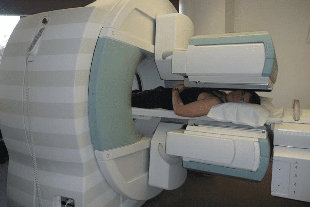
SPECT (Single Photon Emission Computed Tomography) is a nuclear medicine imaging method. It creates detailed 3D images of the body’s internal processes. A SPECT scanner detects gamma rays from the radiopharmaceutical in the body’s tissues.
This information is turned into images. These images give doctors valuable insights into the body’s functions and metabolism.
Radiopharmaceuticals are key in SPECT imaging. They are made to target specific areas or functions in the body. This could be blood flow, metabolic activity, or receptor density.
By choosing the right radiopharmaceutical, doctors can see different parts of the body’s internal processes. This helps them diagnose a variety of health conditions.
The steps to get and process SPECT images are complex. First, the radiopharmaceutical is given to the patient through an intravenous infusion. Then, the SPECT scanner picks up the gamma rays from the radiopharmaceutical.
It takes images from different angles. These images are then turned into a 3D dataset using advanced computer algorithms. This gives a detailed view of the body’s internal structures and functions.
| Step | Description |
| 1 | Administration of radiopharmaceutical |
| 2 | Detection of gamma rays by SPECT scanner |
| 3 | Image reconstruction into 3D dataset |
SPECT scans are key in medical care, giving insights into many health issues. They help diagnose and track diseases like brain, heart, and bone problems.
SPECT scans offer functional info, adding to what CT or MRI show. “Functional imaging lets us see how organs and tissues work,” says a top nuclear medicine expert. This helps doctors spot diseases early and accurately.
SPECT imaging shows blood flow and metabolic activity in the body. It’s great for checking heart health, finding ischemia, and seeing if heart muscle is working. It also helps in diagnosing brain issues like Alzheimer’s or epilepsy.
SPECT scans have many uses in medicine and are getting more. Some main uses are:
A leading nuclear medicine expert, says, “SPECT imaging has changed how we diagnose and treat complex diseases. It gives us vital info for making treatment plans and improving patient care.”

Brain SPECT imaging is a key tool in managing neurological disorders. It helps us understand brain conditions better. This leads to better treatment plans and outcomes for patients.
Brain SPECT scans are very useful for diagnosing and tracking several neurological conditions. Let’s look at some of the main uses:
SPECT scans are important for diagnosing Alzheimer’s disease and other dementias. They show how blood flows to the brain. This helps us spot Alzheimer’s early and plan treatments.
In managing epilepsy, finding the seizure focus is key. Brain SPECT scans help pinpoint this area. This guides doctors in planning surgeries or other treatments.
After a traumatic brain injury, SPECT imaging shows brain activity or perfusion issues. This is important for understanding the injury’s extent. It helps in planning rehabilitation.
Brain SPECT imaging offers several benefits in neurological disorders:
Using Brain SPECT imaging, we get deeper insights into neurological disorders. This leads to more personalized and effective care for our patients.
SPECT scans give us a peek into how the brain works. They help doctors diagnose and treat conditions better.
Parkinson’s disease causes tremors, stiffness, and trouble moving. Dopamine transporter imaging with SPECT scans checks the brain’s dopamine system. This helps doctors diagnose and track Parkinson’s disease.
This method is great for telling Parkinson’s apart from other similar diseases.
SPECT scans are also used for depression and anxiety disorders. They look at brain blood flow to understand these conditions better. This helps doctors create better treatment plans for each patient.
For ADHD and autism spectrum disorder, SPECT scans show brain function and activity. They help doctors understand and monitor these conditions better.
Using SPECT scans, we can better manage psychiatric and movement disorders. This leads to better care for patients.
Cardiac SPECT imaging has changed how we diagnose and treat heart diseases. It shows how well the heart is working and how blood flows through it. This helps doctors make better diagnoses and treatment plans.
Myocardial perfusion imaging with Cardiac SPECT is key for checking coronary artery disease. A stress test is used, where a special drug is given to see how blood flows to the heart. A SPECT scanner then takes pictures of the heart’s blood flow.
The stress test can be done through exercise or medicine. It depends on the patient’s health and if they can exercise. By comparing these images, doctors can spot problems with blood flow in the heart.
Cardiac SPECT imaging is important for finding and checking coronary artery disease. It helps doctors see if parts of the heart are not getting enough blood. This helps decide the best treatment.
Cardiac SPECT is very good at finding coronary artery disease. It also helps figure out who is at higher risk of heart problems.
Cardiac SPECT is used to check if parts of the heart can recover. It looks at how well the heart takes in a special drug. This helps decide if surgery is needed.
It also shows how well the heart is working. This includes how well the heart pumps and moves. This info is key for taking care of the heart.
SPECT scans are key in checking bone health. They help find stress fractures and bone metastases. These scans show how active bones are, helping doctors diagnose and track bone disorders.
Stress fractures are common in athletes and those with repetitive bone stress. Bone SPECT scans are great at finding these fractures when X-rays can’t. They spot areas of high bone activity early, helping avoid more injuries.
Cancer patients often face bone metastases, which hurt their quality of life. Bone SPECT scans find these bone diseases early. This helps doctors plan better treatments, easing pain and preventing worse problems.
CRPS is hard to diagnose and treat. Bone SPECT scans help by showing changes in bone metabolism linked to CRPS. Seeing these changes helps doctors confirm the diagnosis and choose the right treatment.
In summary, bone SPECT scans are vital in many medical situations. They help with stress fractures, bone metastases, and CRPS. Their ability to show bone activity makes them a key part of nuclear medicine today.
We use SPECT scans in cancer care to find primary tumors and see how cancer spreads. It also helps us check if treatment is working. This tool is key in oncology, giving us detailed views of tumor activity and metabolism.
SPECT imaging is great for finding primary tumors and learning about them. It uses special medicines to show how active tumors are. This helps doctors spot aggressive tumors and plan the best treatment.
When cancer spreads to other parts of the body, it’s called metastasis. SPECT scans can spot these sites. This helps doctors stage cancer more accurately and target treatments better.
After starting treatment, SPECT imaging checks how well the cancer is responding. It looks for changes in tumor activity or metabolism. This tells doctors if the treatment is working and if they need to make changes.
| Application | Description | Benefits |
| Primary Tumor Detection | Identifying the original tumor site | Early detection, accurate diagnosis |
| Metastatic Disease Evaluation | Assessing the spread of cancer | Accurate staging, targeted therapy |
| Treatment Response Assessment | Monitoring changes in tumor activity | Adjusting treatment plans, improving outcomes |
In summary, SPECT imaging is a vital tool in cancer diagnosis and management. It gives us important information that helps guide treatment and improve patient care.
SPECT imaging is a key tool for checking how well organs work. It looks at the liver, spleen, kidneys, thyroid, and parathyroid glands. This helps doctors make better diagnoses and treatment plans.
SPECT scans help check the liver and spleen. They see how radiopharmaceuticals move in these organs. This helps find problems like liver cirrhosis and spleen issues.
SPECT scans also check kidney health. They look at blood flow and function in the kidneys. This is great for spotting kidney disease or checking on transplanted kidneys.
SPECT scans are also good for the thyroid and parathyroid. They help find issues like hyperthyroidism and thyroid nodules. They also find problems with the parathyroid glands. This helps doctors treat these conditions better.
Using SPECT scans helps us understand organ function better. As this technology gets better, it will help us learn even more about the body.
Clinical guidelines are key in deciding when to use SPECT scans. Medical professionals follow these guidelines to pick the best tests for patients.
Doctors choose SPECT scans based on clinical guidelines. These guidelines come from research and experience. They help make sure SPECT scans are used right in patient care.
SPECT scans help check how well parts of the body work. For example, in heart care, they check blood flow and how well the heart works.
| Clinical Indication | SPECT Scan Application |
| Cardiac Disease | Myocardial perfusion imaging |
| Neurological Disorders | Brain perfusion and function assessment |
| Cancer Diagnosis | Tumor detection and metastasis evaluation |
Diagnostic algorithms help figure out and manage patient conditions. SPECT scans are a key part of these algorithms.
For heart disease, a test might start with a stress test. Then, a SPECT scan is used if the first test shows something or is unclear.
SPECT scans add to what other imaging does. They give info on how parts of the body work, not just what they look like.
CT or MRI scans show body structure. But, SPECT scans show how active or blood-rich areas are. This helps doctors make better diagnoses.
Using SPECT scans with other imaging helps doctors understand patients better. This leads to better treatment plans.
SPECT scans are a key tool in nuclear medicine. But how do they stack up against other imaging methods? Let’s dive into the differences and similarities between SPECT and other techniques to grasp their roles in healthcare.
SPECT and PET scans both use nuclear medicine. Yet, they have distinct approaches. PET scans offer higher resolution and sensitivity. But SPECT scans are more common and cheaper, making them a good choice for many cases.
The main difference lies in the radiopharmaceuticals used. SPECT uses Technetium-99m, while PET uses Fluorine-18. This affects the information each modality can give. PET is often used for specific tasks like cancer research.
MRI and CT scans give great details of the body’s structure. But they don’t offer the same functional data as SPECT. SPECT is great for showing how body parts work.
In heart imaging, SPECT checks blood flow, while MRI or CT show the heart’s structure. Using all three can give a full picture of a patient’s health.
SPECT is top choice when you need to know how body parts function. It’s perfect for heart or brain issues. It gives detailed info on organ function and blood flow.
In summary, SPECT is a vital tool in medicine. Knowing how it compares to PET, MRI, and CT helps doctors choose the best imaging for each patient.
In recent years, SPECT technology has made big strides. These advancements have made SPECT scans more accurate and useful in medical diagnostics. They help doctors diagnose diseases better in different clinical settings.
Hybrid SPECT/CT systems have been developed by combining SPECT with CT technology. These systems offer both the functional details from SPECT and the anatomical details from CT. This combination gives a deeper understanding of diseases.
| Feature | SPECT | SPECT/CT |
| Functional Information | Yes | Yes |
| Anatomical Detail | No | Yes |
| Diagnostic Accuracy | High | Very High |
New detector technology and algorithms have boosted SPECT image quality. These advancements also allow for more precise measurements of organ function and disease severity. This means doctors can make more accurate diagnoses.
Artificial intelligence (AI) is changing SPECT imaging fast. AI algorithms are being created to help with image analysis. They could make diagnoses faster and more accurate.
SPECT imaging is growing in importance, helping doctors diagnose many health issues. It gives detailed views of how our bodies work. This helps doctors find the right treatments faster.
SPECT scans check blood flow, how cells work, and organ health. They are key in diagnosing brain, heart, and cancer problems. As technology gets better, SPECT imaging will give even clearer pictures of our health.
At our institution, we see how vital SPECT imaging is. It helps us offer top-notch care to patients from around the world. We use SPECT scans to make sure our diagnoses are accurate. This improves patient care and life quality.
A SPECT scan is a test that uses a tiny bit of radioactive material. It creates detailed 3D images of the body’s inside parts and how they work. It does this by catching the gamma rays from the material, which is taken up by certain areas of the body.
Doctors use SPECT scans to find and treat many health issues. This includes brain problems, heart disease, cancer, and bone issues. The scans show how organs and tissues work, helping doctors make the right diagnosis and treatment plans.
SPECT scans are great for finding and managing brain disorders like Alzheimer’s, epilepsy, and brain injuries. They help doctors see how the brain works, spot problems, and check if treatments are working.
SPECT scans have unique benefits compared to other imaging methods. They offer similar info to PET scans but are more available and cheaper. MRI and CT scans show body structure well, but SPECT scans give more info on organ function and metabolism.
SPECT scans are key in finding and managing cancer. They help doctors spot tumors, check for cancer spread, and see how treatments are working. This info helps tailor treatments to each patient’s needs.
Yes, SPECT scans are used a lot in heart imaging. They check blood flow to the heart, find heart disease, and see how well the heart works. This helps doctors treat heart problems and lower the risk of heart attacks.
New SPECT tech, like hybrid SPECT/CT systems, better resolution, and analysis tools, have made it better. These updates mean more accurate and detailed images. This opens up more uses for SPECT scans in medicine.
Artificial intelligence (AI) is helping make SPECT images better. It helps doctors understand complex data and spot things they might miss. AI in SPECT could make diagnoses more accurate and make work easier for doctors.
SPECT scans are usually safe, with little risk or side effects. The radioactive material used is usually okay, and the radiation dose is low. But, like any test, there could be some risks, like allergic reactions or radiation exposure. Your doctor will talk about these with you.
Yandrapalli, S., et al. (2022). SPECT imaging. In StatPearls. Retrieved from https://www.ncbi.nlm.nih.gov/books/NBK564426/
Subscribe to our e-newsletter to stay informed about the latest innovations in the world of health and exclusive offers!