Last Updated on October 22, 2025 by mcelik

Did you know millions of people undergo PET scans every year? Many patients wonder if there are safer or less invasive choices. The search for accurate diagnosis without the potential risks of PET imaging has sparked medical interest in PET scan alternatives imaging options.
PET scans are useful for diagnosing many health issues but have their downsides. They are a key tool in modern medicine, showing how the body’s cells work.
PET scans use a small amount of radioactive tracer injected into the body. This tracer goes to areas with lots of activity, like cancer cells. The scanner then picks up the radiation, making detailed images of the body’s inside.
The tracer, often Fluorodeoxyglucose (FDG), is injected first. Then, the patient waits about an hour for it to spread. After that, they have the scan.
PET scans help find and track many health issues, like cancer and heart disease. They show how active tissues are, helping doctors understand and treat conditions better.
They are used for:
PET scans have some big downsides. One big worry is the radiation, which can increase cancer risk. They also cost a lot and aren’t everywhere.
Other issues include:
Because of the drawbacks of PET scans, other imaging methods are sometimes better. For example, pregnant women or people with certain health issues might not get PET scans because of radiation.
Other options like MRI, CT scans, and ultrasound have their own benefits and limits. The right choice depends on the health question, the patient, and what’s available.

There are many alternatives to PET scans, each with its own benefits and drawbacks. The right choice depends on the medical condition, the patient’s health, and what information is needed.
There are several types of alternative imaging. These include:
Choosing an alternative to PET scans involves several things. These include:
Each imaging method has its own strengths and weaknesses. For example, CT scans are quick and detailed but use radiation. MRI is great for soft tissues without radiation but might not work for patients with metal implants.
Comparing these options is key to finding the best imaging for a situation. It’s not just about how accurate they are but also about safety, comfort, and how they might affect treatment plans.

CT scans are a key alternative to PET scans. They offer great versatility and precision. CT scans combine multiple X-ray measurements from different angles. This creates detailed cross-sectional images of the body.
Standard CT imaging is a basic but powerful tool. It gives clear images of the body’s internal parts. It’s great for looking at organs, bones, and soft tissues.
Its speed and clarity make it vital in emergency care and regular checks.
Contrast-enhanced CT uses a contrast agent to show specific body areas better. This helps spot vascular diseases and some cancers. It makes it easier for doctors to make accurate diagnoses.
Dual-energy CT scans use two X-ray levels to tell different materials apart. Spectral CT goes further, breaking down materials in detail. These methods help find lesions and understand tissue better.
CT angiography focuses on blood vessels. It helps find issues like aneurysms and blockages. It uses contrast agents and timing to show blood flow.
This is great for planning vascular treatments and checking how they go.
CT scans have many benefits. They include:
CT scans are a strong choice for many diagnostic needs, rivaling PET scans.
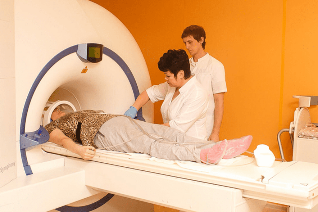
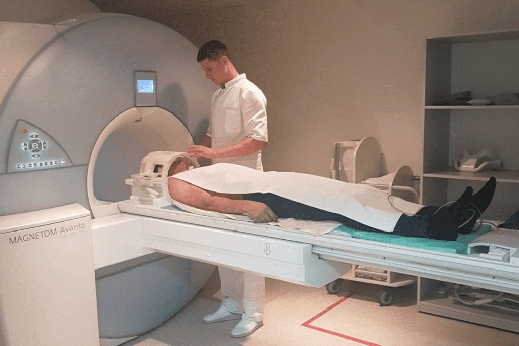
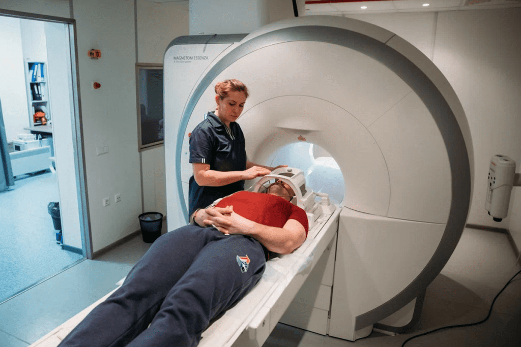
Magnetic Resonance Imaging (MRI) is a strong tool for diagnosing. It’s great because it can see inside the body without using harmful radiation. This makes it a good choice for both patients and doctors.
Conventional MRI shows detailed pictures of inside the body. It’s very helpful for checking the brain, spine, and other areas. It’s also good at telling different tissues apart.
Functional MRI, or fMRI, looks at brain activity. It tracks blood flow to see how the brain works. This makes it a great tool for brain checks and a good choice instead of PET scans in some cases.
Diffusion-weighted imaging is an MRI that looks at water molecule movement in tissues. It’s very good at finding new strokes and understanding lesions. This makes it a key option instead of PET scans for some needs.
Perfusion MRI checks blood flow to tissues. It helps see how well organs and lesions get blood. This is useful for looking at tumor blood supply and checking treatment success. It shows MRI is a full alternative to PET scans.
In summary, MRI alternatives to PET scans have many uses. They range from basic body scans to detailed brain checks. With tools like MRI, doctors can make better choices without always using PET scans.
Ultrasound is a non-invasive and safe way to see inside the body. It uses sound waves to create detailed images. This makes it a great choice for avoiding radiation.
Standard ultrasound is a common and useful tool. It helps check organs like the liver and thyroid. It’s good for finding problems like cysts and tumors.
It’s also used during pregnancy to watch the baby grow.
Doppler ultrasound looks at blood flow in the body. It helps find issues like blocked blood vessels. It also checks how well organs are working.
Contrast-enhanced ultrasound (CEUS) uses special agents to see blood flow better. It’s great for looking at liver lesions. It’s safer than other scans for people with kidney problems.
Elastography is a new ultrasound method. It checks how stiff tissues are. It helps tell if liver or thyroid nodules are cancerous.
Ultrasound is a good choice for many reasons. It’s safe, non-invasive, and shows images in real-time. It’s perfect for patients who need to avoid radiation.
Nuclear medicine offers more than just PET scans. It includes several imaging techniques that are key for diagnosis. These alternatives have their own benefits and are used in various clinical settings.
SPECT (Single Photon Emission Computed Tomography) scans are a big alternative to PET scans. They use a gamma camera to catch gamma rays from a tracer.
They are great for finding and tracking bone disorders, infections, and some cancers.
Bone scans are a vital nuclear medicine method. They help find bone metastases, diagnose osteoporosis, and check for bone infections or inflammation.
The process starts with injecting a tiny amount of radioactive material, like technetium-99m methylene diphosphonate (Tc-99m MDP). It goes to bone areas that are very active.
Gallium scans use gallium-67 citrate to see certain tumors, infections, and inflammation. They are very useful for finding and tracking diseases like lymphoma and abscesses.
Octreotide scans find neuroendocrine tumors with somatostatin receptors. They use a radiolabeled octreotide, a synthetic somatostatin analogue.
| Imaging Technique | Primary Use | Key Characteristics |
| SPECT Scans | Diagnosing bone disorders, infections, and certain cancers | Uses a gamma camera to detect gamma rays |
| Bone Scans | Detecting bone metastases, osteoporosis, and bone infections | Involves technetium-99m MDP |
| Gallium Scans | Visualizing tumors, infections, and inflammatory conditions | Utilizes gallium-67 citrate |
| Octreotide Scans | Detecting neuroendocrine tumors with somatostatin receptors | Involves radiolabeled octreotide |
Blood tests and biomarkers are becoming more popular as alternatives to PET scans. Advances in lab tests let doctors diagnose and track conditions with blood tests. This might cut down on the need for more invasive or pricey imaging like PET scans.
Tumor markers are substances found in higher amounts in the blood, urine, or tissues of some cancer patients. They help detect cancer, track treatment, and spot recurrence. Examples include:
While not alone in diagnosing, tumor markers offer valuable info. They help guide treatment decisions when used with imaging and clinical assessment.
Inflammatory markers show the body’s inflammation. Common ones are:
These tests help track inflammation in conditions like infections, autoimmune diseases, and some cancers. They might reduce the need for PET scans in some cases.
Genetic testing looks at an individual’s genes for mutations or changes. It’s useful for:
Genetic tests give detailed info. They can sometimes replace or add to what imaging studies like PET scans show.
Liquid biopsies are a non-invasive way to diagnose cancer by analyzing blood. They’re promising for:
Liquid biopsies are a convenient and less invasive option. They might give info similar to PET scans.
For patients needing detailed diagnostic info, tissue sampling is a good alternative to PET scans. These methods get tissue or cell samples from the body for study.
Fine Needle Aspiration (FNA) uses a thin needle to get cell samples from suspicious areas. It’s often used for diagnosing thyroid nodules, lymph nodes, and other accessible lesions.
FNA is minimally invasive and quick, often done without local anesthesia.
Core Needle Biopsy uses a larger needle than FNA to get a tissue sample. This method gives more tissue for study, making it great for diagnosing various cancers and other conditions.
Core Needle Biopsy is useful when you need architectural info about the tissue, which FNA can’t provide.
Surgical Biopsy is a more invasive procedure where a surgeon removes a larger tissue sample, often during an operation. It’s used when other biopsy techniques are unclear or when a larger sample is needed for diagnosis.
Surgical Biopsy can be both diagnostic and therapeutic, as it can remove the entire lesion in some cases.
Image-Guided Biopsies use imaging technologies like ultrasound, CT, or MRI to guide the biopsy needle to the exact location of the suspected lesion. This method increases the accuracy of the biopsy.
Image guidance is very useful for sampling hard-to-reach or non-palpable lesions.
| Biopsy Technique | Invasiveness | Diagnostic Yield |
| Fine Needle Aspiration | Low | Cytological diagnosis |
| Core Needle Biopsy | Moderate | Histological diagnosis |
| Surgical Biopsy | High | Comprehensive histological diagnosis |
| Image-Guided Biopsy | Varies | Precise sampling of lesions |
Tissue sampling techniques offer various options for diagnosing and monitoring medical conditions. They often provide detailed info that complements or replaces the need for PET scans.
Endoscopic evaluations are a good choice instead of PET scans for diagnosing. They let doctors see inside the body. This helps find many medical problems.
Colonoscopy lets doctors look inside the colon and rectum. It’s great for finding polyps, cancer, and other issues in the lower gut. It’s often a good choice instead of PET scans for colon problems.
A study in the Journal of Clinical Gastroenterology showed colonoscopy works well for early colorectal cancer. This might mean fewer PET scans are needed.
Bronchoscopy lets doctors check the airways and lungs. It’s used for lung cancer, infections, and other lung issues. It can take tissue samples for biopsies, which might avoid the need for PET scans.
“Bronchoscopy has become an indispensable tool in pulmonary medicine, allowing for both diagnosis and treatment of various lung conditions.” – A Pulmonologist
Laparoscopy uses a thin, lighted tube with a camera through small cuts in the belly. It’s for looking at the organs inside the belly. It’s a good choice instead of PET scans for looking at the belly.
| Endoscopic Procedure | Primary Use | Diagnostic Benefits |
| Colonoscopy | Examining the colon and rectum | Detects polyps, cancer, and other abnormalities |
| Bronchoscopy | Examining the airways and lungs | Diagnoses lung cancer, infections, and respiratory conditions |
| Laparoscopy | Examining abdominal organs | Diagnoses and treats conditions like endometriosis and liver disease |
Thoracoscopy lets surgeons see inside the chest. It’s for diagnosing and treating lung and chest problems. It might mean using PET scans less for chest issues.
In conclusion, endoscopic evaluations are good alternatives to PET scans. Each has its own uses and benefits. Knowing about these options helps doctors choose the best test for their patients.
Cardiac-specific alternatives to PET imaging are key in diagnosing heart conditions. These options offer tailored diagnostic capabilities for patients.
Stress tests are a common alternative to PET scans. They monitor the heart’s activity under physical stress, through exercise or medication. This helps spot issues with blood flow to the heart muscle.
Stress tests are non-invasive and provide immediate results. They assess how well the heart performs under stress.
An echocardiogram uses ultrasound waves to create heart images. It shows the heart’s structure and function, like chambers, valves, and blood vessels.
Echocardiography is great for checking heart valve function and detecting abnormalities in heart movement.
Cardiac MRI offers detailed images of the heart’s structure and function. It’s excellent for assessing the heart’s anatomy and detecting scar tissue.
The advantages of cardiac MRI include its high resolution and lack of ionizing radiation.
Coronary angiography injects a contrast agent into the coronary arteries. It’s great for spotting blockages or narrowing in the arteries.
Coronary angiography is often used with other tests to fully understand cardiac health.
Brain imaging has grown beyond PET scans. Now, techniques like functional MRI and EEG are more common. These new methods give detailed views of the brain’s structure and function.
MRI is key in brain imaging because it shows detailed brain pictures. Brain MRI techniques help find tumors, blood vessel problems, and diseases like Alzheimer’s.
Regular MRI shows soft tissues in the brain well. But, advanced MRI like diffusion-weighted imaging gives more info on brain function and problems.
Functional MRI (fMRI) tracks blood flow to show brain activity. It’s great for planning surgeries and studying brain functions. fMRI spots important brain areas for speech and movement.
“Functional MRI has revolutionized the field of neuroscience by allowing us to map brain function with unprecedented detail.” – A Neuroscientist
SPECT scans use a radioactive tracer to see brain function. SPECT scans help diagnose Alzheimer’s and some mental health issues.
They show blood flow and activity in brain areas. This helps check neurological conditions.
EEG records brain electrical activity through scalp electrodes. It’s key for finding and tracking seizures, sleep issues, and brain problems.
EEG is great for live monitoring. It catches brief brain events.
When we talk about diagnostic imaging, knowing about radiation levels is key. Different imaging methods use different amounts of ionizing radiation. This affects patient safety and the choice of imaging tool.
Imaging methods vary in radiation exposure. For example, CT scans use more radiation than X-rays. A CT scan’s radiation dose can be between 2 to 10 millisieverts (mSv), based on the scan and body part. On the other hand, a chest X-ray has about 0.1 mSv.
Long-term radiation exposure is a big worry, mainly for those needing many scans. The ALARA (As Low As Reasonably Achievable) rule helps doctors lower radiation while getting good images. This rule is very important for kids and young adults, as they are more vulnerable to radiation.
Not all scans use radiation. MRI (Magnetic Resonance Imaging) and ultrasound don’t. MRI uses a strong magnetic field and radio waves for detailed images. Ultrasound uses sound waves. These options are great for those who can’t handle radiation or need many scans.
Choosing the right scan is about weighing benefits against risks, like radiation. Knowing how much radiation each scan uses helps doctors make better choices for their patients.
Looking into alternatives to PET scans means understanding the costs. The cost of diagnostic imaging can affect patient choices and healthcare systems.
Insurance coverage for PET alternatives varies a lot. For example, CT scans and MRI are often covered by most insurance plans for many reasons. But, how much is covered can depend on the insurance company and the patient’s policy.
| Imaging Modality | Average Cost | Typical Insurance Coverage |
| CT Scan | $400-$1,000 | 80%-100% |
| MRI | $600-$1,500 | 80%-100% |
| Ultrasound | $100-$500 | 80%-100% |
Patients face different out-of-pocket costs based on their insurance and the imaging choice. For instance, a CT scan might be fully covered, but an MRI could cost more due to deductibles or co-pays.
Factors influencing out-of-pocket expenses include:
Cost-effectiveness analysis compares the costs of PET alternatives with their effectiveness and patient outcomes. This helps healthcare providers and patients make better choices.
A detailed analysis shows which imaging options are the best value. It balances cost with how well they diagnose and help patients.
Choosing the right alternative to PET scans is complex. Doctors must think about many factors. They look at the benefits and limits of different imaging tools to find the best one for each patient.
The decision-making process is thorough. Doctors look at the patient’s medical history and current health. They also think about what tests or treatments the patient has had before.
Key considerations in the clinical decision-making process include:
Patient-specific factors are very important. These include the patient’s age, health, and any medical conditions they have.
| Patient Factor | Consideration |
| Age | Pediatric or geriatric patients may require special consideration due to sensitivity to radiation or contrast agents. |
| Underlying Conditions | Patients with kidney disease may need to avoid certain contrast agents, while those with claustrophobia may require alternative imaging modalities. |
| Previous Treatments | Patients who have undergone previous surgeries or treatments may require imaging modalities that can accommodate these factors. |
The type and stage of the disease are key. Different imaging tools work better for different conditions.
For example: MRI or CT scans are often used for cancer. They can show detailed images of soft tissues.
The availability of imaging tools affects the choice. Things like where facilities are, what equipment they have, and insurance coverage matter.
Healthcare professionals make informed choices based on these factors. This helps them find the best alternative to PET scans for each patient.
Finding a PET scan alternative can be tough. But, there are many imaging options like CT scans, MRI, ultrasound, and nuclear medicine. These can help diagnose problems effectively.
It’s important to know what each option can do. Things like how much radiation it uses, the cost, and how accurate it is matter a lot. These factors help decide which test is best.
When looking at pet scan alternatives, patients and doctors can work together. This team effort helps find the best test for each person. It makes sure patients get the care they need.
To navigate imaging options well, you need to understand all the diagnostic tools. Knowing this helps make better health choices. It leads to better health outcomes for everyone.
You can use CT scans, MRI scans, ultrasound, and nuclear medicine scans like SPECT. Bone scans and tissue sampling techniques are also options.
Your doctor will pick the best test based on your condition and what they need to know. They’ll also think about radiation and cost.
Yes, you can try MRI, ultrasound, or some nuclear medicine scans with lower radiation.
CT scans show detailed body structures. PET scans show how tissues and organs work. Sometimes, CT scans are a good choice, like with contrast.
Yes, MRI can replace PET for brain scans. It uses advanced techniques to show brain function and activity.
SPECT and PET scans are both nuclear medicine tests. SPECT uses a different tracer and is often used for the heart and bones.
Blood tests can give important info about some conditions. But, they’re not always a direct swap for PET scans, which show images.
Doctors look at your condition, what they need to know, your health, radiation, and cost. These factors help them choose the best test.
For heart issues, you can try stress tests, echocardiograms, cardiac MRI, or coronary angiography.
Different tests have different radiation levels. CT scans and some nuclear medicine scans have radiation. MRI and ultrasound don’t. Think about radiation when choosing a test.
Yes, ultrasound and some blood tests might be cheaper. The cost depends on the test, insurance, and what you need for your care.
Yes, tests like colonoscopy, bronchoscopy, and laparoscopy can be alternatives. They let doctors see and sample tissues directly.
Ciliberto, M., et al. (2013). Comparison between whole-body MRI and Fluorine-18-Fluorodeoxyglucose PET/CT in oncology: A systematic review. Insights into Imaging, 4(6), 625-639. https://pmc.ncbi.nlm.nih.gov/articles/PMC3794875/
Subscribe to our e-newsletter to stay informed about the latest innovations in the world of health and exclusive offers!