Last Updated on October 22, 2025 by mcelik

Every year, 1.8 million are diagnosed with lung cancer. Lung nodules may be cancerous. A PET scan aids diagnosis by showing cell activity. Understanding PET scan black spots meaning is crucial, as these spots often indicate cancer due to high glucose uptake. This helps determine if lung nodules are malignant or benign.
PET scanning technology has changed medical imaging a lot. It shows how cells work. This tool is key in modern medicine, mainly in fighting cancer.
A PET scan is a test that uses a special sugar to find diseases. It injects a tiny bit of fluorodeoxyglucose (FDG) into you. Then, a PET scanner makes pictures of where the sugar goes.
PET scans are different from Computed Tomography (CT) and Magnetic Resonance Imaging (MRI). They show how things work, not just what they look like. This makes PET scans great for finding cancer.
PET scans work by looking at how fast cells use sugar. Cancer cells use more sugar than healthy ones. So, PET scans can spot cancer early.
As technology gets better, PET scans are more important. They help doctors see how organs work. This helps make treatments better and safer.

PET scans find cancer cells by looking at how they use energy. Cancer cells use more energy than normal cells. This difference shows up on PET scans.
Cancer cells use more glucose than normal cells. This is a key sign of cancer. It’s because cancer cells grow and divide fast, needing more energy.
The process involves:
Fluorodeoxyglucose (FDG) is a glucose-like substance used in PET scans. It shows how cells use glucose. Cancer cells take up more FDG, making them visible on scans.
FDG in PET scans helps find cancer cells by their energy use. It’s not just about where they are in the body.

Standardized Uptake Values (SUVs) measure how much FDG cells take up. SUVs help tell if a lesion is cancerous by comparing its FDG uptake.
Key aspects of SUVs include:
Understanding SUVs helps doctors read PET scans better. This leads to better care for patients.
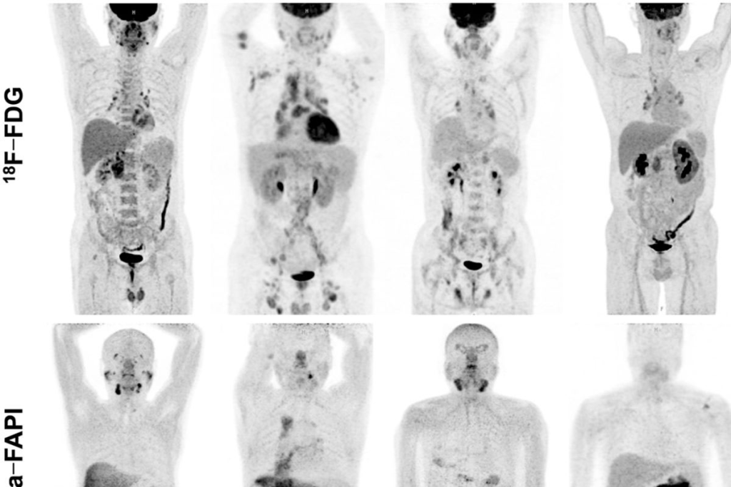
Black spots on PET scans can mean different things about your health. They show up because of how cells work and the colors used in the scan. These scans help find diseases, like cancer, by showing how active cells are.
PET scans use colors to show how active cells are. Bright colors mean cells are very active, often a sign of cancer. Knowing this color scale is key for correct diagnosis.
The colors range from black (low activity) to white (high activity). There are also shades of gray and other colors, depending on the tracer used.
Dark or black spots on PET scans mean low cell activity. This can be because of:
Figuring out why these spots appear needs a detailed look at the scan. This is often done with other tests too.
Reading PET scans means knowing the difference between normal and abnormal activity. Understanding the patient’s history and overall health is very important.
| Characteristic | Normal Findings | Abnormal Findings |
| Metabolic Activity | Expected levels for tissue type | Unexplained high or low activity |
| Symmetry | Symmetrical uptake | Asymmetrical uptake |
| Clinical Context | Consistent with patient’s history | Inconsistent or unexpected |
By looking closely at PET scan images and the patient’s health, doctors can make better diagnoses. This helps them plan the best treatment.
When we talk about PET scans for lung nodules, we look at their sensitivity, specificity, and what they can’t do. These scans are key in figuring out if a lung nodule might be cancer. They help doctors make important decisions.
The sensitivity of a PET scan shows how well it spots lung nodules. Specificity shows how well it misses them. Research shows PET scans vary in how well they detect lung nodules.
Sensitivity and Specificity of PET Scans
| Study | Sensitivity (%) | Specificity (%) |
| Gould et al. (2001) | 96.8 | 77.8 |
| Herder et al. (2006) | 93 | 80 |
| Li et al. (2012) | 95.5 | 83.3 |
PET scans have a problem with small nodules. They can’t always find nodules smaller than 8-10 mm.
PET scans can sometimes say a nodule is cancer when it’s not. They can also miss cancer that’s there. False positives can happen with inflammation, and small or slow-growing tumors can cause false negatives.
Understanding PET scans, and what low uptake means, is key in medical imaging. Low uptake shows areas that don’t take in much of the tracer. This means they have lower metabolic activity.
Many things can cause low metabolic activity. This includes benign conditions or tissues that naturally have lower activity. For example, some lung nodules might not be as active as others, showing lower uptake on PET scans.
Factors influencing low metabolic activity:
Several benign conditions can show low uptake on PET scans. These include:
Knowing about these conditions helps in accurately reading PET scan results.
Low uptake often means a condition is benign. But, sometimes it can hint at a concern. For instance, slow-growing tumors might not show high activity, yet they could be malignant.
| Condition | Typical Uptake Pattern | Clinical Implication |
| Benign Nodules | Low uptake | Generally not concerning |
| Slow-growing Tumors | Low to moderate uptake | May be malignant |
| Inflammatory Conditions | Variable uptake | Needs clinical correlation |
In conclusion, understanding low uptake on PET scans is complex. It requires knowing the causes and the clinical context. By looking at the factors that affect metabolic activity and knowing about benign conditions, doctors can make better decisions.
PET scans show high metabolic activity in lung nodules, which means we need to take a closer look. This activity is often linked to cancer because cancer cells use more glucose. But, it’s important to remember that not all high activity is cancer.
Standardized Uptake Values (SUVs) measure metabolic activity in lung nodules. An SUV above 2.5 might suggest cancer, but it’s not a sure sign. This value is just a guideline.
Doctors look at SUVs, nodule size, patient history, and symptoms together. They use all this information to make a diagnosis.
High metabolic activity can also mean non-cancerous conditions. Inflammation, infections, and some benign tumors can show high uptake on PET scans. For example, sarcoidosis can cause high SUVs.
Studies show that PET scan uptake intensity might link to cancer aggressiveness. Tumors with higher SUVs might be more aggressive. But, this link isn’t always true and must be seen with other diagnostic results.
| SUV Value | Malignancy Likelihood | Clinical Consideration |
| Low | Monitor or further diagnostic tests | |
| 2.5-4.0 | Moderate | Biopsy or close follow-up |
| >4.0 | High | Strong consideration for malignancy, biopsy recommended |
It’s key to understand what high metabolic activity in lung nodules means for diagnosis and treatment. PET scans are helpful but just one part of the diagnostic process. Clinical evaluation and other imaging are also important.
PET scans are useful but have some big limitations for lung nodule diagnosis. It’s key to know these to understand PET scan results well. This helps in making the best care decisions for patients.
PET scans struggle to spot small lung nodules. The resolution of PET scans is limited, around 4-6 mm. This means tiny nodules might not show up or be misjudged. This can cause false negatives, missing small cancers.
Detecting small lung nodules is tough for PET scans because of its low resolution.” This shows we need other imaging or follow-up scans for small nodules.
PET scans look for cell activity to find cancer. But slow-growing cancers may not show much activity. This makes them hard to find with PET scans. It can lead to false negatives, missing cancers, because they don’t take up glucose as expected.
“Some lung cancers, like well-differentiated or slow-growing ones, might not take up much FDG. This can cause false-negative PET scans.”
– Journal of Thoracic Oncology
PET scans can also give false positives due to inflammation. Infections, granulomatous diseases, and other inflammatory issues can look like cancer. This can cause worry and more tests.
To deal with these issues, doctors use PET scans with other tests like CT scans. They also look at the patient’s history and risk factors.
Mixing PET and CT scans gives a clearer view of lung nodules. It shows both their metabolic activity and structure.
PET/CT fusion brings together the best of both worlds. It gives doctors a detailed look at lung nodules. This helps them tell if a nodule is likely to be cancerous or not.
Key benefits of PET/CT fusion include:
Linking anatomy and metabolism is key for lung nodule assessment. PET/CT fusion pinpoints active areas in nodules. This gives doctors important clues about their nature.
| Characteristics | PET Scan | CT Scan | PET/CT Fusion |
| Metabolic Information | High | Low | High |
| Anatomical Detail | Low | High | High |
| Diagnostic Accuracy | Moderate | Moderate | High |
Understanding PET/CT reports is complex. Radiologists must link metabolic and anatomical data for accurate diagnoses.
Key elements to consider when reading PET/CT reports include:
A PET scan for lung nodules needs special preparation for the best results. Knowing what to do can make patients feel more at ease.
Before a PET scan, patients get clear instructions. These include:
On the day of the PET scan, here’s what happens:
The procedure starts with a small injection of a radioactive tracer, usually FDG (fluorodeoxyglucose), into a vein. This tracer goes to areas with high metabolic activity, like cancer cells.
After the tracer is absorbed, usually in 30-60 minutes, you’ll lie on a scanning table. This table slides into a PET scanner. The scan is painless and lasts about 30 minutes.
After the PET scan, you can usually go back to your normal activities. But it’s wise to:
By following these steps and knowing what to expect, patients can have a smooth and effective PET scan for their lung nodule check-up.
It’s important to know about PET scan safety, mainly about radiation. PET scans use small amounts of radioactive tracers. These tracers help show how the body works.
A typical PET scan with Fluorodeoxyglucose (FDG) has a dose of 7-10 mSv. This amount can change based on the scan type and the person’s size.
Comparing PET scan radiation to other scans helps understand it better. The average yearly background radiation is 3 mSv. A chest CT scan is about 7 mSv, and a chest X-ray is 0.1 mSv.
| Imaging Modality | Typical Effective Dose (mSv) |
| PET Scan (FDG) | 7-10 |
| Chest CT Scan | 7 |
| Chest X-Ray | 0.1 |
| Annual Background Radiation | 3 |
Medical places have strict rules to lower radiation. They use the least amount of tracer needed and improve scanning methods. Patients are told how to reduce their exposure after the scan, like drinking more water.
Knowing about PET scan radiation and safety steps helps patients make better choices for their health.
Doctors decide on a PET scan for lung nodules based on size and patient risk. They look at both the nodule’s characteristics and the patient’s health. This helps them figure out if a PET scan is needed.
Nodules larger than 8-10 mm or with unusual features on scans might get a PET scan. Nodules greater than 8-10 mm in diameter are often checked with PET scans because they could be cancerous.
The shape of the nodule on CT scans is also important. Nodules with spiculated margins, irregular shapes, or those that are growing are seen as more suspicious. They might need a PET scan for more checks.
Patient risk factors play a big role in deciding on a PET scan. People with a history of smoking, exposure to carcinogens, or a family history of lung cancer are at higher risk. They might get a PET scan for nodule checks.
After a PET scan, follow-up imaging plans depend on the scan results. Nodules with low activity might be checked with regular CT scans. But those with high activity might need quicker action, like a biopsy or treatment.
The integration of PET scan results with clinical risk factors and other imaging findings is key for deciding the best treatment for patients with lung nodules.
Doctors consider the nodule’s characteristics and the patient’s risk when deciding on a PET scan. This ensures patients get the right care for their situation.
Several methods are used to check lung nodules, aside from PET scans. These methods help figure out what the nodules are.
CT and HRCT scans are key for lung nodule checks. CT scans show detailed lung images, spotting nodules and their details. HRCT scans give even more detailed info, like density and shape, helping tell if nodules are bad or not.
CT scans are great for watching how nodules grow. This helps doctors guess if they might be cancer. CT scans with contrast agents also show how blood flows through nodules, giving more clues.
MRI is used for lung nodule checks, but less often than CT scans. It’s good when avoiding radiation or checking the chest area.
MRI can show how tissues work, like with diffusion-weighted imaging. But, it’s not usually the first choice for lung nodule checks because it’s not as good at showing lung details.
Biopsies are key for lung nodule diagnosis. Fine-needle aspiration biopsy (FNAB) or core needle biopsy (CNB) under CT guide take tissue samples for lab tests.
The right biopsy method depends on the nodule’s spot, size, and the patient’s health. Biopsy results can confirm what the nodule is, helping decide treatment.
In summary, PET scans are important for lung nodule diagnosis. But, CT, HRCT, MRI, and biopsies are also vital. Together, they help get a clear diagnosis and guide treatment plans.
Advances in PET scanning are changing how we find and treat lung cancer. The field has grown a lot in recent years. This has made lung cancer detection more accurate and effective.
New tracers are being developed to go beyond Fluorodeoxyglucose (FDG). Scientists are looking into tracers that show more about tumor biology. For example, Fluorothymidine (FLT) is being studied for its role in showing tumor growth.
Modern PET scanners have better resolution thanks to new technology. Better detector materials and algorithms are making images clearer. This is key for spotting and understanding small lung nodules early.
Artificial Intelligence (AI) is being used to help with PET image analysis. AI can point out important areas, measure tracer levels, and even suggest diagnoses. This teamwork between humans and AI is expected to make diagnoses more accurate and quicker.
The future of using PET technology for lung cancer detection is bright. Ongoing research aims to make it even better. These advancements will likely help improve patient care.
PET scans are key in finding and managing lung cancer. They show where in the lungs there’s high activity, helping spot cancerous nodules.
When used with CT scans, PET scans give a clearer picture of lung nodules. This helps doctors tell the difference between harmless and harmful nodules.
But, PET scans have their limits. Things like how big a nodule is, its glucose use, and inflammation can affect their results. Knowing these can help make a correct diagnosis and plan treatment.
In summary, PET scans are essential in lung cancer diagnosis. They offer a special view of lung nodules’ activity. By combining PET scan data with other tests, doctors can make better choices. This leads to better care for patients with lung cancer.
A PET scan looks at how cells use energy. It helps find lung nodules by spotting areas that use a lot of energy, which might be cancer.
“PET scan black spots meaning” refers to dark areas on a scan. These spots show low energy use. Knowing about these spots helps doctors find out if something is normal or not.
PET scans find cancer by showing how cells use energy. FDG is a special substance that shows up in cancer cells because they use a lot of energy.
SUVs measure how much FDG is used by tissues. They help doctors see if lung nodules are likely to be cancer.
PET scans are very good at finding cancer in lung nodules. But, they might not work as well for small nodules or if there’s inflammation.
Low uptake can mean a nodule is not cancerous. But, it can be a worry for small nodules or in people at high risk of lung cancer.
Using PET and CT together gives a clearer picture. It shows how energy use matches up with the body’s structure, helping doctors understand lung nodules better.
PET scans are great for finding cancer because they look at energy use. But, they might miss small cancers or have false positives due to inflammation.
Before a PET scan, follow the instructions given. The scan involves getting a tracer, waiting, and then scanning. After, you might need to follow up.
PET scans use low amounts of radiation. Safety steps, like using the least amount of tracer, help keep radiation risks low.
Doctors suggest PET scans based on nodule size, look, and patient risk. They might also consider follow-up scans.
Other methods include CT, MRI, and biopsies. These options give different information and can be used with PET scans to diagnose lung nodules.
New PET tech includes better tracers and AI for scan analysis. These advancements improve how well PET scans can find lung cancer.
Subscribe to our e-newsletter to stay informed about the latest innovations in the world of health and exclusive offers!
WhatsApp us