Last Updated on November 27, 2025 by Bilal Hasdemir
Every year, 1.8 million PET scans are done in the U.S. They help doctors diagnose and treat many health issues. But, what is a PET scan, and does it check the head?

A PET scan, or Positron Emission Tomography scan, is a tool for doctors. It uses a special drug to see how tissues and organs work in the body. PET scans are often linked to cancer, but they also help with brain and nervous system problems.
How much of the body a PET scan covers can change. Some scans look at just the brain, while others check the whole body. Knowing what a PET scan does is key for those getting ready for it.
Key Takeaways
- PET scans are a vital diagnostic tool used in various medical conditions.
- The procedure involves using a radioactive tracer to image body parts.
- PET scans can be used to diagnose and monitor cancer and neurological disorders.
- The scope of a PET scan can vary, including or excluding the head.
- Understanding the specifics of a PET scan is essential for patient preparation.
What is a PET Scan and How Does it Work?
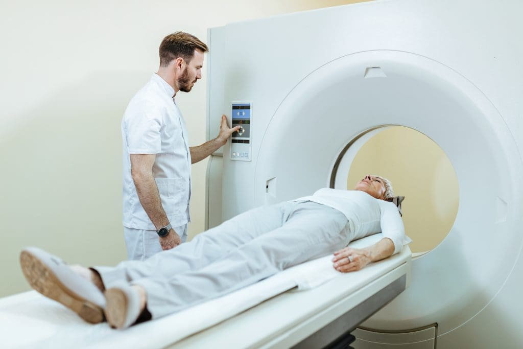
Learning about PET scans starts with the basics of Positron Emission Tomography. A PET scan is a way to see how active different parts of the body are. It helps doctors understand what’s going on inside us.
Definition and Basic Principles of Positron Emission Tomography
PET scans use a tiny bit of radioactive tracer. This tracer goes into the body and sticks to areas that are very active, like cancer cells. The PET scan machine picks up the signals from this tracer. It then makes detailed pictures of what’s inside us.
The Science Behind Metabolic Imaging
PET scans work by showing how different parts of the body work. They don’t just look at structures like X-rays do. They show how active each part is. This is really helpful for finding and treating diseases like cancer and heart problems.
Types of Radioactive Tracers Used
The main tracer used is FDG (Fluorodeoxyglucose). It’s a sugar molecule with a radioactive tag. Cancer cells take up more of this sugar than normal cells, so they show up on the scan. Other tracers are used for different things, like checking the heart or finding certain tumors.
Knowing how PET scans work helps doctors make better diagnoses. They can then plan the best treatment for each patient.
The Anatomy of a PET Scanner Machine
Exploring a PET scanner’s anatomy shows its complex design and innovation. It’s a high-tech device that uses advanced tech and precise engineering. It creates detailed images of the body’s metabolic activities.
Key Components and Technology
A PET scanner has several important parts, like a gantry, detectors, and a computer. The gantry holds the detectors, which are set up in a ring or multiple rings around the patient. These detectors catch the gamma rays from the radioactive tracer, making detailed images of metabolic activity possible.
Key technologies in PET scanning include:
- Coincidence detection: This tech helps the scanner spot gamma rays emitted at the same time, improving image clarity.
- Attenuation correction: This feature adjusts for the loss of signal due to gamma rays being absorbed or scattered by body tissues.
- Advanced reconstruction algorithms: These algorithms enhance image quality by reducing noise and artifacts.
How PET Scanners Detect Metabolic Activity
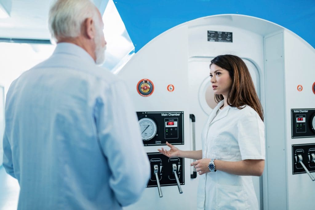
PET scanners find metabolic activity by tracking a radioactive tracer, usually Fluorodeoxyglucose (FDG). Cells with high metabolic rates, like cancer cells, take up more FDG. This leads to more gamma rays and shows up as “hot spots” on the PET scan images.
Evolution of PET Scanning Equipment
PET scanner technology has grown a lot, with better detector materials, gantry designs, and image algorithms. Today’s PET scanners, like PET/CT and PET/MRI, can do more by combining functional and anatomical imaging.
Research keeps improving PET scanner tech. It’s all about better detectors and image methods to enhance quality and speed up scans.
Standard Coverage Areas in a PET Scan
The area covered in a PET scan can change a lot. PET scans are flexible tools used for different body parts or the whole body. This depends on what the doctor needs to check.
Full-Body vs. Regional PET Scans
A full-body PET scan looks at the body from the skull to the mid-thigh. It’s often used for cancer staging and tracking treatment. A regional PET scan looks at a specific area, like the brain or heart.
Doctors choose between full-body and regional scans based on what they need to know.
“PET scans offer a unique window into the body’s metabolic activity, allowing for early detection and monitoring of various conditions.”
Does the Head Get Included by Default?
Whether the head is scanned depends on the doctor’s question. For cancer staging, the head might not be scanned if there’s no brain cancer suspicion. But for brain issues or brain cancer suspicion, the head is scanned.
Customizing Scan Regions Based on Clinical Needs
PET scans can be adjusted to fit the patient’s needs. Doctors can choose which areas to scan based on the patient’s condition. This makes sure the scan gives the most useful information for care.
In summary, PET scans can be adjusted to meet each patient’s needs. Whether it’s a full-body scan or a focused one, the aim is to get accurate info for treatment decisions.
PET Scans of the Brain and Head: Specific Applications
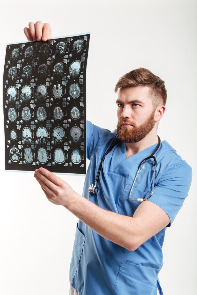
PET scans of the brain and head are key in neurology. They show how the brain works and what it’s doing. This helps doctors find and treat many brain problems.
Neurological Conditions Diagnosed with PET
PET scans help find and track many brain diseases. This includes Alzheimer’s and Parkinson’s. They spot where the brain is not working right, so doctors can act fast.
Brain Tumor Detection and Monitoring
PET scans are vital for finding and watching brain tumors. They tell doctors what kind of tumor it is and how serious it is. This helps doctors plan the best treatment.
Epilepsy and Seizure Disorder Evaluation
PET scans also help with epilepsy and seizures. They find where in the brain seizures start. This lets doctors target treatments for better results.
In short, PET scans are a big help in brain health. They help doctors understand and treat many brain issues. This includes finding and tracking brain tumors and helping with epilepsy and seizures.
The Role of FDG in PET Scan Imaging
Fluorodeoxyglucose (FDG) is key in PET scan imaging. It shows where the body’s cells are most active. This helps doctors see and study different body processes.
How Radioactive Glucose Works in the Body
FDG goes into cells that use a lot of glucose, like cancer cells or brain neurons. Inside these cells, it builds up and sends out positrons. The PET scanner catches these positrons to make detailed images of cell activity.
Why Brain Activity Shows Clearly on PET Scans
The brain needs a lot of glucose to work. So, FDG PET scans are great for seeing brain activity. They show areas of the brain that are very active, like those involved in thinking or affected by disease.
Alternative Tracers for Specialized Brain Imaging
Even though FDG is common, other tracers are used for certain brain scans. For example, tracers that find amyloid plaques help diagnose Alzheimer’s. These tracers give more details that are important for understanding and treating brain diseases.
In short, FDG is essential for PET scan imaging. It helps doctors see how active different parts of the body are, including the brain. Its ability to spot high glucose use makes it very useful in medical and research fields.
PET Scan vs. CT Scan: Key Differences
Understanding the differences between PET scans and CT scans is key for accurate diagnosis. Both are used to see inside the body, but they do different things. They give different kinds of information.
Structural vs. Functional Imaging
CT scans mainly show the body’s structure, like organs and bones. They’re great for finding problems like tumors or fractures. PET scans, on the other hand, show how the body works. They’re good for spotting metabolic issues, like cancer or neurological problems.
Radiation Exposure Comparison
Both PET and CT scans use ionizing radiation. But, the kind and amount of radiation are different. CT scans use X-rays, while PET scans use a radioactive tracer. Knowing these differences helps understand the risks and benefits of each.
When Each Scan Type is Preferred for Head Imaging
Choosing between PET and CT scans for the head depends on the situation. CT scans are fast and good for acute injuries or detailed images. PET scans are better for certain brain conditions, like tumors or epilepsy, because they look at metabolic activity. This helps doctors decide the best scan for each patient.
PET Scan vs. MRI: When Each is Recommended
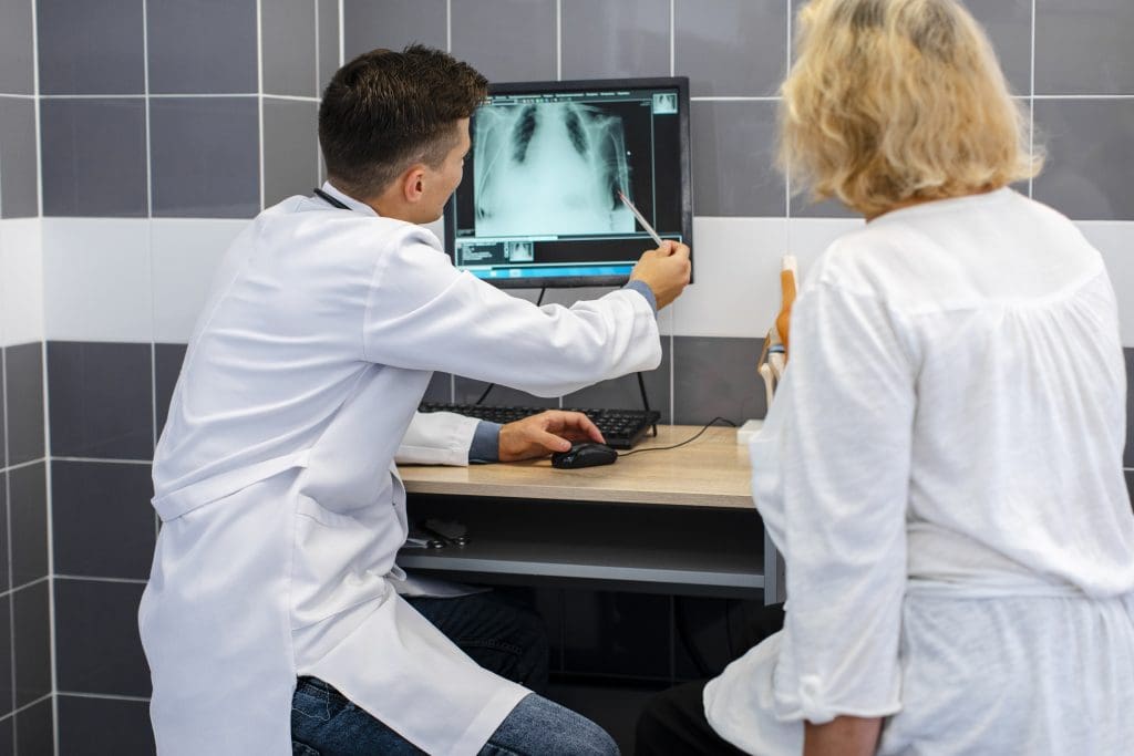
It’s important to know the differences between PET scans and MRI for brain checks. Both are key in diagnosing brain issues but serve different needs. They offer unique benefits.
Comparing Imaging Technologies for Brain Assessment
PET scans and MRI look at the brain but in different ways. PET scans are great for checking how active the brain is. They’re best for finding cancer or brain disorders where activity changes.
PET Scan Strengths: PET scans show how the brain works, like its metabolic rate and blood flow. They’re key for spotting Alzheimer’s and some cancers.
Strengths and Limitations for Neurological Diagnosis
MRI, on the other hand, gives detailed pictures of brain structures. It’s best for seeing structural problems, like injuries or tumors.
MRI Strengths: MRI’s clear images without radiation are a big plus. It’s often the first choice for many brain issues, thanks to its detailed views.
a neuroradiologist, says, “MRI is unmatched for showing soft tissue in the brain. It’s essential for diagnosing.”
“The combination of PET and MRI offers a complete view of the brain’s structure and function. It boosts diagnostic accuracy.” Neurologist
Combined PET-MRI Applications
PET-MRI combines PET and MRI into one scan. This is a big step forward in brain imaging. It lets you see both metabolic and anatomical details at the same time.
Benefits of PET-MRI: Using PET and MRI together gives a fuller picture of brain health. It helps doctors make better diagnoses and treatment plans.
The PET-CT Combination: Enhanced Diagnostic Power
PET and CT scans together have changed how we see inside the body. They mix the metabolic activity from PET scans with the detailed images from CT scans. This makes a powerful tool for doctors to diagnose better.
Why Doctors Often Order Combined Scans
Doctors choose PET-CT scans for a more accurate diagnosis. The PET scan shows where cells are active, like cancer. The CT scan gives detailed images of the body’s inside. Together, they help find and track cancer, and see how treatments work.
Medical experts say, “PET-CT scans have made cancer diagnosis and treatment planning much better.”
“The combination of PET and CT scans allows for precise localization of metabolic activity, which is key for good treatment planning.”
Benefits for Head and Brain Imaging
PET-CT scans are great for looking at the head and brain. They help doctors spot and track brain tumors, epilepsy, and Alzheimer’s disease. The scan shows both the brain’s structure and how it works, helping plan treatments better.
- Improved diagnostic accuracy
- Enhanced understanding of brain anatomy and function
- Better treatment planning for neurological conditions
Reading Fused PET-CT Images
Reading PET-CT images needs special training. Doctors compare the PET and CT parts to find abnormal activity. This helps them diagnose and track many conditions, from cancer to brain disorders.
Key advantages of fused PET-CT images include:
- Improved diagnostic accuracy
- Enhanced understanding of disease extent and spread
- Better treatment planning and monitoring
PET Scan for Cancer Detection and Staging
PET scans are key in cancer detection and staging. They show how cancer cells work by looking at their sugar use. This helps find cancer cells because they use more sugar than normal cells.
How Cancer Cells Appear on PET Scans
Cancer cells use more sugar than normal cells because they grow fast. A PET scan uses a sugar tracer that lights up where cancer is. This makes cancer cells show up as bright spots on the scan.
Brain Cancer Evaluation with PET Technology
PET scans are very important for brain cancer. They help doctors know if a tumor is new or if it’s a side effect of treatment. They also help find the best spot for a biopsy.
Monitoring Treatment Response in Head and Neck Cancers
PET scans check if head and neck cancers are getting better with treatment. They look at how the cancer’s activity changes over time. This helps doctors choose the best treatment for each patient.
In short, PET scans are very useful in fighting cancer. They help find, stage, and check how well treatments work. This is true for many cancers, like brain and head and neck cancers.
Preparing for a PET Scan That Includes the Head
Getting ready for a PET scan of the head needs some special steps. A PET scan is a detailed tool that shows how tissues work, like in the brain.
Dietary and Activity Restrictions
Before a PET scan of the head, you might need to follow certain diet rules. You might have to fast for a while and avoid sugary foods and drinks. These can change how your body uses glucose and affect the scan’s results. You might also need to not be too active before the scan.
Special Considerations for Brain Imaging
For brain scans, you should remove any metal items like hairpins or jewelry. They could mess with the scan. You should also avoid caffeine and some medicines that might change brain activity.
What to Tell Your Doctor Before the Procedure
Tell your doctor about any health issues, allergies, or medicines you take before the PET scan. This helps make sure the scan is safe and the results are right.
The Complete PET Scan Procedure Step by Step
The PET scan procedure is a detailed process that helps understand the body’s metabolic activities. It’s key for spotting and managing health issues like cancer and neurological problems.
Before the Scan: Tracer Injection and Uptake Period
The first step is injecting a radioactive tracer, usually FDG (fluorodeoxyglucose), into the patient’s blood. This tracer goes to areas with high activity, like tumors or inflamed tissues. After the injection, the patient waits for 30 to 60 minutes for the tracer to spread.
During this time, it’s important for patients to stay calm and not move too much. This helps the tracer spread evenly.
During the Scan: Positioning and Image Acquisition
After the tracer spreads, the patient lies down on a table that slides into the PET scanner. The scanner picks up the gamma rays from the tracer, creating detailed images of the body’s activity. The scan takes 30 to 60 minutes, depending on the area and the scan type.
It’s vital for patients to stay very quiet and not move during the scan. This ensures the images are clear and accurate.
After the Scan: Recovery and Follow-up
Right after the scan, patients can usually go back to their normal day. The tracer leaves the body through urine and feces over a few hours. Drinking lots of water helps flush it out.
The images are then checked by a radiologist. The results are shared with the patient during a follow-up meeting.
In summary, the PET scan procedure is a series of steps from injecting the tracer to reviewing the images. Knowing this helps patients prepare and understand the scan’s importance.
How Long Does a PET Scan Take?
Many patients wonder how long a PET scan lasts. Knowing how long it takes is key to getting ready for this test.
Typical Duration for Whole-Body Scans
A whole-body PET scan usually takes 30 minutes to 1 hour. But getting ready and waiting for the tracer to work can take hours.
Time Factors for Head-Specific PET Scans
Head-specific PET scans are quicker, lasting 20 to 30 minutes. This is because they focus on a smaller area.
Factors That May Extend Scan Duration
Several things can make a PET scan last longer, including:
- The need for extra images or delayed scans
- The patient’s condition, like trouble staying calm
- Technical problems with the scanner
Patients should follow all instructions before the scan. This helps make the process smoother and faster.
Understanding PET Scan Results and Images
Getting the results of a PET scan can be tricky, like when it’s about the brain. PET scans show detailed images of how cells work. This helps doctors find and track different health issues.
How to Interpret PET Scan Images of the Brain
Understanding PET scan images of the brain needs a deep grasp of the tech and brain cell activity. PET scan images are studied by experts. They look for spots where cell activity is off.
The images use colors to show activity levels. Color coding is key. It helps spot “hot spots” where activity is high.
Color Coding and “Hot Spots” in Brain Scans
“Hot spots” in PET scans mean areas with more tracer, showing high activity. This could mean tumors, inflammation, or other issues.
- Red and yellow areas show high activity.
- Blue and green areas show low activity.
Timeframe for Receiving and Discussing Results
How long it takes to get PET scan results varies. But usually, it’s a few days. Talking about the results with a doctor is important. It helps understand what the scan means and what to do next.
Learning about PET scan results and images helps patients understand their health better. It also helps them know their treatment options.
Potential Side Effects and Risks of PET Scans
It’s important to know the risks and side effects of PET scans. These scans are usually safe, but there are some risks and side effects. Understanding these can help patients prepare for the procedure.
Radiation Exposure Considerations
PET scans use a small amount of radiation from a radioactive tracer. The dose is low, but it’s key to think about the total radiation over time. Patients should talk to their doctor about their risk factors.
Allergic Reactions and Other Risks
Some people might have allergic reactions to the tracer or other parts of the scan. Common signs include hives, itching, and trouble breathing. People with diabetes or kidney disease should also talk to their doctor before getting a PET scan.
Safety Profile Compared to Other Imaging Methods
It’s important to compare PET scans to other imaging methods. PET scans give unique information that other methods can’t. But, the radiation from PET scans is something to consider against their benefits.
When Doctors Specify Head PET Scans
Head PET scans are key in diagnosing many neurological and psychiatric issues. They show how the brain works, helping doctors understand and treat complex problems.
Neurological Conditions Requiring PET Imaging
Doctors use head PET scans for conditions like brain tumors, epilepsy, and neurodegenerative diseases. PET imaging checks the brain’s activity, spotting issues.
For example, PET scans help find out how active brain tumors are. This info helps doctors choose the best treatment. In epilepsy, scans show where seizures start, helping plan surgery.
Psychiatric Applications of Brain PET Scans
Head PET scans also help in psychiatry. They study brain function in mental health issues like depression, schizophrenia, and substance abuse.
PET imaging shows how these conditions affect the brain. This helps doctors find better treatments.
Dementia and Alzheimer’s Diagnosis
Head PET scans are key in diagnosing dementia and Alzheimer’s. They spot brain changes and atrophy linked to these diseases.
Using FDG-PET, doctors see where Alzheimer’s affects the brain. This helps them tell it apart from other dementias. It’s vital for the right care.
In summary, head PET scans are essential for diagnosing many conditions. They offer detailed brain information, making them vital in today’s medicine.
Conclusion: The Importance of PET Scans in Head and Brain Diagnostics
PET scans are key in brain diagnostics and head imaging. They show how brain tissues work. This is important for spotting and managing brain issues like tumors, epilepsy, and dementia.
In medical practice, PET scans give doctors important info. They show where brain activity is off, helping doctors make treatment plans. Using PET scans with CT and MRI scans makes them even more useful.
PET scans have many uses in brain diagnostics. They help find and check brain cancers. As technology gets better, PET scans will play an even bigger role. This means doctors can give better care to patients.
FAQ
What is a PET scan, and how does it work?
A PET scan is a test that shows how active your body’s cells are. It uses a special tracer that lights up areas where cells are working hard, like tumors. This helps doctors see what’s going on inside you.
Does a PET scan typically include the head?
It depends on what your doctor needs to see. Some scans look at the whole body, while others focus on the head. Your doctor will decide based on your health needs.
What is the difference between a PET scan and a CT scan?
A PET scan shows how active your cells are. A CT scan shows the structure of your body. PET scans help find cancer and other diseases. CT scans help find structural problems.
How long does a PET scan take?
The time it takes varies. Whole-body scans can take 30-60 minutes. Head scans are quicker. The type of tracer and your body’s rate also play a part.
What are the possible side effects and risks of a PET scan?
PET scans are mostly safe but involve some radiation. You might feel allergic to the tracer or anxious. Your doctor will talk about the risks and benefits with you.
How do I prepare for a PET scan that includes the head?
To get ready, you might need to fast or avoid certain foods. Tell your doctor about any allergies or health issues. You’ll also need to arrive early to fill out paperwork.
What is the role of FDG in PET scan imaging?
FDG is a special sugar that cancer cells love. It helps doctors see where cancer might be. This is how PET scans find tumors and other problems.
How do I interpret PET scan images of the brain?
A radiologist or specialist will look at your scan. They find areas that are not working right. Your doctor will then explain what it means for you.
When are PET scans used to diagnose neurological conditions?
PET scans help find and track brain diseases like Alzheimer’s and Parkinson’s. They show where the brain is not working right. This helps doctors make treatment plans.
Can PET scans be used to detect cancer in the head and neck?
Yes, PET scans are great for finding cancer in the head and neck. They light up cancer cells. This helps doctors see how well treatments are working and if cancer is coming back.


