Last Updated on November 27, 2025 by Bilal Hasdemir
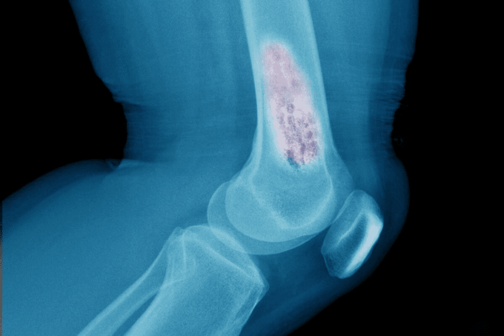
At Liv Hospital, we use bone scintigraphy, also known as radionuclide bone scanning. This method helps us diagnose and track bone-related issues like arthritis and cancer. Many patients ask, “what does a bone scan show?” It reveals areas where the bone is not functioning correctly, giving us clues about conditions such as fractures, infections, cancer spread, and other bone abnormalities. This detailed imaging helps us understand bone health and plan appropriate treatments.
A bone scan can find the reason for bone pain that doesn’t make sense. It can also spot damage from infections or other bone problems. Our goal is to offer top-notch healthcare and support to patients from around the world.
Key Takeaways
- Bone scintigraphy helps diagnose and monitor bone-related conditions.
- It reveals areas of abnormal bone metabolism or turnover.
- A bone scan can identify the cause of unexplained bone pain.
- It detects damage caused by infection or other bone conditions.
- Our hospital provides extensive international patient support.
Understanding Bone Scans: A Complete Overview
A bone scan, also known as bone scintigraphy, is a special imaging technique. It helps diagnose and monitor bone diseases. This tool gives important info about bone health, helping doctors spot many conditions.
Definition and Purpose of Bone Scintigraphy
Bone scintigraphy uses small amounts of radioactive materials. It highlights areas of bone activity. This helps detect conditions like cancer, infection, and arthritis.
It uses radionuclides that build up in bone tissue. This lets doctors see bone activity. It’s key for spotting bone disease and tracking its progress.
Types of Radionuclide Bone Scanning
There are different types of bone scanning, each using different radioactive materials. The most common uses Technetium-99m labeled diphosphonates, like Tc-MDP (Methylene Diphosphonate).
| Type of Radionuclide | Application | Characteristics |
| Tc-MDP | Bone scanning for various bone diseases | High sensitivity for detecting bone metastases and other bone abnormalities |
| Tc-HMDP | Bone scanning, particular for bone metastases | Similar to Tc-MDP but with slightly different biodistribution |
When Bone Scans Are Recommended
Bone scans are recommended for many reasons. They help diagnose and monitor cancer, like bone metastases. They also check for bone diseases like Paget’s disease and bone infections.
They’re useful for looking at bone trauma or stress fractures too. Doctors decide on a bone scan based on symptoms, medical history, and other test results.
How Bone Scans Work: The Science Behind the Procedure
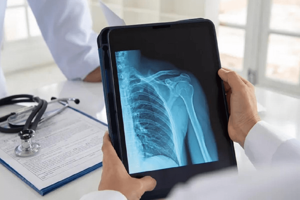
Bone scans use nuclear medicine to check bone health. They highlight areas of abnormal bone activity. This helps doctors understand bone health better.
Radiotracers and Nuclear Imaging Fundamentals
Bone scans rely on radiotracers, substances that emit radiation. These substances go to areas of high bone activity. Technetium-99m methylene diphosphonate (Tc-99m MDP) is often used because it binds to bone.
“Radiotracers in bone scans show metabolic activity in bones. This gives insights that other scans can’t.” Imaging specialists note, explaining the science behind bone scans.
Bone Metabolism and Radiotracer Uptake
Bone metabolism is a constant process of breaking down and building bone. Conditions like arthritis or cancer change this process. The radiotracer used in scans shows these changes.
The amount of radiotracer taken up shows how active the bone is. This helps doctors find and diagnose bone-related issues.
The Role of Gamma Cameras in Image Capture
Gamma cameras are key in capturing images from bone scans. They detect the gamma radiation from the radiotracer. This creates images showing where the radiotracer is most active.
Modern gamma cameras, like those with SPECT or SPECT/CT, give detailed images. These images help doctors make more accurate diagnoses.
New medical technology has made bone scans better. SPECT/CT combines functional and anatomical information. This gives a full picture of bone health.
The Bone Scan Procedure: What to Expect
Getting a bone scan might seem scary, but knowing what to expect can make it easier. We’ll walk you through each step, from getting ready to after the scan. This way, you’ll feel more at ease and informed.
Preparation Requirements Before the Scan
Before your bone scan, there are a few things to do. Take off any jewelry or metal objects that might get in the way. Wear comfy, loose clothes. You might need to change into a gown.
- Tell your doctor about any medicines you’re on.
- Let them know about any allergies, like to contrast dye.
- If you’re expecting or breastfeeding, talk to your doctor about it.
The Injection and Waiting Period
A bone scan starts with a small injection of radioactive material into your arm. This material goes to your bones, showing where there’s unusual activity. You’ll wait a few hours for it to spread.
While waiting, you can do your usual things. But remember to drink lots of water.
The Scanning Process and Duration
The scanning part takes about 30 to 60 minutes. You’ll lie on a table that slides into a big camera. This camera takes pictures from different angles.
Post-Procedure Care and Considerations
After the scan, you can go back to your normal day right away. The material will leave your body through urine and feces. Drink lots of water to help get rid of it. The radiation is very low, and it’s gone in a few days.
Always follow any special instructions from your doctor or the radiology team. If you have questions or worries, ask your medical team.
What Does a Bone Scan Show? Detecting Abnormalities
Bone scans are key in understanding bone health. They help doctors find bone-related problems. This test is a diagnostic imaging tool.
Normal vs. Abnormal Bone Scan Results
Bone scan results can be normal or abnormal. Normal results mean the radiotracer spreads evenly in the bones. Abnormal results show uneven radiotracer uptake, pointing to different conditions.
Abnormal results can mean many things, like fractures, infections, or cancer. But, not all abnormal results are cancer. They can also show other bone issues.
Hot Spots and Cold Spots: What They Indicate
Bone scans show “hot spots” and “cold spots.” Hot spots mean more radiotracer, showing active bone areas. This could be due to fractures, infections, or tumors. Cold spots have less radiotracer, suggesting issues like avascular necrosis or bone lesions.
Common Conditions Detected Through Bone Scans
Bone scans can find many bone-related issues. Some common ones include:
| Condition | Description |
| Fractures | Bone scans can spot fractures, even if X-rays can’t. |
| Infections | Osteomyelitis and other bone infections are detectable. |
| Cancer | Bone scans can find both primary and metastatic cancer. |
These conditions affect bone health a lot. Bone scans are a great tool for diagnosing and tracking these issues.
Bone Scans for Arthritis Detection and Evaluation
Bone scans are a key tool in diagnosing arthritis. They help us understand joint health and how the disease progresses. This information helps doctors make better decisions for their patients.
Types of Arthritis Visible on Bone Scans
Bone scans can spot different types of arthritis. This includes osteoarthritis, rheumatoid arthritis, and other inflammatory joint diseases. They show joint inflammation and changes in bone metabolism.
In rheumatoid arthritis, bone scans show increased activity in affected joints. This is due to inflammation and bone turnover. This info is vital for tracking disease activity and treatment success.
Joint Inflammation Assessment
Bone scans are great for checking joint inflammation. They find areas of high bone activity, showing where inflammation is happening. This is key for diagnosing arthritis.
“Bone scintigraphy provides a unique window into the metabolic activity of bone, allowing us to assess the extent of joint inflammation and monitor changes over time.”
– A Nuclear Medicine Specialist
Monitoring Disease Progression and Treatment Response
Bone scans are essential for tracking arthritis and treatment success. By comparing scans over time, doctors can see how the disease is changing. This helps them adjust treatment plans as needed.
| Arthritis Type | Bone Scan Findings | Clinical Implication |
| Osteoarthritis | Increased uptake in joint margins | Indicative of degenerative changes |
| Rheumatoid Arthritis | Symmetric uptake in multiple joints | Suggests active inflammation |
| Psoriatic Arthritis | Uptake in joints and entheses | Reflects inflammatory and proliferative changes |
As shown in the table, different types of arthritis show unique patterns on bone scans. This helps in diagnosis and treatment.
Using bone scans helps us better understand arthritis. This leads to more effective treatments and better patient outcomes.
Will a Bone Scan Show Arthritis? Sensitivity and Specificity
Bone scans are a key tool in diagnosing diseases. But, they can’t always show arthritis. It depends on the type of arthritis and how far it has progressed.
Inflammatory vs. Degenerative Arthritis on Scans
Arthritis is a broad term for joint diseases. It includes inflammatory and degenerative types. Inflammatory arthritis, like rheumatoid arthritis, causes joint inflammation. Degenerative arthritis, such as osteoarthritis, is due to cartilage wear and tear.
Bone scans can spot both types of arthritis. But, they’re better at catching inflammatory arthritis. This is because inflammatory arthritis shows up as hot spots on the scan due to inflammation and bone changes.
On the other hand, degenerative arthritis might show up as slightly warmer spots. But these spots are usually less intense than those seen in inflammatory arthritis.
Comparison with Other Imaging Modalities for Arthritis
Besides bone scans, other imaging tools are used to diagnose arthritis. X-rays are good for seeing joint damage and degeneration. MRI and CT scans give detailed views of soft tissues and bones, helping to diagnose and track arthritis.
| Imaging Modality | Sensitivity for Arthritis | Specificity for Arthritis | Additional Information |
| Bone Scan | High for inflammatory arthritis | Moderate | Shows metabolic activity |
| X-ray | Low to Moderate | High for bone damage | Good for assessing joint space narrowing |
| MRI | High | High | Excellent for soft tissue evaluation |
| CT Scan | Moderate to High | High for bone details | Good for detecting bone erosions |
Limitations in Arthritis Diagnosis
Bone scans have their limits in diagnosing arthritis. They can show activity in conditions other than arthritis, like infections or tumors. Also, early or mild arthritis might not be seen on a bone scan. So, doctors often need to use other tests to confirm a diagnosis.
In summary, bone scans are helpful in diagnosing arthritis, mainly for inflammatory types. But, they should be part of a broader diagnostic approach. This includes considering clinical findings and possibly other imaging tests.
Bone Scans in Cancer Detection and Staging
Bone scans are key in finding cancer and seeing how far it has spread to bones. They help spot both bone cancers and cancer that has spread to bones. This info is vital for planning treatment.
Primary Bone Cancers Visible on Scans
Bone scans can find different types of bone cancers. These include osteosarcoma, Ewing’s sarcoma, and chondrosarcoma. They work because these cancers use a lot of energy, showing up on scans.
Metastatic Cancer Detection Capabilities
Bone scans are great at finding cancer that has spread to the bones. They’re very good at spotting cancer from breast, prostate, and lung cancers. Radiology experts say they’re a key tool for seeing how far cancer has spread.
Patterns of Cancer Presentation on Bone Scans
Cancer can show up in different ways on bone scans. It might look like hot spots, spread out, or even cold spots. The way it looks can tell doctors a lot about the cancer.
Role in Cancer Staging and Treatment Planning
Bone scans help figure out how far cancer has spread to bones. This info is key for choosing the right treatment. For example, if cancer is in many bones, doctors might choose treatments that target the whole body.
| Cancer Type | Typical Bone Scan Findings | Clinical Significance |
| Breast Cancer | Multiple focal areas of increased uptake | Indicates metastatic disease, guiding systemic therapy |
| Prostate Cancer | Osteoblastic metastases with intense uptake | Aids in staging and assessing treatment response |
| Osteosarcoma | Intense localized uptake | Helps in assessing primary tumor activity and possible metastases |
Bone scans give detailed info on bone involvement. They’re essential for managing cancer patients. They help doctors make the best treatment plans.
What Cancers Can a Bone Scan Detect? Sensitivity for Different Malignancies
Bone scans are key in finding different cancers. They help doctors see if cancer has spread to bones. This info is vital for planning treatment and understanding how far the cancer has spread.
Breast Cancer Metastases
Breast cancer often spreads to bones. Bone scans are great at finding this spread. They show where bone activity is high, which means cancer might be there. Studies show bone scans work well for finding breast cancer in bones, like the spine and pelvis.
Prostate Cancer Spread to Bone
Prostate cancer also often goes to bones. Bone scans are very helpful in spotting this. They’re good at finding cancer in bones, thanks to their high sensitivity. This helps doctors keep track of how the cancer is growing and if treatments are working.
Lung Cancer and Other Common Metastatic Sources
Lung cancer also spreads to bones, and bone scans can find it. Other cancers, like kidney and thyroid, can also be found in bones with bone scans. While the scans might not catch every case, they’re a big help in managing cancer.
Primary Bone Malignancies
Bone scans can also spot cancers that start in bones. Tumors like osteosarcoma show up on scans. But, other tests like MRI are often needed for a closer look. Bone scans help doctors diagnose primary bone cancers, working alongside other tests.
To show how well bone scans work, let’s look at a table:
| Cancer Type | Sensitivity of Bone Scans | Common Sites of Metastasis |
| Breast Cancer | High | Axial skeleton, ribs |
| Prostate Cancer | High | Pelvis, spine, ribs |
| Lung Cancer | Moderate to High | Spine, ribs, pelvis |
| Primary Bone Malignancies | Variable | Depends on tumor type and location |
The table shows bone scans work differently for each cancer. Knowing this helps doctors understand scan results better. It’s key for making the best care plans for patients.
Differentiating Arthritis from Cancer on Bone Scans
Bone scans can show similar results for arthritis and cancer, making diagnosis hard. Both conditions change bone metabolism, which bone scans detect.
Overlapping Patterns and Diagnostic Challenges
Arthritis and cancer can look alike on bone scans. They both show “hot spots” where the bone is more active.
- Arthritis: Shows symmetrical uptake, mainly in joints affected by arthritis.
- Cancer: Presents with focal or asymmetrical uptake, not just in joints.
Key Distinguishing Features
Despite challenges, there are features that help tell arthritis from cancer on bone scans.
- Distribution: Arthritis affects joints symmetrically, while cancer is more random or asymmetric.
- Intensity: Cancerous lesions may have more intense uptake than arthritis.
- Location: Arthritis mainly involves joints, but cancer can affect any bone.
When Additional Testing Is Necessary
Sometimes, more tests are needed to confirm a diagnosis. This might include:
- CT or MRI scans: For detailed images of bones and tissues.
- PET scans: To check metabolic activity and distinguish between benign and malignant.
- Biopsy: To get a definitive diagnosis by examining tissue samples.
By using bone scan results, clinical info, and extra tests when needed, doctors can make accurate diagnoses. This helps in creating effective treatment plans.
Advanced Bone Scintigraphy Technologies and Machines
Modern bone scintigraphy technologies have changed how we diagnose and treat bone diseases. These advancements have made diagnosis more accurate and patient care better.
SPECT and SPECT/CT Imaging
SPECT and SPECT/CT imaging have improved bone scintigraphy. SPECT gives three-dimensional images, showing more about bone health. SPECT/CT adds CT images, giving both function and anatomy, making diagnosis more precise.
SPECT/CT imaging is great for complex cases like finding cancer spread or bone cancer extent. It helps tell if a lesion is benign or malignant, guiding treatment.
PET Bone Scans and Hybrid Imaging
PET bone scans are a big step forward in bone scintigraphy. They use Fluorodeoxyglucose (FDG) to see bone metabolism. Hybrid imaging, like PET/CT and PET/MRI, combines PET’s metabolic info with CT or MRI’s anatomy, giving a full view of bone issues.
PET bone scans are key in cancer care, spotting cancer spread and checking treatment success. They show tumor metabolism, vital for staging and treatment planning.
Modern Bone Scintigraphy Machine Capabilities
Today’s bone scintigraphy machines are more sensitive, detailed, and fast. These improvements mean doctors can get high-quality images quickly, helping with fast diagnosis and treatment.
| Technology | Key Features | Clinical Applications |
| SPECT/CT | 3D imaging, functional and anatomical information | Detecting metastases, assessing bone cancers |
| PET/CT | High sensitivity, metabolic activity assessment | Oncology, detecting cancer spread, treatment response |
| PET/MRI | Excellent soft tissue contrast, functional information | Assessing tumor extent, monitoring treatment response |
Future Directions in Bone Scanning Technology
The future of bone scintigraphy looks bright, with new research and tech advancements. New radiotracers, better detectors, and AI in image analysis are on the horizon.
These new developments will make bone scintigraphy even more valuable for managing bone diseases.
Interpretation of Bone Scan Results by Healthcare Professionals
Understanding bone scan results is a detailed task. It combines scan data with the patient’s medical history and symptoms. This requires a deep knowledge of nuclear medicine.
The Role of Nuclear Medicine Specialists
Nuclear medicine specialists are key in reading bone scan results. They have the skills to spot abnormalities and explain what they mean. A study on NCBI shows their importance in accurate interpretations.
These specialists do several things:
- They look at bone scan images for any unusual activity.
- They match scan results with the patient’s medical history and symptoms.
- They suggest possible causes based on the scans.
- They recommend more tests if needed.
Integrating Clinical Information with Scan Findings
Reading bone scan results is more than just looking at images. It’s about combining scan data with the patient’s health history. This helps understand the condition fully.
For example, a cancer patient might show many spots of increased activity on the scan. The specialist must think about metastasis and compare with other tests like CT or MRI.
Multidisciplinary Approach to Result Interpretation
Getting bone scan results right often needs teamwork. Specialists from different fields work together. This ensures all information is considered for a complete picture.
Teamwork brings many benefits:
- It makes diagnosis more accurate.
- It leads to better care by looking at everything together.
- It helps plan treatment more effectively.
Communication of Results to Patients
After understanding bone scan results, it’s important to share them with the patient. This means explaining the results clearly and discussing what they mean. It’s also about what to do next.
Good communication helps patients understand and follow treatment plans. It also makes them happier with their care.
Conclusion: The Value of Bone Scans in Modern Diagnostics
At Liv Hospital, we understand how important bone scans are in today’s medicine. We’ve looked at what bone scans are, how they work, and their uses. This includes spotting and managing issues like arthritis and cancer.
Bone scans give us detailed views of bone health. This helps doctors diagnose and keep track of conditions better. With cutting-edge bone scintigraphy, we offer care that’s both precise and tailored to each patient.
Bone scans are key in today’s medical world. They’re a gentle way to find problems without surgery. We keep using these tools to give top-notch care to our patients from around the globe.
FAQ
What is a bone scan, and how does it work?
A bone scan, also known as bone scintigraphy, is a test that uses a small amount of radioactive material. It helps see how bones work and find bone-related issues. The test injects a radiotracer into your blood. This radiotracer goes to active bone areas. Then, a gamma camera captures images of where it goes.
Does a bone scan show arthritis?
Yes, a bone scan can spot arthritis, like rheumatoid arthritis. It checks for joint inflammation and tracks how the disease changes. But, bone scans might not always be 100% accurate. Other tests might be needed to confirm a diagnosis.
What cancers can a bone scan detect?
Bone scans can find cancers that have spread to bones, like from breast, prostate, or lung cancer. They can also find cancers that start in bones. How well bone scans work for different cancers varies. But, they’re great for finding osteoblastic metastases.
How does a bone scan differentiate between arthritis and cancer?
It’s hard to tell arthritis from cancer on a bone scan because they can look similar. But, the scan’s details can help tell them apart. More tests might be needed to be sure of a diagnosis.
What is the role of nuclear medicine specialists in interpreting bone scan results?
Nuclear medicine specialists are key in understanding bone scan results. They use their knowledge to spot problems and share results with patients and doctors.
What are the latest advancements in bone scintigraphy technology?
New tech in bone scintigraphy includes SPECT and SPECT/CT imaging, PET bone scans, and hybrid imaging. These advancements make bone scans more accurate. Modern machines have better features, and research keeps improving bone scanning.
How do I prepare for a bone scan?
Preparation for a bone scan is simple. You might need to remove jewelry and follow hydration and diet rules. Your healthcare provider or nuclear medicine team will give you specific instructions.
What should I expect during and after the bone scan procedure?
During the bone scan, you’ll get a radiotracer injection and then imaging with a gamma camera. It’s painless and takes about 30 minutes to an hour. Afterward, you can go back to normal activities. The radiotracer will leave your body over time.
References
- Van den Wyngaert, T., Strobel, K., Kampen, W. U., Bielack, S., Busch, R., Chiti, A., ¦ & Palmer, J. (2016). The EANM practice guidelines for bone scintigraphy. European Journal of Nuclear Medicine and Molecular Imaging, 43(9), 1723“1738. https://www.ncbi.nlm.nih.gov/pmc/articles/PMC4932135/
- Zhao, Z., Zhang, Y., & Wang, L. (2020). Deep neural network based artificial intelligence assisted bone scintigraphy interpretation in detecting bone metastasis. Scientific Reports, 10, 13622. https://www.nature.com/articles/s41598-020-74135-4


