Last Updated on November 27, 2025 by Bilal Hasdemir

When someone is diagnosed with cancer, they often have to go through many tests. One of these tests is the bone scan, which checks if cancer has spread to the bones. Many patients wonder how long does a bone scan last ” typically, the whole process can take 1 to 4 hours. This scan uses a special tracer to show where cancer cells are in the bones.
At Liv Hospital, we use the newest and most accurate methods for bone cancer scans. We make sure our tests are reliable and patient-focused. Knowing how long a bone scan lasts helps patients prepare and feel more comfortable during the process. This careful approach helps us find out if cancer has spread to the bones and how it’s progressing.
Key Takeaways
- A bone scan is a diagnostic tool used to detect bone metastases in cancer patients.
- The process involves injecting a radioactive tracer to highlight abnormal cell activity.
- The entire procedure can take 1 to 4 hours from injection to image acquisition.
- Bone scans are effective in detecting primary cancer and cancer that has spread to the bones.
- Patients can expect a painless procedure with minimal discomfort or side effects.
Understanding Bone Scans for Cancer Detection

A radionuclide bone scan, also known as a whole body nuclear bone scan, is a test used to find bone metastases in cancer patients. It involves injecting a small amount of radioactive material into the bloodstream. This material gathers in the bones, showing areas of abnormal activity.
What Is a Radionuclide Bone Scan?
A radionuclide bone scan is a tool that spots areas of high bone activity. It’s great for finding bone metastases, as cancer cells can change bone structure. Studies show these scans are good at finding bone metastases and are key in cancer staging.
The process involves several steps:
- A small amount of radioactive tracer is injected into the bloodstream.
- The tracer gathers in the bones over time.
- A gamma camera captures the radiation from the tracer, making bone images.
Role in Cancer Diagnosis and Staging
Bone scans are key in cancer diagnosis and staging. They help see how far cancer has spread. By spotting bone metastases early, doctors can plan the right treatment. The scan also helps track how well treatment is working and if bone health changes.
Key benefits of bone scans in cancer diagnosis include:
- Early detection of bone metastases.
- Accurate staging of cancer.
- Monitoring of treatment response.
Medical experts say bone scans are vital for cancer patients. They provide important info for treatment decisions. We see how important this tool is for our patients’ care.
How Long Does a Bone Scan Last: Complete Timeline
Knowing how long a bone scan takes is important for those getting ready for it. The whole process, from start to finish, can take several hours. We’ll go through the timeline to help you know what to expect.

Pre-Scan Preparation Time
Before the scan, patients usually do some prep work. They might arrive early to fill out papers and get comfortable. The American Cancer Society says patients should plan to spend some time before the scan. But, how much time varies.
Radiotracer Injection and Uptake Period
At the facility, a radiotracer is injected into a vein. This material, a bit radioactive, goes to the bones. It helps the scan show active bone areas. The time it takes for the material to absorb can be 1 to 3 hours.
“The radiotracer uptake period is a critical component of the bone scan process, as it directly affects the quality of the images obtained during the scan.”
Actual Scanning Duration
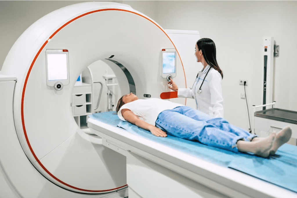
After the material absorbs, the scanning starts. You’ll lie on a table that moves into a big machine. The scan takes 30 to 60 minutes, depending on the scan type and body area. A whole body scan usually takes 30 to 45 minutes.
The whole process, from injection to scan, can take 1 to 4 hours. It’s key to plan ahead and stay hydrated. Every person’s experience is different, based on the facility and health status.
Types of Bone Scans and Their Duration
Bone scans are not a one-size-fits-all tool for cancer detection. They come in different types to meet various needs. We use different imaging methods to get detailed diagnostic info.
Standard Whole Body Nuclear Bone Scan
A Standard Whole Body Nuclear Bone Scan is often used in cancer diagnosis. It involves injecting a small amount of radioactive material, like technetium-99m, into the bloodstream. The scan takes about 30 to 60 minutes.
During this time, the patient lies on a table. A gamma camera captures images of the whole skeleton.
Multi-Phase Bone Scans
Multi-Phase Bone Scans take images at different times after the radiotracer injection. This helps spot different bone conditions. These scans can take longer, often several hours.
SPECT and SPECT/CT Imaging
SPECT (Single Photon Emission Computed Tomography) and SPECT/CT imaging offer detailed images. SPECT/CT combines SPECT’s functional info with CT’s anatomical details. These scans can last 30 minutes to an hour or more.
Knowing about the different bone scans and their times helps patients get ready. Each scan has its own benefits and is picked based on the patient’s needs.
The Bone Scan Procedure: Step-by-Step
The bone scan procedure has several key steps. These steps include preparation and the actual scanning. They help get accurate results for cancer diagnosis. Knowing each step can make patients feel more ready and less worried.
Patient Preparation Guidelines
Before a bone scan, patients must follow certain guidelines. This ensures the procedure goes well. We suggest that patients:
- Remove any jewelry or metal objects that could interfere with the scan.
- Wear comfortable, loose-fitting clothing.
- Inform their doctor about any medications they are currently taking.
- Be prepared to drink plenty of water after the radiotracer injection to help flush out the tracer.
Good preparation is key for quality images. A study on NCBI shows that careful preparation and handling of the radiotracer are vital for accurate results.
The Injection Process
The bone scan starts with a small amount of radioactive material, called a radiotracer, injected into a vein. This material is drawn to active bone areas, like cancer metastases.
The injection takes a few minutes and is usually easy for patients. After, patients wait a few hours for the radiotracer to spread through their bones.
Positioning and Image Acquisition
After waiting, the patient is placed on a scanning table. The bone scan images are then taken with a gamma camera. This process is painless and takes about 30-60 minutes.
During scanning, patients must stay very quiet. This ensures clear and accurate images. The gamma camera captures the radiotracer’s spread in the bones. A radiologist then looks at these images to find any abnormal bone activity.
| Procedure Step | Duration | Description |
| Radiotracer Injection | A few minutes | Injection of radioactive material into a vein. |
| Waiting Period | 2-4 hours | Allowing the radiotracer to distribute throughout the bones. |
| Scanning | 30-60 minutes | Capturing images using a gamma camera. |
“Proper patient preparation and handling of the radiotracer are key for quality bone scan images.”
Source: NCBI
Understanding the bone scan process helps patients prepare better. It reduces anxiety and makes the experience smoother.
Interpreting Bone Scan Results
Understanding bone scan results is key. We need to know what’s normal and what’s not. Radiologists help us figure this out by looking at the scan patterns.
Normal vs. Abnormal Bone Scan Patterns
A normal bone scan shows even tracer uptake. This means the bones are working right. But, an abnormal scan shows spots where the tracer is more or less. This could mean cancer or other problems.
Normal bone scans have even tracer spread. Abnormal scans have “hot spots” where activity is high. These spots can be from cancer, injury, or infection.
Understanding “Hot Spots” in Cancer Diagnosis
“Hot spots” on a bone scan mean high bone activity. In cancer, these spots can show where cancer has spread to bones. But, they can also mean other things like fractures or infections.
We look closely at hot spots to understand them. We use this info along with other tests to make a correct diagnosis.
False Positives and Their Causes
False positives happen when scans show cancer when there isn’t any. This can be because of joint disease, fractures, or inflammation. It’s important to check the scan with the patient’s history and other tests to avoid mistakes.
Knowing why false positives happen helps us get better at reading scans. This way, we can give our patients the best care possible.
Comparing Bone Scans to Other Imaging Methods
Different imaging techniques have their own strengths in finding bone metastases. Knowing these differences is key for accurate diagnosis. Doctors often use a mix of imaging methods to fully understand the disease.
CT Bone Scans: Duration and Differences
A CT bone scan uses X-rays to show detailed bone and tissue images. It doesn’t catch early metabolic changes like nuclear medicine scans do. But, it gives more detailed anatomy.
CT bone scans take a few minutes to finish. Sometimes, a contrast agent is used to make certain areas clearer.
Key differences between CT bone scans and nuclear medicine bone scans include:
- CT scans provide more detailed anatomical images
- Nuclear medicine scans detect metabolic changes earlier
- CT scans are generally quicker
MRI vs. Nuclear Medicine Bone Scans
Magnetic Resonance Imaging (MRI) gives high-resolution images of soft tissues and bones. It’s great for seeing marrow involvement and soft tissue issues. But, nuclear medicine bone scans are better at catching bone metabolism changes.
Doctors often use MRI and nuclear medicine scans together. MRI gives detailed views of specific areas. Bone scans show the whole skeletal system.
PET Scans for Bone Metastases
Positron Emission Tomography (PET) scans, like those with F-Fluorodeoxyglucose (FDG), are key for finding bone metastases. They combine functional and anatomical details, making them great for cancer staging.
The advantages of PET scans include:
- High sensitivity for detecting metabolically active cancer cells
- Ability to assess the entire body in a single scan
- Combining functional and anatomical information
Understanding each imaging method’s strengths and weaknesses helps doctors choose the best approach. This ensures accurate diagnosis and effective treatment plans.
Factors That May Extend Bone Scan Duration
Bone scan duration can vary a lot. Knowing what affects it can help plan better.
Patient-Related Factors
How well a patient is can change how long a bone scan takes. Some medical conditions might need more prep or extra scans.
- Physical Limitations: Patients with mobility issues might take longer to get into position.
- Medical Conditions: Kidney disease, for example, can change how the body handles the scan’s tracer.
Technical Considerations
Technical problems or the need for advanced scans can also make bone scans longer.
| Technical Factor | Impact on Scan Duration |
| Equipment Calibration | May need extra time before starting the scan. |
| SPECT/CT Imaging | Can make scans longer because of the detailed images. |
Additional Imaging Requirements
At times, more images are needed for a full diagnosis. This might mean more scans or different imaging types.
- Multi-Phase Bone Scans: These scans take images at different times and can make the scan longer.
- SPECT Imaging: Offers detailed images and is often used with CT scans for better results.
In summary, many things can affect how long a bone scan takes. These include the patient’s health, technical needs, and extra imaging. Knowing these can help plan and manage the scan better.
Post-Scan Care and Radiation Considerations
After your bone scan, we help you with important care steps. We also talk about radiation exposure. Knowing these details is key for your safety and the scan’s success.
Radiation Exposure and Safety
A bone scan uses a small amount of radiation. This is because of the radiotracer used. Even though the radiation is safe, it’s good to know the risks. Research shows the benefits of bone scans in cancer diagnosis are more important than the risks of radiation (Source: First web source).
We take strict safety steps to lower radiation exposure. The radiotracer used in bone scans breaks down fast. We suggest drinking lots of water after the scan to help get rid of the radiotracer.
Post-Procedure Instructions
After your bone scan, there are important steps to follow. It’s best to keep drinking lots of fluids to get rid of the radiotracer. Most people can go back to their usual activities right after the scan. But, it’s wise to avoid being close to pregnant women and young kids for about 24 hours.
Some people might feel a little discomfort where the injection was given. If you notice any odd symptoms or have worries, reach out to your healthcare provider. They can offer advice and reassurance based on your situation.
By following these instructions and knowing about radiation safety, you can reduce any risks from your bone scan.
Conclusion
Learning about bone scans helps patients get ready for their tests. Studies show bone scans are key in finding and treating cancer. They help doctors see if cancer has spread to bones and plan treatments.
Bone scans are important for spotting cancer in bones. They tell doctors how far the cancer has spread. Knowing how bone scans work helps patients and doctors make better treatment plans.
We know how vital bone scans are in finding cancer. We aim to give top-notch care and support to patients. Our goal is to help patients get the best care possible during their diagnosis.
FAQ
What is a bone scan, and how is it used in cancer diagnosis?
A bone scan is a test that uses a tiny amount of radioactive material. It’s injected into the blood to find bone metastases. This test is key in cancer diagnosis, showing if cancer has spread to bones.
How long does a bone scan typically last?
A bone scan usually takes 30 minutes to an hour to scan. But, getting ready and the radiotracer to take effect can take hours.
What are the different types of bone scans, and how do they differ?
There are many types of bone scans, like standard whole body scans and SPECT/CT imaging. Each type has its own time and use, helping to see different bone health aspects.
How do I prepare for a bone scan?
To get ready for a bone scan, remove jewelry and metal objects. Tell your doctor about any allergies or health issues. We give detailed instructions for a smooth process.
What is the difference between a normal and abnormal bone scan?
A normal scan shows even radiotracer distribution. An abnormal scan has “hot spots,” showing possible bone metastases. Our radiologists carefully look at the results for accurate diagnoses.
How does a bone scan compare to other imaging methods, such as CT or MRI?
Bone scans are great for finding bone activity. CT and MRI give more detailed body pictures. PET scans are also used, often with bone scans.
What are the risks associated with radiation exposure from a bone scan?
The radiation from a bone scan is low. We take steps to reduce risks. Tell your doctor about any concerns and follow instructions for safety.
How long does it take to get the results of a bone scan?
Getting bone scan results can take time. Our team works fast to give accurate results. We’ll talk about them with you and your doctor to plan next steps.
Can I undergo a bone scan if I have certain medical conditions or implants?
If you have certain conditions or implants, tell your doctor before the scan. We’ll check your situation and decide the best way to keep you safe and get accurate results.
What is the role of SPECT/CT imaging in bone scans?
SPECT/CT imaging combines SPECT’s function with CT’s anatomy. It gives a full view of bone health. This is great for finding and understanding bone metastases.
How does a whole body nuclear bone scan work?
A whole body nuclear bone scan uses a radiotracer in the blood. It highlights abnormal bone areas. The scan shows the whole body, giving a detailed bone health check.
References
- Al-Ibraheem, A., Garcia Vicente, A. M., & Rager, O. L. (2023). Nuclear medicine imaging for bone metastases assessment. Frontiers in Medicine. https://www.frontiersin.org/journals/medicine/articles/10.3389/fmed.2023.1320574/full


