Last Updated on November 27, 2025 by Bilal Hasdemir

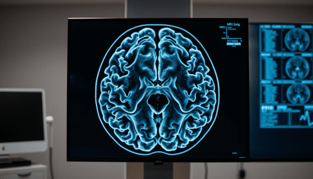
Recent breakthroughs in brain scanning technology have changed the game in neuroimaging. Now, we have tools like the Connectome 2.0 MRI and the BrainPET 7T insert. They let us see tiny brain details with amazing clarity.
The arrival of 7T MRI is a big deal. It gives us better images and helps diagnose brain issues more accurately. The National Institutes of Health (NIH) says these improvements are key to tackling complex brain diseases and finding new treatments.
These new brain scanner technologies are changing what we can do in neuroscience and healthcare. Places like Liv Hospital are leading the way with their focus on quality, innovation, and trust in their patients.
Key Takeaways
- Advances in brain scanning technology enable high-resolution MRI scans.
- The Connectome 2.0 MRI and BrainPET 7T insert offer unprecedented clarity.
- 7T MRI provides superior spatial resolution and diagnostic accuracy.
- These breakthroughs improve understanding and treatment of neurological conditions.
- Institutions like Liv Hospital support these advancements.
The Current State of Brain Scanner Technology

The world of brain imaging is changing fast with new MRI tech. These new tools help fix old MRI problems. They’ve been key in brain imaging for years.
Traditional MRI Limitations in Neurological Imaging
Old MRI systems can’t show details well, which is a big problem for brain health. Traditional MRI technology can’t see the tiny details needed for accurate diagnoses. It’s hard to spot the small changes in brain structures with standard MRI.
New MRI tech is a big step up from the old systems. Here’s a table showing the main differences:
| Feature | Traditional MRI | High-Resolution MRI |
| Resolution | Limited detail | Up to 3.5 million pixels per scan |
| Neural Structure Visualization | General overview | Detailed microscopic visualization |
| Clinical Application | General diagnosis | Precise diagnosis and treatment planning |
The Clinical Need for Microscopic Brain Visualization
Doctors need to see the brain in detail to help patients. High-resolution MRI is way better than old MRI. It shows details that help understand brain problems.
These new tools are a huge improvement. They let us see the brain in a new way. This means we can diagnose and treat brain diseases better.
What Makes High-Resolution MRI Revolutionary
High-resolution MRI has changed how we see complex brain structures. It’s not just a small step forward. It’s a big leap in brain imaging technology.
Technical Foundations of Advanced Brain Imaging
High-resolution MRI’s power comes from big technical steps. New tech like parallel transmission and deep learning has boosted image quality.
Parallel transmission speeds up data gathering and boosts image sharpness with multiple channels. Deep learning cuts down on noise and makes images clearer.
Visualizing the Previously Invisible Neural Structures
High-resolution MRI can now show neural structures we couldn’t see before. It reveals detailed images of neural fibers and tiny changes in brain tissue.
| Feature | Traditional MRI | High-Resolution MRI |
| Resolution | Limited to millimeter scale | Micrometer scale |
| Neural Structure Visibility | Poor definition of small structures | Clear visualization of neural fibers and small structures |
| Diagnostic Accuracy | Limited by resolution | Enhanced diagnostic capabilities |
Other new tech, like wearable OPM-MEG, lets us map brain activity without hurting anyone. It works with MRI’s detailed images to give us a full picture.
Breakthrough #1: Connectome 2.0 MRI System
Researchers and clinicians now have a new tool for studying the brain with the Connectome 2.0 MRI system. This technology lets them see neural structures with single-micron precision. This is a big step up from older MRI systems.
The Connectome 2.0 MRI system is a big deal in brain imaging. A top neuroscientist, said it’s a game-changer for understanding brain networks. It lets us see details we couldn’t before, opening up new ways to study and diagnose.
Single-Micron Precision Neural Fiber Mapping
The Connectome 2.0 MRI system can map neural fibers with single-micron precision. This means we can see brain anatomy more accurately. It’s key for spotting small changes in the brain linked to neurological disorders.
- Detailed mapping of neural fibers
- Enhanced visualization of brain anatomy
- Improved accuracy in diagnosing neurological conditions
Clinical Applications in Neurological Disorders
The Connectome 2.0 MRI system has many uses in treating neurological disorders. It helps us understand diseases like Alzheimer’s, Parkinson’s, and multiple sclerosis better. This is thanks to the detailed maps of neural fibers it provides.
“The ability to map neural fibers with such high precision will revolutionize our understanding of neurological diseases and potentially lead to more effective treatments.”
As we keep improving with the Connectome 2.0 MRI system, we’ll see better care and treatments for neurological conditions.
Breakthrough #2: BrainPET 7T Insert Technology
The BrainPET 7T insert technology is a big step up in brain imaging. It lets us see both the structure and metabolism of the brain at the same time. This gives us a deeper look into how the brain works and what might be wrong with it.
This technology combines two types of imaging. It helps doctors diagnose and track complex brain conditions better. Studies show it could make diagnoses more accurate and help patients get better faster.
Simultaneous Structural and Metabolic Brain Imaging
The BrainPET 7T insert technology lets us get both structural and metabolic info at once. It uses PET and MRI together. This way, we can see the brain’s anatomy and how it functions in detail.
Experts say this is a big step forward in brain imaging. It gives us new ways to understand brain function and disease. This can lead to better care for brain disorders.
Clinical Advantages of Combined Imaging Modalities
The BrainPET 7T insert technology has many benefits for doctors. It gives them both structural and metabolic info. This helps them diagnose conditions more accurately and track how diseases progress.
For example, in diseases like Alzheimer’s, this technology shows both structural changes and metabolic activity. This info is key for making treatment plans that really work.
Also, it fits with Liv Hospital’s goal of using the latest academic protocols. This means patients get the best care possible. For more on PET scanner tech, check out Northeastern University’s news page.
Breakthrough #3: Wearable OPM-MEG Brain Scanners
Wearable OPM-MEG brain scanners are a big step forward in brain imaging. They are flexible and accurate. These devices can track brain activity without touching the brain, making it easier to monitor under different conditions.
Non-Invasive Brain Activity Mapping in Natural Conditions
The wearable OPM-MEG tech lets researchers and doctors study the brain in a more natural way. Unlike old MEG systems, these scanners are flexible. They can track brain activity even when patients are moving or doing everyday tasks.
This is great for understanding how the brain works in real life. For example, a study showed OPM-MEG can track brain activity in kids without needing them to stay very quiet. This makes the data more accurate.
Applications in Pediatric and Movement Disorder Patients
The OPM-MEG tech is very promising for kids. Their brains are always changing, and these scanners can track that without needing them to stay very quiet. This helps a lot in studying developmental disorders and improving diagnosis.
Also, people with movement disorders like Parkinson’s disease get a lot from these scanners. They can see how the brain works when they move. This helps doctors find better ways to treat these conditions.
In short, wearable OPM-MEG brain scanners are changing how we study the brain. They let us see brain activity in real-life situations. This is super helpful for research and helping patients, both kids and those with movement disorders.
Modern Brain Scanner Systems with Ultra-High Pixel Density
The introduction of ultra-high pixel density in brain scanners is a big step forward. It’s changing how we study and diagnose brain diseases.
3.5 Million Pixels Per Scan: Technical Achievement
Now, MRI scans can have up to 3.5 million pixels per scan. This is a huge leap in technology. It lets doctors see brain details more clearly than ever before.
This is thanks to better magnetic fields, faster gradients, and advanced algorithms. Together, they create images that are incredibly sharp.
Clinical Impact of Enhanced Image Detail
The effects of high-resolution brain scans are huge. They help doctors spot and track diseases like Alzheimer’s, multiple sclerosis, and brain tumors more accurately. This means better care for patients.
Also, these scans help scientists understand brain disorders better. This could lead to new treatments and better health outcomes for people.
Breakthrough #4: AI-Enhanced Brain Imaging Analysis
AI is changing neurology with amazing accuracy. It uses machine learning to look at MRI scans. This makes diagnosing neurological disorders much better.
Machine Learning Algorithms for High-Resolution MRI
Machine learning is making MRI scans better. It spots things we can’t see. This leads to more accurate diagnoses.
AI learns from big datasets of MRI scans. It gets better at finding neurological problems over time.
Radiologist-AI Collaboration in Neurological Diagnosis
Radiologists and AI work together in brain imaging. AI quickly analyzes data, but radiologists add their expertise. This makes diagnoses more accurate and fast.
This teamwork makes diagnosing neurological issues better. Radiologists check AI’s findings. This ensures diagnoses are both correct and trustworthy.
| Benefits of AI-Enhanced Brain Imaging | Description |
| Improved Diagnostic Accuracy | AI algorithms can detect patterns and anomalies that may not be visible to the human eye. |
| Increased Efficiency | AI can process and analyze large amounts of data quickly, reducing diagnosis time. |
| Enhanced Collaboration | Radiologists and AI systems work together to improve diagnosis accuracy and reliability. |
Breakthrough #5: Portable High-Definition Brain Scanning
A big step in brain scanning tech is making it portable. This change lets more people get high-quality MRI scans. It’s a big win for advanced brain care.
Miniaturization of High-Resolution MRI Technology
Turning MRI tech into small, portable devices is a huge win. It’s all about keeping the image quality high while making the equipment smaller. This portable high-definition tech is set to change how we diagnose brain issues.
To make MRI tech smaller, engineers have worked on magnets, gradient systems, and RF coils. These parts are key for clear MRI images. By improving them, makers can create portable MRI systems that match the quality of the big ones.
| Technological Component | Traditional MRI | Portable MRI |
| Magnet Technology | High-field strength | Optimized for portability |
| Gradient Systems | High-performance | Compact design |
| RF Coils | High-sensitivity | Adapted for portability |
Expanding Access to Advanced Neurological Care
Portable brain scanning tech is making advanced care more available. It’s great for places where big MRI machines can’t go. This includes remote areas, emergency services, and sports medicine.
With portable MRI, doctors can give better care. This means better treatment plans for patients. It shows how places like Liv Hospital focus on the latest care and put patients first.
The effect of portable brain scanning on care is huge. It’s moving towards care that’s more for the patient. This makes care better overall.
Breakthrough #6: Real-Time Functional Brain Microscopy
Researchers and clinicians can now see neural dynamics clearly with real-time functional brain microscopy. This technology lets us watch neural activity at the microscopic level in real-time. It gives insights we couldn’t get before.
Dynamic Visualization of Neural Activity at Microscopic Level
Real-time functional brain microscopy shows dynamic visualization of neural activity. It gives a detailed look at how neurons work together and react to different things. This is key for understanding complex brain processes and finding problems.
This tech uses advanced imaging to show the brain’s tiny details. It helps us understand neural circuits better, both in health and disease.
Applications in Neurosurgical Planning and Research
This technology has many uses, like in neurosurgical planning. Surgeons can now see exactly where they’re working in the brain. This lowers the chance of harming important brain parts.
In research, it’s a big help for studying brain diseases. Scientists can see how diseases spread at a tiny level. This helps them create better treatments.
| Application | Benefit |
| Neurosurgical Planning | Enhanced precision in surgical procedures |
| Neurological Research | Detailed understanding of disease progression |
| Clinical Diagnosis | Early detection of neurological disorders |
As real-time functional brain microscopy gets better, it will change neurology a lot. It will help doctors diagnose and treat brain diseases better.
Breakthrough #7: Integrated Multimodal Brain Scanning Platforms
Integrated multimodal brain scanning platforms are a big step up in neuroimaging. They use different imaging technologies together. This gives a full picture of how the brain works and looks.
Combining Multiple Imaging Technologies in Single Sessions
Putting many imaging types into one platform is a game-changer. It lets you get different brain data at the same time. This has many benefits:
- Enhanced Diagnostic Accuracy: Mixing structural, functional, and metabolic images helps doctors make better diagnoses.
- Improved Patient Experience: Patients need less time and make fewer trips to the imaging center.
- Comprehensive Brain Health Assessment: These platforms give a complete view of brain health. This helps doctors spot complex brain issues better.
Holistic Brain Health Evaluation
Being able to use many imaging types at once lets us see the brain in a new way. It shows how different parts of the brain work together. This is hard to see with just one type of imaging.
Key benefits of this holistic view include:
- Early Detection of Neurological Disorders: Seeing the brain in full can spot problems sooner.
- Personalized Treatment Planning: The detailed info from these platforms helps tailor treatments better.
- Advanced Research Capabilities: They open up new ways to study the brain, letting scientists dive deeper into brain functions.
In summary, these platforms are a big leap in brain imaging. They combine many imaging types for a better understanding of brain health. This leads to more accurate diagnoses and better treatments.
Conclusion: The Future of High-Resolution Brain Imaging
The future of brain imaging is changing fast, thanks to new MRI technology. These updates help doctors diagnose and track neurological diseases better. This brings hope to both patients and doctors.
New brain scanner tech is on the horizon. It will make diagnoses more accurate and treatments more effective. High-resolution MRI is getting better, leading to more advanced brain scanning tools. These will help us understand and treat neurological issues better.
AI and multimodal imaging are set to change how we diagnose neurological diseases. With ongoing improvements in MRI and other imaging, the future of brain care looks bright. Doctors will have better tools to help patients, leading to better health outcomes.
FAQ
What is the significance of high-resolution MRI in neurological diagnosis?
High-resolution MRI has changed how we diagnose neurological issues. It lets us see tiny brain structures clearly. This makes diagnosing and tracking neurological conditions more accurate.
What are the limitations of traditional MRI systems?
Old MRI systems can’t show details well. This has led to the creation of new brain scanner technologies. These new tools aim to improve what we can see.
What is the Connectome 2.0 MRI system, and how does it work?
The Connectome 2.0 MRI system is a new technology. It maps neural fibers with great precision. This has big implications for diagnosing and studying neurological disorders.
What is BrainPET 7T insert technology, and what are its clinical advantages?
BrainPET 7T insert technology is a big step forward. It lets us see both structure and metabolism at the same time. This helps in diagnosing and tracking complex neurological conditions.
What are wearable OPM-MEG brain scanners used for?
Wearable OPM-MEG brain scanners map brain activity without surgery. They’re great for patients who need imaging in their natural state. This includes kids and those with movement disorders.
How does AI-enhanced brain imaging analysis improve diagnostic capabilities?
AI-enhanced brain imaging uses machine learning to analyze MRI scans. It helps doctors diagnose neurological disorders better. This is thanks to the teamwork between radiologists and AI systems.
What is the significance of portable high-resolution MRI technology?
Portable high-resolution MRI technology makes advanced neurological care more accessible. It’s perfect for places where big MRI machines can’t go.
What is real-time functional brain microscopy used for?
Real-time functional brain microscopy shows how the brain works in real time. It’s useful for planning surgeries and for research into the brain.
What are integrated multimodal brain scanning platforms?
Integrated multimodal brain scanning platforms use different imaging technologies together. They give a full picture of brain health. This helps in diagnosing and treating better.
How will future advancements in brain scanner technology impact neurological care?
Future brain scanner tech will make diagnosing and treating neurological conditions better. It will help us understand and treat these conditions more effectively. This includes using high definition MRI and other advanced tools.
References
- Feinberg, D. A., et al. (2023). Next-generation 7 Tesla MRI scanner designed for ultra-high resolution neuroscience imaging. Nature Methods, 20, 1234“1242. https://www.nature.com/articles/s41592-023-02068-7
- Forschungzentrum Jülich. (2025). BrainPET 7T: A new era in PET/MRI neuroimaging. https://www.fz-juelich.de/en/inm/inm-4/forschung/pet-imaging/technical-aspects-of-mr-pet/brainpet-7t


