Last Updated on October 21, 2025 by mcelik

X-ray of Brain Tumor: Why Advanced Imaging Works Better
Diagnosing a brain tumor requires accurate and reliable imaging. While an xray of brain tumor may provide some basic information, it’s not the best tool for detecting brain abnormalities.
At Liv Hospital, we use advanced imaging technologies like CT scans and MRI, which offer far more detail and clarity than standard X-rays. These methods help us identify tumors, assess their size, and plan precise treatments.
Understanding the role of each imaging technique is essential for accurate diagnosis. That’s why we rely on advanced scans ” ensuring trust, ethics, and patient safety in every step of care.

Exploring brain tumors, we find key facts and stats that shape this condition. These are abnormal cell growths in the brain, ranging from benign to malignant. Knowing the basics is vital for both patients and healthcare teams.
A brain tumor grows out of control in the brain. These can be primary, starting in the brain, or metastatic, coming from elsewhere. The exact cause is often unknown, but genetics and environment play a role.
“Diagnosing a brain tumor is a life-changing event,” say doctors. “Knowing the tumor type is key to choosing the right treatment,” they add.
About 1 million people worldwide live with a primary brain tumor. The five-year survival rate for malignant tumors is around 33%. Survival rates differ based on tumor type, location, and overall health.
Brain tumors fall into several categories based on their origin and characteristics. Here are some common ones:
Each brain tumor type has its own challenges and needs a specific treatment plan.

X-rays are used to check brain tumors, showing both strengths and weaknesses. They are not the main tool for finding brain tumors. Yet, they have a special place in medical history and certain uses.
X-rays send beams through the body, which different tissues absorb in various ways. For brain checks, X-rays can see the skull and spot issues like fractures. But, they struggle to see soft tissues like tumors.
Key aspects of X-ray imaging for brain assessment include:
X-rays are good at finding changes in bone and calcifications in brain tumors. Some tumors can cause bone changes or calcifications seen on X-rays.
For example, some brain tumors like meningiomas can show calcifications on X-rays. This helps doctors plan more tests.
X-rays were key in early brain and skull diagnosis. Before CT and MRI, they were vital for seeing inside the skull.
“The discovery of X-rays by Wilhelm Conrad Röntgen in 1895 revolutionized medical imaging, opening a window into the human body without surgery.” –
A History of Medical Imaging
Though X-rays are not as common today for brain tumors, they remain important in some medical situations.
X-ray technology has its limits when it comes to finding brain tumors. It struggles with sensitivity, dealing with soft tissue tumors, and comparing to other imaging methods.
X-rays are not as good as CT scans and MRI for finding tumors. CT scans give detailed images that spot tumors early. MRI is better at showing soft tissue, helping to find and understand brain tumors.
Soft tissue tumors in the brain are hard to see with X-rays. X-rays work best for bones, not soft tissues. So, tumors that don’t show up on X-rays might be missed. MRI, on the other hand, is great at showing soft tissue issues.
Even with their flaws, X-rays might be used in some cases. They can spot calcifications in tumors or bone erosion. But, they’re usually a first step. More detailed tests like CT and MRI follow to get a clearer picture.
In summary, X-rays have their uses in medical imaging but are limited for brain tumors. CT and MRI are better for finding and understanding brain tumors.
Many new imaging techniques have changed how we diagnose brain tumors. These advanced methods give doctors a clearer view of tumors. This helps them plan better treatments.
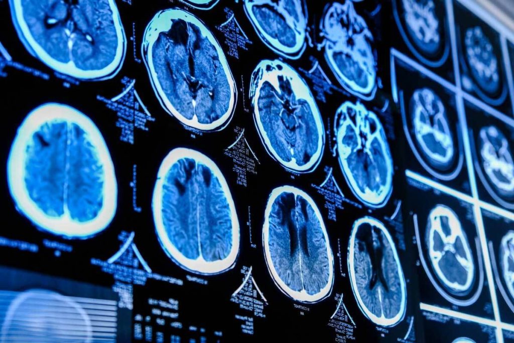
CT scans use X-rays to make detailed images of the brain. They show the size, location, and type of tumors. CT scans are very helpful in emergency cases when symptoms come on suddenly. They spot changes in bones and soft tissues related to tumors.
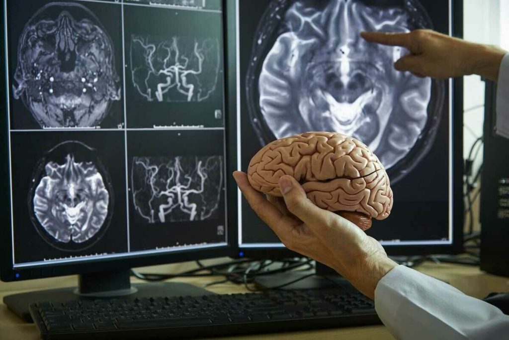
MRI uses magnetic fields and radio waves to create detailed brain images. MRI is great for seeing soft tissue tumors. It shows how big a tumor is and where it is in the brain.
PET scans use tiny amounts of radioactive tracers to see how tumors work. PET scans help figure out how aggressive a tumor is and if treatments are working. They give insights into how tumors grow, helping doctors plan better treatments.
Each imaging method has its own benefits and drawbacks. CT scans are fast and good for emergencies. MRI gives detailed images of soft tissues. PET scans show how tumors work. Using all these methods together gives the best view of brain tumors. This helps doctors make the best treatment plans.
Diagnosing a brain tumor starts with noticing early signs. Understanding this journey and the role of radiology is key.
Brain tumors can show up in many ways. Symptoms like headaches, seizures, and changes in thinking or mood are common. It’s important to see a doctor right away if you notice these signs.
Some symptoms that might make you visit the doctor include:
When symptoms suggest a brain tumor, a thorough check-up begins. This includes a detailed medical history and physical exam. Doctors look at how well you think, move, and sense things.
The first steps may include:
Doctors pick the right imaging tests based on the first check-up. The choice depends on the tumor’s likely location and type, and the patient’s health.
Here’s a table showing what influences the choice of imaging tests:
| Imaging Modality | Best for | Key Features |
| CT Scan | Emergency situations, detecting calcifications | Quick, sensitive to acute hemorrhage |
| MRI | Soft tissue tumors, detailed brain anatomy | High resolution, versatile sequences |
| PET Scan | Metabolic activity, tumor grading | Functional imaging, useful for tumor staging |
Radiology plays a key role in diagnosing brain tumors, providing vital information for treatment.
Understanding brain tumor imaging results is key in patient care. When we get these results, knowing what they mean is vital. It helps us decide the next steps in diagnosis and treatment.
Radiologists use X-rays, CT scans, MRI, and PET scans to check brain tumors. Each type gives different info. For example, X-rays are great for spotting calcifications and bone changes. On the other hand, MRI shows detailed images of soft tissue tumors.
We look at tumor size, location, and how it reacts to contrast to make a diagnosis.
We know the strengths and limits of each imaging type. For instance, X-rays might not catch soft tissue tumors well. But they’re good for bone structure changes.
Diagnostic reports are full of important info about your condition. We break down these reports to help you grasp your diagnosis. They usually talk about tumor size, location, and if it’s reacting to contrast.
“The radiologist’s interpretation of imaging studies is a critical step in the diagnostic process, providing essential information for treatment planning.”
” Expert in Neuroradiology
It can be tough to understand these reports, but it’s key for making informed decisions. We work with patients to explain their reports clearly.
Tumor size, location, and characteristics are very important. They help us figure out the diagnosis and treatment plan. The size can show how aggressive the tumor is. The location can affect symptoms and if surgery is possible.
| Tumor Characteristic | Significance |
| Size | Indicates how aggressive the tumor might be and guides treatment. |
| Location | Affects symptoms, if surgery is possible, and treatment planning. |
| Enhancement Patterns | Helps us understand how the tumor behaves and if it’s likely to be cancerous. |
By studying these characteristics, we can fully understand the tumor. Then, we can create a treatment plan that fits the patient’s needs.
Diagnosing brain tumors needs more than just imaging. X-rays, CT scans, and MRIs give us a first look. But, we need more steps to confirm the tumor type and plan treatment.
A biopsy is key for getting a tumor sample. It helps tell if the tumor is cancerous and what type it is. This info is vital for treatment planning and understanding the tumor’s genetics.
There are several biopsy methods. Stereotactic biopsy uses imaging to find the tumor. Open biopsy is done during surgery. The choice depends on the tumor’s location and size.
Lab tests on biopsy samples are very important. They look for biomarkers that show cancer or tell us about the tumor’s behavior. Biomarkers help predict how the tumor will react to treatments.
Advanced lab techniques like molecular profiling and genetic testing give deep insights. This info helps create a treatment plan that fits the patient’s needs.
Diagnosing brain tumors involves a team of experts. This team includes neurologists, neurosurgeons, radiologists, pathologists, and oncologists. They work together to understand the diagnosis and plan treatment.
This team approach ensures a thorough diagnosis and effective treatment. It also means ongoing support and care during treatment.
New technologies are changing how we find and treat brain tumors. These advancements make diagnosis more accurate and care better.
New tools like high-field MRI and advanced CT scans are making a big difference. They give clearer images of tumors, helping doctors plan better treatments.
High-field MRI can spot smaller tumors better. This is key for catching tumors early and treating them fast.
Artificial intelligence (AI) is helping doctors understand neuroimaging results better. AI can look at lots of data fast, spotting things humans might miss.
AI in brain tumor diagnosis makes things faster and more accurate. This means doctors can start treatments sooner, which can help patients more.
The future of brain tumor diagnosis looks bright with new tech. We’ll see even better AI and more ways to look at tumors together.
Following global standards in neurological care is very important. It ensures patients get the best care everywhere.
By following these standards, doctors can keep their care up to date with the latest research.
| Diagnostic Tool | Advantages | Limitations |
| X-ray | Quick, widely available | Limited soft tissue detail |
| CT Scan | Fast, good for emergencies | Radiation exposure |
| MRI | High soft tissue detail, no radiation | Expensive, not suitable for all patients |
When we compare tools like CT scans and X-rays for brain tumors, we see their pros and cons. X-rays are fast but don’t show soft tissues well. CT scans are better for details but use radiation.
The right tool depends on the situation and what’s needed for the patient. As we keep improving, the debate between CT and X-ray will stay important for doctors.
Diagnosing brain tumors needs a detailed approach. This includes using different imaging methods and tests. We’ve looked at X-rays and their limits, and the benefits of Gadolinium-enhanced magnetic resonance imaging (MRI). Knowing these details helps patients and doctors make better choices for treatment.
Good radiology is key for finding out what kind of tumor it is and how big it is. This info helps plan treatment. Tests like MRI and CT scans give important details about the tumor. A team effort in diagnosis, including biopsies and lab tests, is vital for accurate results.
Using the newest in neuroimaging and tests, doctors can create treatment plans that work best for each patient. It’s important to see a doctor right away if symptoms don’t go away or get worse. This ensures the best care and treatment.
X-rays have been used to spot changes in bone or calcifications linked to brain tumors. But, they’re not the best for soft tissue tumors. This makes them less useful for a full brain tumor check.
X-rays struggle to find soft tissue tumors. They’re not as good as newer imaging methods. This limits their use in brain tumor diagnosis.
Modern tools like CT scans, MRI, and PET scans are key for brain tumor diagnosis. They offer better views of soft tissue tumors than X-rays.
Doctors pick tests based on symptoms, medical history, and initial checks. The choice depends on the tumor’s type, location, and the patient’s health.
Radiologists check tumor size, location, and type. They also look at changes in brain tissue. They use CT, MRI, and PET scans for a full diagnosis.
Biopsies are key for confirming brain tumor diagnosis and treatment planning. They involve examining a tumor sample to determine its type and characteristics.
AI is helping in brain tumor diagnosis by improving image analysis. AI algorithms help spot tumors and patterns in scans.
Signs include headaches, seizures, nausea, vomiting, and changes in thinking or movement. If you notice these, see a doctor right away.
International standards ensure quality care worldwide. Following these standards is vital for accurate diagnosis and best treatment outcomes.
Subscribe to our e-newsletter to stay informed about the latest innovations in the world of health and exclusive offers!
WhatsApp us