Last Updated on November 27, 2025 by Bilal Hasdemir

CT vs Ultrasound: Which Imaging Method Is Right for You?
Understanding the differences between CT and ultrasound is key for accurate and safe diagnosis. At Liv Hospital, patient safety and precision come first. We make sure you get the right test for the best possible results.
Both CT scans and ultrasounds are powerful diagnostic tools, but they work differently. CT scans use X-rays to create highly detailed images, ideal for detecting small injuries, internal bleeding, and certain cancers.
Ultrasounds, in contrast, use sound waves to show what’s happening inside the body in real time ” without any radiation exposure.
Knowing when to choose CT ultrasound helps ensure accurate diagnosis and effective treatment planning.
Key Takeaways
- CT scans provide detailed, cross-sectional images using X-rays.
- Ultrasounds use high-frequency sound waves for real-time visualization.
- The choice between CT scans and ultrasounds depends on the specific diagnostic needs.
- CT scans are ideal for identifying small abnormalities and certain cancers.
- Ultrasounds offer a safer option without radiation exposure.
The World of Medical Imaging

Medical imaging is key in today’s healthcare. It changes how we diagnose and treat diseases. We use different imaging methods to see inside the body. This helps us find problems, track disease, and plan treatments.
Diagnostic Imaging’s Role in Modern Healthcare
Diagnostic imaging is vital in healthcare. It lets us look inside the body without surgery. CT scans and ultrasounds are top choices, each with its own strengths.
- CT scans give detailed pictures of the brain, spine, and organs.
- Ultrasounds use sound waves to show the inside of the body. They’re great for looking at organs and checking on babies during pregnancy.
Overview of Common Imaging Modalities
There are more imaging methods like X-rays, MRI, and PET scans. Each has its own use and benefits.
- X-rays mainly show bones and find breaks or objects inside.
- MRI gives clear pictures of soft tissues. It’s key for brain, spinal cord, and joint issues.
- PET scans check how active body cells are. They help find and track cancer, brain problems, and heart disease.
Knowing about these imaging methods is key for doctors. It helps them make the best choices for patients.
What is a CT Scan?

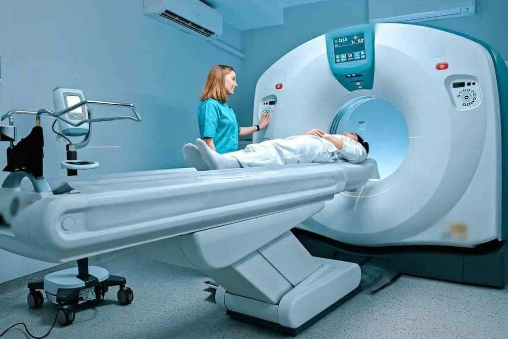
A CT scan is a high-tech medical tool that uses X-rays to show detailed images of the body’s inside. It combines many X-ray images from different angles to give a full view of organs, bones, and blood vessels. This is very helpful in emergencies to check injuries and find things like internal bleeding or tumors.
How Computed Tomography Technology Works
CT scans use a rotating X-ray machine to take images from various angles. A computer then puts these images together to show detailed cross-sections of the body’s inside. This whole process is fast and doesn’t hurt, taking just a few minutes.
Cross-Sectional Imaging Capabilities
CT scans show the body’s inside structures in great detail. This is very useful for finding problems with organs, bones, and blood vessels. For example, they can spot tumors, fractures, and vascular diseases very accurately.
Common Applications of CT Scanning
CT scans are used in many ways in medicine. They help check injuries, find diseases like cancer, and guide treatment plans. They are used for CT angiography to see blood vessels, body CT to look at organs and tissues, and CT enterography to check the small intestine.
| Application | Description | Benefits |
| CT Angiography | Visualizes blood vessels | Helps diagnose vascular diseases |
| Body CT | Examines organs and tissues | Detects diseases and injuries |
| CT Enterography | Inspects the small intestine | Diagnoses intestinal conditions |
Knowing how CT scans work and their uses is key to understanding their role in medicine. For a comparison with other imaging methods, like ultrasound, see our detailed guide on the differences between ultrasound and CT.
What is an Ultrasound?
Ultrasound technology has changed how we diagnose diseases. It uses sound waves to see inside the body without surgery. This makes it great for checking and tracking many health issues.
Understanding Sonography and Sound Wave Technology
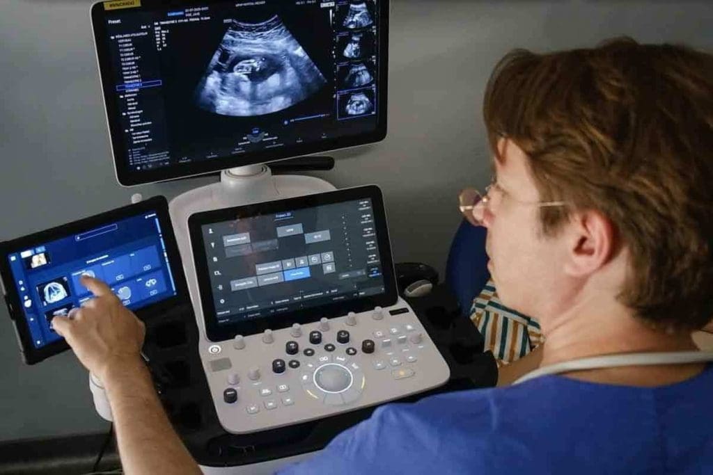
Ultrasound, or sonography, uses sound waves to see inside the body. A device called a transducer sends sound waves into the body. These waves bounce back and are turned into images on a screen.
The sound waves used in ultrasound are between 2 and 15 megahertz. This range helps us see different parts of the body well. We can see things like the thyroid gland and the liver and kidneys.
Real-Time Visualization Benefits
Ultrasound is great because it shows what’s inside the body in real-time. Doctors can see how organs move and work. This is very helpful for checking blood flow, watching how a baby grows during pregnancy, and helping with procedures.
Real-time ultrasound lets us:
- Check how organs like the heart and liver work
- Look at blood flow and find problems with blood vessels
- Help guide needles during biopsies or other procedures
- Watch how a baby grows and find issues early
Types of Ultrasound Examinations
There are many types of ultrasound tests, each for different reasons. Some common ones are:
- Abdominal Ultrasound: Looks at organs like the liver, gallbladder, and kidneys.
- Obstetric Ultrasound: Important for watching how a baby grows during pregnancy.
- Doppler Ultrasound: Uses the Doppler effect to check blood flow and find blood vessel problems.
- Musculoskeletal Ultrasound: Helps check muscle, tendon, and ligament injuries.
A leading medical journal says, “Ultrasound is a key tool in medicine today. It’s safe and works well for diagnosing and tracking many health issues.”
In summary, ultrasound is a big step forward in medical imaging. It’s safe, non-invasive, and very useful. Its ability to show what’s happening inside the body in real-time makes it a vital tool in healthcare today.
CT Ultrasound: Fundamental Technology Differences
It’s important to know how CT scans and ultrasounds work. They use different methods to create images of the body. Let’s look at what makes them unique.
X-Ray Based vs. Sound Wave Based Imaging
CT scans and ultrasounds are different in how they make images. CT scans use X-rays to see inside the body. Ultrasounds use sound waves for real-time images.
CT scans are great for seeing bones and solid organs. They use X-rays for detailed images. Ultrasounds are better for soft tissues like the liver and kidneys. They don’t use radiation.
Image Generation and Processing Comparison
How images are made is different for CT scans and ultrasounds. CT scans move an X-ray tube around the patient. They then use algorithms to make images. Ultrasounds send sound waves into the body and catch the echoes to make images in real-time.
Here’s a comparison of how they make images:
| Characteristics | CT Scans | Ultrasounds |
| Imaging Technology | X-ray based | Sound wave based |
| Image Detail | High-resolution, cross-sectional images | Real-time images, less detailed |
| Radiation Exposure | Yes | No |
Digital Reconstruction Methods
Digital reconstruction is key for both CT scans and ultrasounds. CT scans use algorithms to make images from X-ray data. Ultrasounds process sound wave echoes to create images in real-time.
Difference #1: Image Quality and Precision
Medical imaging quality is key in diagnosis. The clarity of images from CT scans and ultrasounds affects their usefulness. This directly impacts how well they can help doctors diagnose.
CT Scan Detail and Resolution Capabilities
CT scans are known for their clear images. They show detailed views of the body’s inside. This is great for spotting small issues like tumors or fractures.
Key advantages of CT scan image quality include:
- High-resolution images with fine detail
- Ability to detect small abnormalities
- Precise measurements for diagnostic accuracy
Ultrasound Image Characteristics
Ultrasounds give real-time images. They’re great for watching moving parts like the heart or a fetus. They don’t use harmful radiation, making them safe.
Notable characteristics of ultrasound imaging include:
- Real-time visualization of moving structures
- Non-invasive and radiation-free
- Portability and accessibility for point-of-care applications
Comparing Diagnostic Accuracy for Different Conditions
CT scans and ultrasounds work differently for different health issues. CT scans are better for finding internal injuries or conditions like appendicitis. Ultrasounds are better for checking on a baby during pregnancy or looking at the heart.
A comparison of diagnostic accuracy for different conditions is as follows:
| Condition | CT Scan Accuracy | Ultrasound Accuracy |
| Internal Injuries | High | Moderate |
| Fetal Development | Limited | High |
The right choice between CT scans and ultrasounds depends on the situation. Knowing what each can do helps doctors give the best care.
Difference #2: Radiation Exposure and Safety
When it comes to diagnostic imaging, safety is key. The main difference between CT scans and ultrasounds is radiation exposure. This choice can greatly affect patient safety.
CT Scan Radiation Levels and Associated Risks
CT scans use X-rays to create detailed images. This involves ionizing radiation, which can raise cancer and genetic mutation risks. The dose from a CT scan is measured in millisieverts (mSv). It depends on the scan type, body part, and technology used.
While the dose is controlled, it’s a concern, mainly for those needing many scans.
Ultrasound’s Radiation-Free Advantage
Ultrasounds, on the other hand, don’t use ionizing radiation. They use sound waves to create images. This makes ultrasounds safer for those sensitive to radiation or needing repeated scans.
It’s great for pregnant women and children, where radiation is a big concern.
Safety Considerations for Special Populations
For pregnant women, children, and those needing many scans, safety is even more important. Ultrasounds are safer because they don’t use radiation. But, CT scans might be needed for accurate diagnoses.
The choice between CT scans and ultrasounds depends on the situation. Healthcare providers must consider each patient’s needs and the safety of each option. This way, they can make the best choice for diagnosis and safety.
Difference #3: Cost Factors of CT vs US
When looking at medical imaging, the cost between CT scans and ultrasounds matters a lot. The price can change based on the equipment, where it’s done, and insurance. These factors all play a part in the final cost.
Equipment and Facility Expenses
CT scans need fancy and pricey equipment. This includes the CT scanner and the setup to run it. Also, the cost of running a facility adds up, making CT scans more expensive.
Ultrasound tech is cheaper and easier to move around. This makes it more affordable for use in different places. It’s great for quick checks or in areas far from big hospitals.
Patient Billing and Insurance Coverage
The price for patients depends on their insurance. CT scans often have higher deductibles and copays because they cost more.
Insurance plans vary in what they cover. Knowing what your plan covers helps you figure out how much you’ll pay. It’s important to check your insurance details.
Long-Term Economic Considerations
Looking at the long-term, CT scans might seem pricey. But, they can lead to better treatments. This could save money in the long run.
Ultrasounds are cheaper and good for ongoing checks. Using them for long-term monitoring can save a lot of money. This is true for conditions that need regular scans.
Difference #4: Accessibility and Portability
Medical imaging tech’s accessibility and portability are key in healthcare. When we look at CT scans and ultrasounds, we see big differences. These differences affect how they’re used in different healthcare places.
Fixed vs. Mobile Imaging Solutions
CT scans need big, fixed machines. This limits where they can be used in hospitals. On the other hand, ultrasounds are very portable. They can be used at the bedside or in other places. This is a big plus in emergencies where quick imaging is needed.
Point-of-Care Ultrasound Benefits
Ultrasound’s portability makes it great for point-of-care ultrasound. Doctors can do scans right at the patient’s side or in other places. This helps care by cutting down on moving patients and making quick decisions.
Emergency and Remote Setting Applications
In emergencies, ultrasound’s mobility is very useful. It helps doctors quickly check and diagnose patients. Also, in places with few resources, portable ultrasound is a lifesaver. It brings imaging to areas that might not have it.
The differences in how easy to move and use CT scans and ultrasounds matter a lot for patient care. Knowing these differences helps doctors choose the best imaging for each situation. This improves care quality and speed.
Difference #5: Clinical Applications and Limitations
CT scans and ultrasounds are key tools in diagnosing diseases. They serve different purposes and have their own limits. Knowing these differences helps doctors make better choices.
Superiority of CT Scans in Certain Conditions
CT scans are great for checking complex injuries from accidents. They show detailed images of internal injuries, like bleeding and organ damage. They can spot small problems, like tumors, that ultrasounds might miss.
For example, CT scans are better at finding appendicitis by looking at the appendix and nearby tissues. They’re also used to see how far cancer has spread.
Preference for Ultrasound in Specific Scenarios
Ultrasounds are best for watching how a fetus grows during pregnancy. They offer live images of the fetus and can find any issues. They’re also good for checking soft tissue injuries and helping with injections.
Ultrasounds are also used to check blood flow and find blood clots. They’re easy to move around and are cheaper, making them great for emergencies and remote areas.
In summary, CT scans and ultrasounds each have their own uses and limits. Understanding these helps doctors choose the right tool for each situation. This leads to better care for patients.
Difference #6: Speed and Patient Experience
CT scans and ultrasounds differ in technology and how they affect patients. Knowing their differences in speed, preparation, and comfort is key.
Procedure Duration Comparison
CT scans are usually quicker, taking just a few minutes. This is great in emergencies where time is critical. Ultrasounds are also fast but might take longer because the technician needs to move the probe around.
CT scans are faster because they’re simple. The patient just moves through the scanner. Ultrasounds need more interaction, as the technician must place the probe correctly.
Preparation Requirements
CT scans require little prep, like removing metal items and possibly drinking a contrast agent. Ultrasound prep can vary, like fasting for an abdominal exam.
Knowing what to expect can make the experience better. It reduces anxiety and ensures the procedure goes well.
Comfort and Convenience Factors
Comfort during the procedure is important. CT scans might cause claustrophobia because the patient lies on a table that slides into the scanner. Ultrasounds are often more comfortable because they involve a gel and a probe, allowing for more interaction.
Ultrasounds are also more convenient because they can be done in many places. This is useful in some clinical situations.
Difference #7: Recent Technological Advances
Medical imaging is getting better, thanks to new tech. This means better care and more accurate diagnoses. CT scans and ultrasounds have seen big changes, changing how we see inside the body.
Low-Dose CT Innovations
Low-dose CT scans are a big deal. They use less radiation but keep the image quality high. A study in the Journal of the American College of Radiology found they cut radiation by up to 50% without losing accuracy.
“Low-dose CT scans are a huge step forward,” says Expert. “They’re great for patients who need many scans.”
High-Resolution Ultrasound Developments
Ultrasound tech has also improved a lot. Now, ultrasounds can show tiny details clearly. This helps doctors make better diagnoses.
Artificial Intelligence Integration in Both Modalities
AI is being used in CT and ultrasound scans. AI helps doctors spot problems faster and more accurately. This tech is making diagnosis better and faster.
- AI-assisted image analysis
- Enhanced diagnostic accuracy
- Improved workflow efficiency
These advances show a bright future for medical imaging. With better CT scans, ultrasounds, and AI, we’re moving towards safer and more precise care.
Conclusion: Choosing Between CT Scan and Ultrasound
Choosing the right imaging test is key for accurate diagnosis and treatment. We’ve looked at the main differences between CT scans and ultrasounds. These include image quality, radiation, cost, and how easy they are to get.
The choice between a CT scan and an ultrasound depends on many things. These include the patient’s needs and the specific question being asked. Healthcare providers use this information to make the best imaging choice for patients.
It’s important to know what each test can do well. CT scans give detailed images and are great for complex cases. Ultrasounds are safe, non-invasive, and cheaper for some needs.
When deciding between CT and ultrasound, we must consider their pros and cons. This ensures patients get the best care for their specific needs. Making this choice carefully is essential for quality patient care.
FAQ
What is the main difference between a CT scan and an ultrasound?
A CT scan uses X-rays to create detailed images. An ultrasound uses sound waves for real-time images.
Is a CT scan better than an ultrasound for diagnosing internal injuries?
CT scans are better for complex injuries because of their detailed images. Ultrasounds are good for soft tissue injuries.
Does a CT scan expose patients to more radiation than an ultrasound?
Yes, CT scans use X-rays, which are ionizing radiation. Ultrasounds don’t use radiation, making them safer for pregnant women and kids.
Are CT scans more expensive than ultrasounds?
CT scans are usually more expensive because of the advanced technology and expertise needed. But costs can vary based on location and insurance.
Can ultrasounds be used in emergency settings?
Yes, ultrasounds are portable and can be used in emergencies or remote areas. They’re great for quick, on-the-spot diagnoses.
How do CT scans and ultrasounds compare in terms of diagnostic accuracy?
CT scans are good at finding small issues. Ultrasounds show real-time images, which is helpful for moving parts. The best choice depends on the condition being checked.
What are the benefits of low-dose CT scans?
Low-dose CT scans use less radiation. This makes them safer for people who need multiple scans or are sensitive to radiation.
How has artificial intelligence impacted CT and ultrasound technology?
Artificial intelligence has improved how images are analyzed and diagnosed in both CT scans and ultrasounds. This has made diagnostic imaging better overall.
Can ultrasounds be used for pregnancy monitoring?
Yes, ultrasounds are often used to check on fetal development during pregnancy. They’re safe and provide real-time images.
What is the difference between ultrasonography and CT scan?
Ultrasonography uses sound waves to create images. CT scans use X-rays to make detailed images.
Is ultrasound better than CT scan for certain medical conditions?
The choice between ultrasound and CT scan depends on the condition. Ultrasounds are better for soft tissue and blood flow. CT scans are better for small abnormalities and complex injuries.
What is the role of diagnostic imaging in modern healthcare?
Diagnostic imaging is key in healthcare. It helps doctors diagnose and treat conditions effectively. Tools like CT scans and ultrasounds are essential in medical practice.
References
- van Randen, A., Laméris, W., van Es, H. W., Kiewiet, J. J., van Heesewijk, H. P., ten Hove, W., … & Boermeester, M. A. (2011). A comparison of the accuracy of ultrasound and computed tomography in common diagnoses causing acute abdominal pain. European Radiology, 21(7), 1535-1545. https://www.ncbi.nlm.nih.gov/pmc/articles/PMC3101356/
- Wertz, J. R., Cofield, S., Trautman, R., & de Virgilio, C. (2018). Comparing the Diagnostic Accuracy of Ultrasound and CT for Acute Cholecystitis. Journal of Vascular and Interventional Radiology, 29(8), 1163-1167.e1. https://pubmed.ncbi.nlm.nih.gov/29702020/


