Last Updated on November 27, 2025 by Bilal Hasdemir
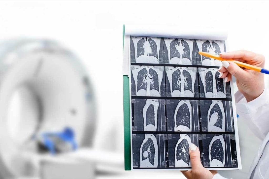
At Liv Hospital, we use top-notch medical imaging to help patients. A CT of chest with contrast is a key tool. It helps us see inside the chest clearly, spotting many issues.
A chest CT scan with contrast uses X-rays to show detailed images of the chest’s contents. The contrast dye makes it easier to spot problems like pulmonary embolism and lung cancer.
This advanced tech helps us give accurate diagnoses. Then, we create treatment plans that meet each patient’s needs.
Key Takeaways
- A chest CT scan with contrast is a diagnostic tool that enhances visualization of thoracic structures.
- It is used to diagnose various conditions, including pulmonary embolism and lung cancer.
- The use of intravenous contrast dye highlights areas of concern, aiding in accurate diagnosis.
- Liv Hospital employs advanced medical imaging technologies for effective treatment planning.
- A chest CT scan with contrast provides detailed cross-sectional images of organs and tissues.
The Fundamentals of CT of the Chest With Contrast
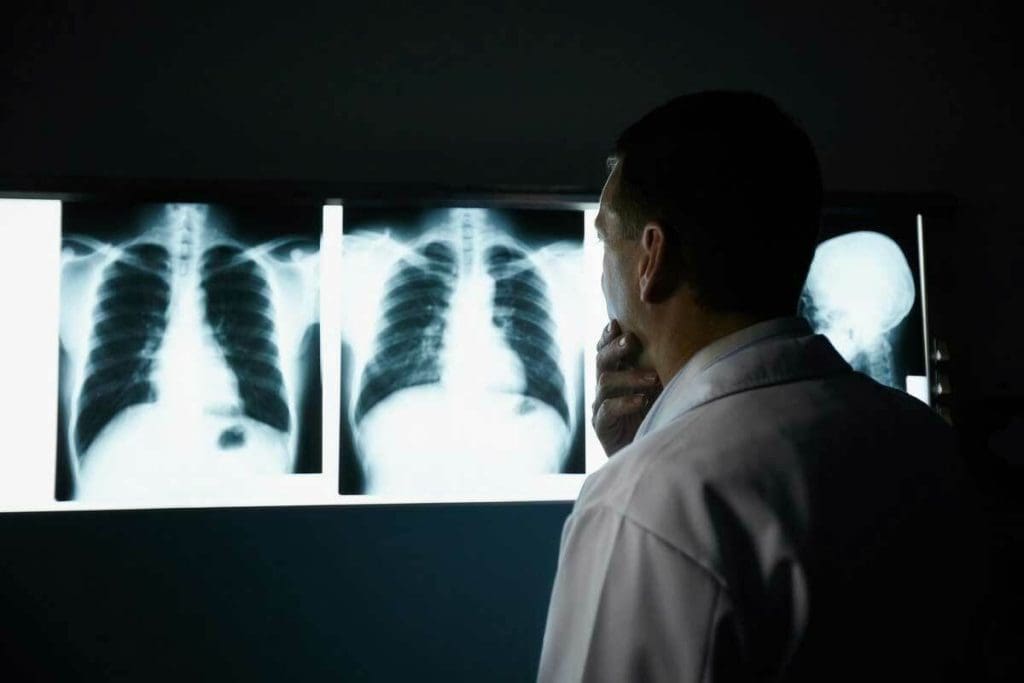
Understanding a CT chest with contrast is key to diagnosing chest problems. We’ll explore how it works and its benefits in medical imaging.
Definition and Basic Principles
A CT scan of the chest with contrast is a detailed imaging test. It uses X-rays to show the chest’s organs, blood vessels, and tissues. The “contrast” part means a special dye makes images clearer.
During a CT chest scan with contrast, a dye is given through a vein. This dye, iodine, absorbs X-rays and shows up bright on images. It makes blood vessels and some tissues stand out.
CT scans work by moving an X-ray machine around the body. It takes pictures from many angles. Then, these images are turned into detailed pictures of the body’s inside.
Key components of a CT chest with contrast include:
- X-ray technology to capture images
- Contrast agent to enhance visualization
- Advanced computer software to reconstruct images
How Intravenous Contrast Enhances Visualization
The intravenous contrast agent is crucial for seeing thoracic structures clearly. It highlights areas of concern, making it easier to spot tissues, organs, or blood vessels. This is vital for accurate diagnosis.
For example, in suspected pulmonary embolism, the contrast agent lights up lung blood vessels. This makes it easier to see clots.
The contrast agent also helps tell different tissues and abnormalities apart. This is why CT chest scans with contrast are so important in medicine.
Benefits of using intravenous contrast in CT chest scans include:
- Improved detection of vascular abnormalities
- Enhanced visualization of tumors and lesions
- Better differentiation between various tissue types
CT Chest W Wo Contrast: Understanding the Differences
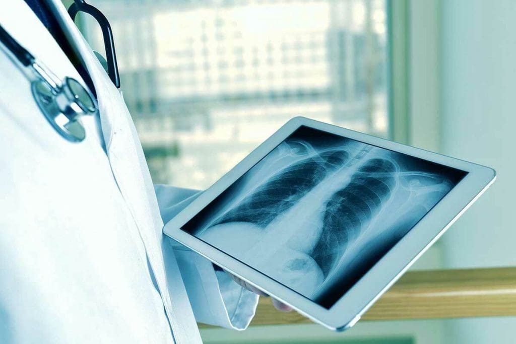
When we diagnose chest conditions, knowing the difference between CT chest scans with and without contrast is key. CT scans help us see the chest area, like the lungs, heart, and blood vessels. Whether we use contrast depends on the condition we’re checking for.
Visualization Capabilities Comparison
CT scans without contrast are used for finding things like kidney stones, lung nodules, or lung cancer screening. But, CT scans with contrast show more detail of blood vessels and some organs. This makes it easier to spot problems like pulmonary embolism or to see the blood flow around tumors.
The contrast agent, usually iodine-based, makes blood vessels and certain tissues stand out.
Clinical Scenarios for Each Approach
Choosing between a CT chest scan with or without contrast depends on the situation. For example, if someone might have a pulmonary embolism, a CT scan with contrast is better. It helps see the blood vessels in the lungs better.
- CT scans without contrast are often used for lung cancer screening and detecting lung nodules.
- CT scans with contrast are preferred for assessing vascular structures, detecting pulmonary embolism, and evaluating the extent of certain tumors.
Knowing these differences helps us choose the right test for each patient. This ensures we give the best care possible.
The Technology and Methodology Behind Chest CT With Contrast
The tech behind Chest CT scans with contrast has changed how we diagnose diseases. We use top-notch CT scanning gear to get clear images. These images are key to makingg accurate diagnoses.
Modern CT Scanning Equipment
Today’s CT scanners have advanced technology for quick and precise scans. They mix X-rays and computer tech to show detailed chest images. High-resolution imaging is key in thoracic imaging. It helps spot small issues that might not show up on lower-quality images.
Types of Contrast Agents Used in Thoracic Imaging
Contrast agents are crucial for better CT images. In chest imaging, iodine-based contrast agents are often used. They make blood vessels and other chest structures stand out. The right contrast agent depends on the imaging needs and the patient’s health history.
There are many contrast agents, each with its own uses. Choosing the right one is key to the best image quality and patient safety.
- Iodine-based contrast agents are great for showing vascular structures.
- Gadolinium-based contrast agents, mainly used in MRI, have specific uses in CT.
- New contrast agents are being developed to improve CT scan diagnostics.
Understanding Chest CT scans with contrast shows the advanced tech in today’s imaging. It also highlights the need for the right imaging plan for each patient.
Patient Experience: Preparation and Procedure for CT Scan Chest With Contrast
Getting ready for a CT scan chest with contrast can make you feel more at ease. We’ll walk you through each step, from getting ready to recovering, to make your experience smooth.
Pre-Scan Preparation Guidelines
Before your CT scan, there are a few things to do. Take off any metal objects like jewelry or glasses. Also, avoid wearing clothes with metal parts. You might need to fast for a few hours or skip some medications. Always follow what your healthcare provider or radiology department tells you.
Also, tell us about any allergies or medical conditions, especially if you’re allergic to contrast dye or have kidney issues. This helps us keep you safe during the scan.
During the Procedure: What to Expect
During the scan, you’ll lie on a table that moves into a big, doughnut-shaped machine. The whole thing usually takes just a few minutes. You might need to hold your breath for a bit to get clear pictures.
Our radiology team will help you through it. They’ll give you contrast dye through an IV. This might make you feel warm or taste metallic for a bit.
Post-Scan Recovery and Results Timeline
After the scan, you can usually go back to your normal activities right away. But if you’ve been given sedatives, you might need someone to drive you home.
The radiologist will look at your scan and send the results to your doctor. You’ll likely hear back in a few days. Your doctor will then talk to you about what the scan showed and what to do next.
At our place, we aim to make your CT scan experience as comfortable and supportive as possible. If you have any questions or worries, just ask our team for help.
Pulmonary Applications: What Lung CT With Contrast Can Reveal
CT scans with contrast have changed how we diagnose lung diseases. They give us detailed views of lung health. This helps us spot problems early and treat them well.
Lung Parenchyma and Nodule Assessment
Lung CT with contrast is key for checking lung tissue and nodules. Contrast enhancement makes it easier to see nodules and what they look like. This is important for figuring out if they are harmful.
- Nodule size and location
- Presence of calcification or fat
- Enhancement patterns with contrast
- Relationship to surrounding structures
This info helps us tell if a nodule is likely to be cancerous. It guides how we should treat it.
Interstitial Lung Disease Patterns
CT scans with contrast are also great for looking at interstitial lung disease (ILD). Even though high-resolution CT (HRCT) without contrast is often used first, contrast CT adds more details. We look for signs like:
- Ground-glass opacities
- Reticular changes
- Honeycombing
- Traction bronchiectasis
These signs help us figure out what type of ILD someone has and how severe it is.
Pleural Effusions and Chest Wall Abnormalities
CT scans with contrast are also good for checking pleural effusions and chest wall issues. Contrast enhancement helps us understand what the pleural effusions are and if there are any problems. We can see things like:
- Pleural thickening or enhancement
- Presence of loculations or septations
- Involvement of adjacent lung parenchyma
- Chest wall invasion or metastasis
This detailed look is to planningning the right treatment.
Vascular Evaluation Using Thoracic CT Scan With Contrast
Contrast-enhanced thoracic CT scans show us the blood vessels in great detail. They help us find and treat complex vascular diseases. These scans are key to checking how well our blood vessels are working.
Pulmonary Embolism Detection
Pulmonary embolism (PE) is a serious condition that needs quick diagnosis and treatment. CT scans with contrast are the best way to find PE. They let us see the blood vessels in the lungs and spot any blockages.
- High sensitivity and specificity for diagnosing PE
- Ability to detect alternative diagnoses
- Guiding treatment decisions, including anticoagulation therapy
Aortic and Great Vessel Assessment
The aorta and great vessels are very important for our heart health. Thoracic CT scans with contrast help us check these areas for problems like aneurysms, dissections, and stenosis.
Key benefits include:
- Accurate measurement of aortic diameter
- Detection of intimal flaps in aortic dissections
- Assessment of branch vessel involvement
Coronary Artery Visualization
Coronary artery disease is a big problem worldwide. While coronary angiography is the usual go-to, coronary CT angiography is becoming a good non-invasive option. It helps us see how blocked the arteries are and how much plaque there is.
The advantages of coronary CT angiography include:
- Non-invasive assessment of coronary arteries
- Detection of coronary artery stenosis and plaque characteristics
- Guiding revascularization decisions
Oncological Applications of CT Scan Thorax With Contrast
In oncology, CT scans with contrast are key for finding and understanding cancers in the thorax. They make tumors stand out, helping doctors plan treatments. This is crucial for getting the right diagnosis and treatment.
Primary Lung Cancer Detection and Characterization
Contrast-enhanced CT scans are great for spotting lung cancer. They help tell tumors apart from other tissues. This lets doctors know the tumor’s size, where it is, and if it’s spreading.
“…the diagnostic accuracy for lung cancer detection was significantly higher with contrast-enhanced CT compared to non-contrast CT.”
- Tumor size and location
- Relationship with adjacent structures
- Presence of necrosis or cavitation
- Potential vascular involvement
Tumor Margin Delineation and Vascular Involvement
Knowing the tumor’s edges is key to surgery planning. Contrast-enhanced CT scans show how the tumor touches nearby tissues. They also check if it’s touching big blood vessels. This info is key for cancer staging and treatment planning.
Metastatic Disease and Lymph Node Evaluation
CT scans with contrast are also vital for spotting cancer spread and lymph node involvement. They help find big lymph nodes and see if they’re connected to the tumor. This helps doctors stage cancer right and choose the best treatment.
We count on contrast-enhanced CT scans for managing thoracic cancers. They help find and understand tumors, their edges, and if cancer has spread. These scans are crucial in oncology.
Infectious and Inflammatory Conditions Visualized by CT Thorax With Contrast
CT scans with contrast are key in spotting infections and inflammation in the chest. The contrast makes it easier to see what’s going on inside, helping doctors make accurate diagnoses.
We use CT scans with contrast to check for infections and inflammation in the chest. This tool is especially helpful in seeing how bad diseases like pneumonia and COVID-19 are.
Pneumonia and COVID-19 Imaging Features
CT scans with contrast are great for spotting pneumonia, including COVID-19. They show up areas of consolidation, ground-glass opacities, and sometimes fluid in the lungs. COVID-19 pneumonia often shows up as bilateral ground-glass opacities.
A study on the National Center for Biotechnology Information shows CT scans are key in diagnosing and tracking COVID-19.
Contrast helps see blood vessels and spot problems like blood clots. These can happen in severe COVID-19 cases.
Mediastinal Masses and Inflammation
Mediastinal masses and inflammation can also be checked with Ca T thorax with contrast. Mediastinitis, an inflammation of the mediastinum, can be spotted by looking for widened spaces, fluid, or abscesses on CT scans. Contrast helps see how far the inflammation goes and any complications.
CT scans with contrast also help figure out what mediastinal masses are. They can tell if a mass is cystic or solid and how it relates to nearby tissues. This info is important for deciding on treatment, like biopsies or surgery.
Safety Considerations and Potential Risks of CT With Contrast Chest
CT scans with contrast are useful for diagnosing, but they carry risks. It’s important to know that the benefits usually outweigh the risks for most patients.
Allergic Reactions and Contraindications
One major concern is allergic reactions to the contrast agent. These can be mild or severe. People with allergies, especially to iodine or previous agents, are at higher risk. We check each patient’s history before giving contrast.
Severe kidney disease and certain thyroid conditions are also contraindications. Patients need to share their full medical history, including allergies or conditions, with their doctor.
| Risk Factor | Description | Precautionary Measures |
| Allergic Reaction | Reaction to contrast agent, ranging from mild to severe | Assess allergy history, monitor during and after scan |
| Kidney Disease | Contrast can impair kidney function in patients with severe kidney disease | Assess kidney function before scan, consider alternative imaging |
| Thyroid Conditions | Certain thyroid conditions may be contraindications for contrast use | Evaluate thyroid function and history before administering contrast |
Radiation Exposure Concerns
Radiation exposure is another key concern. CT scans use more radiation than X-rays, which can raise cancer risk. We aim to keep radiation doses low while still getting good images.
We use new CT technology to lower radiation doses. We also plan each scan carefully to balance radiation exposure and benefits.
Knowing the risks helps patients decide on CT scans with contrast. We aim to provide a safe and effective diagnostic experience. We balance the benefits of contrast-enhanced imaging with the risks.
Conclusion: The Diagnostic Value of Contrast-Enhanced Chest CT in Clinical Practice
We’ve seen how important contrast-enhanced chest CT scans are in diagnosing many thoracic conditions. These scans give detailed images that help doctors make accurate and quick diagnoses. This leads to better care for patients.
These scans are key in healthcare today. They let doctors see complex body parts and find problems clearly. Using contrast agents makes these scans even better. They help doctors see lung details, nodules, and blood vessels more clearly.
The use of contrast-enhanced chest CT scans has changed pulmonology a lot. They help find things like blood clots, lung cancer, and lung diseases. As medical tech gets better, these scans will keep being crucial for patient care.
To wrap it up, contrast-enhanced chest CT scans are very valuable. They give doctors the info they need to make good treatment plans. So, these scans are a big part of modern medicine. They will keep being important for top-notch patient care.
FAQ
What is a CT chest scan with contrast, and how does it differ from a CT scan without contrast?
A CT chest scan with contrast uses a special dye to show more details. It’s different from a CT scan without contrast, which just uses the natural differences in body tissues. The dye makes it easier to see problems.
What are the benefits of using contrast in a CT chest scan?
Using contrast in a CT chest scan helps doctors see more clearly. It shows blood vessels, tumors, and other issues better. This is key to finding problems like lung cancer and blood clots.
How is the contrast agent administered during a CT chest scan?
The contrast dye is given through a vein in your arm. A small tube is used to inject it before the scan. This makes sure the dye spreads right for the best pictures.
What are the potential risks or side effects associated with CT chest scans with contrast?
There are some risks, like allergic reactions and kidney problems. But the scan’s benefits usually outweigh these risks. Doctors take steps to keep you safe.
How should I prepare for a CT chest scan with contrast?
Before the scan, you might need to fast and avoid certain medicines. Tell your doctor about any allergies or health issues. They’ll give you specific instructions.
What can I expect during the CT chest scan procedure?
During the scan, you’ll lie on a table that moves into the CT machine. The scan is fast, lasting just a few minutes. You might need to hold your breath for a bit to get clear pictures.
How long does it take to receive the results of a CT chest scan with contrast?
Results time varies by the facility and scan details. Usually, you’ll get them in a few hours to a few days. Your doctor will talk about them with you.
Can a CT chest scan with contrast detect all types of lung diseases?
CT scans with contrast are very good at finding many lung diseases. But, they’re not perfect. Some diseases might need more tests to confirm.
Are there alternative imaging tests to CT chest scans with contrast?
Yes, other tests like chest X-rays, MRI, and PET scans are available. The choice depends on your health and what the doctor needs to see.
How does a CT chest scan with contrast contribute to the diagnosis and treatment of vascular conditions?
CT scans with contrast are key for finding vascular problems like blood clots and aortic tears. They help doctors see how bad the problem is and plan treatment.
What is the role of CT scans with contrast in oncological applications?
CT scans with contrast are vital in cancer care. They help find tumors, see how big they are, and check for spread. This helps doctors plan treatment and stage cancer.
References
- Kaller, M. O. (2023). Contrast Agent Toxicity. StatPearls. https://www.ncbi.nlm.nih.gov/books/NBK537159/


