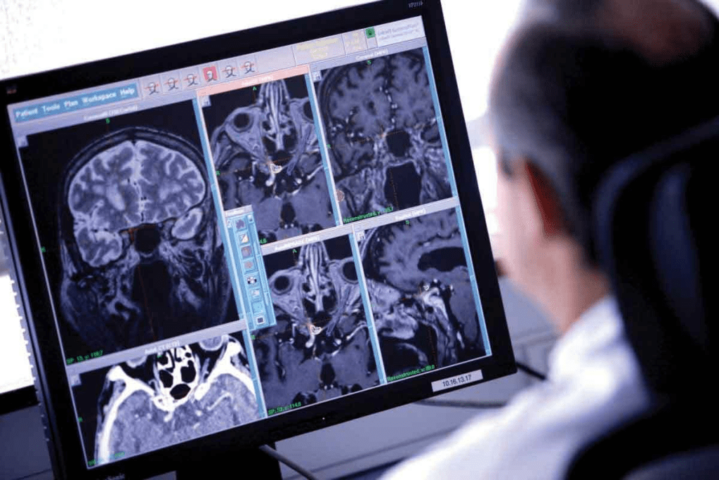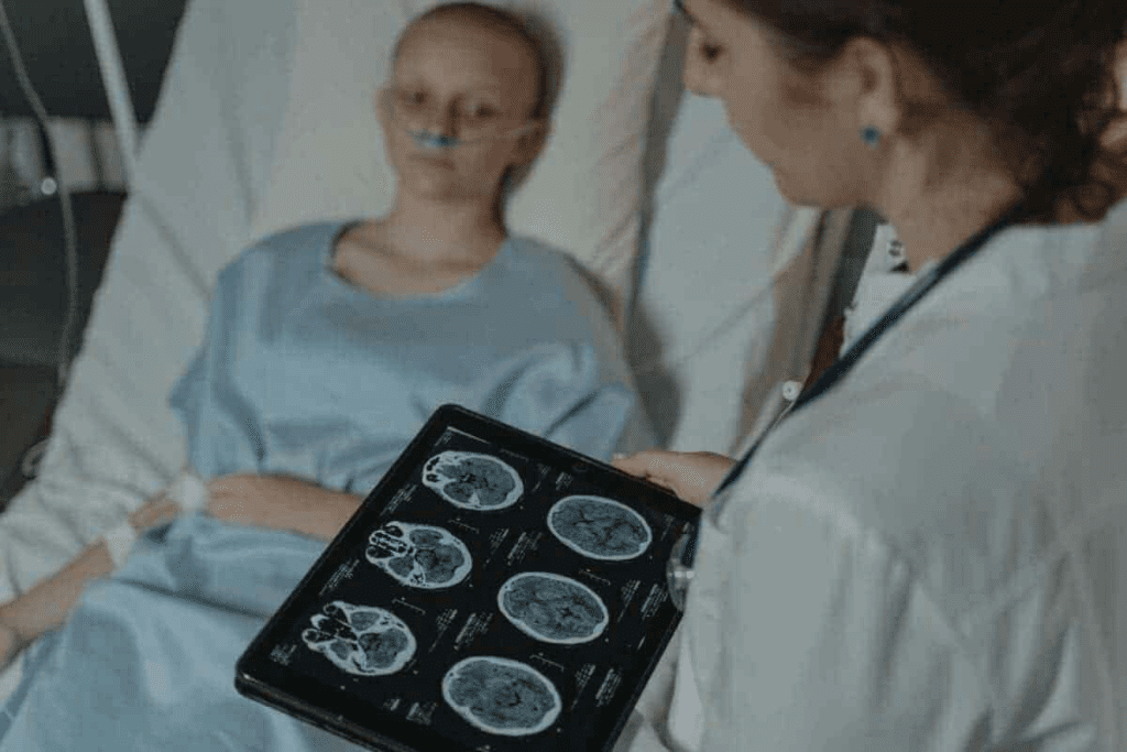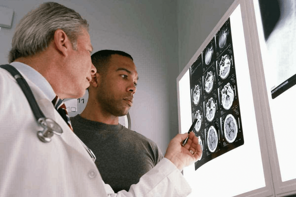
Getting a correct diagnosis is key to treating brain tumors. At Liv Hospital, we follow the latest research to improve care. We use CT scans and MRI scans to find brain tumors. Each has its own role and benefits.
Deciding between a CT scan and an MRI for brain tumor diagnosis can be tough. We’ll look at the main differences between these scans. This can greatly affect your diagnosis and treatment.
Key Takeaways
- CT scans are faster and provide a good view of the overall structure.
- MRI scans offer a clearer view of soft tissues and do not expose patients to radiation.
- MRI scans provide a three-dimensional view of the brain, allowing for precise analysis.
- The choice between CT and MRI depends on the specific needs of the patient.
- Accurate diagnosis is critical for effective treatment planning.
Brain Tumors and the Critical Role of Imaging

Imaging is key in finding and treating brain tumors. We use advanced methods to see the brain’s details and spot problems.
Types of Brain Tumors and Diagnostic Challenges
Brain tumors can be harmless or dangerous. They are hard to find because of the brain’s complex layout. Primary brain tumors start in the brain, and metastatic brain tumors come from other places. Knowing the type is important for treatment.
A neurological exam checks the brain’s parts. It works with imaging to learn about the tumor and its effects.
- Gliomas
- Meningiomas
- Medulloblastomas
These are common brain tumors, each needing its own treatment.
Why Accurate Imaging Is Essential for Treatment Planning
Good imaging is key to planning treatment. It shows the tumor’s size, where it is, and how it affects the brain. This info helps plan surgery, radiation, and chemotherapy.
“Imaging is not just about diagnosis; it’s about guiding treatment and improving patient outcomes.”
Expert in Neuro-Oncology
Using the right imaging, like CT scans and MRI, boosts accuracy. This leads to better treatment plans for each patient.
How CT Scans Detect Brain Tumors

CT scans are key in finding brain tumors. They use X-rays to make detailed images. These images help doctors see if there’s a tumor and what it’s like.
X-ray Technology and Image Formation
CT scans work by using X-rays to show the brain’s inside. X-rays go through the brain at different speeds, depending on the tissue. This helps create detailed images of the brain’s structures.
CT scan technology spots tumors by how they look compared to the brain around them. This is because tumors are different in density and contrast.
Brain CT Scan Procedure and Patient Experience
Getting a brain CT scan is quick and easy. Patients lie on a table that moves into the scanner. They must stay very quiet while it scans.
The whole thing usually takes just a few minutes. Getting ready and settled in might take a bit longer. We aim to make it as comfortable as possible for everyone.
We focus on making our patients’ experience better at our facility. We use the latest CT scan tech and put patients first. This helps us give accurate diagnoses and treatment plans for brain tumors.
How MRI Scans Visualize Brain Tumors
MRI technology has greatly improved how we detect and study brain tumors. It gives us detailed images that were hard to get before. At our institution, we’re proud to offer top-notch care. We use the newest medical tech to help our patients.
Magnetic Resonance Technology Explained
MRI scans use strong magnets and radio waves to show us what’s inside our bodies. This tech lets us see soft tissues, like brain tumors, very clearly. It works by using a strong magnetic field to line up hydrogen nuclei in our bodies.
Then, it uses radio waves to disturb this alignment. As the nuclei go back to their original state, they send out signals. These signals help create detailed images.
The main reason MRI works so well is its ability to tell different soft tissues apart. This is really important in brain imaging. The brain’s complex structures need high-quality images for accurate diagnoses.
Brain MRI Procedure and Patient Experience
Getting a brain MRI is pretty simple, but you might need to get ready first. You’ll likely have to take off any metal items and stay very quiet during the scan. The whole thing is safe and doesn’t use harmful radiation.
At our place, we really focus on making sure patients are comfortable and supported. Our team works hard to make sure you know what’s happening and feel okay during the whole thing.
A top doctor once said, “MRI is now a key tool for finding and treating brain tumors. It gives us amazing details and insights into these tough conditions.” This shows how important MRI tech is in today’s healthcare.
Difference #1: Image Resolution and Tumor Visualization
It’s key to know how CT scans and MRI differ in showing brain tumors. The clear images these tools give help doctors plan treatments better. This affects how well patients do.
CT Scan Capabilities for Detecting Brain Tumors
CT scans use X-rays to show the brain. They’re fast and good for first checks. But, they might miss some soft tissue tumors that RI can see.
- CT scans are great in emergencies when quick checks are needed.
- They spot calcifications and bone issues in some tumors.
- But, they don’t show soft tissues as well as an MRI does.
MRI’s Superior Soft Tissue Contrast for Tumor Delineation
MRI is top-notch for seeing soft tissues. This makes it key for finding and understanding brain tumors. Its clear images help doctors see tumor edges and what’s around them.
MRI’s advantages include:
- It shows soft tissues well, helping spot tumor types and edges.
- It gives detailed views of tumors and what’s nearby.
- It shows things CT scans can’t, like some tumor features.
Comparative Visual Examples of the Same Tumor
MRI shows the tumor and its area in more detail than CT scans. This detail is vital for doctors to plan treatments.
“MRI’s clear soft tissue images are essential in brain cancer care. They help doctors diagnose and plan treatments more accurately.”
Expert Opinion
In summary, CT scans and MRI both help in finding brain tumors. But MRI’s better images and soft tissue view make it more important for seeing and understanding tumors.
Difference #2: Radiation Exposure and Long-term Safety
When it comes to diagnosing brain tumors, the amount of radiation from CT scans and MRI is key. Both patients and doctors need to know about radiation exposure. This helps in making the best choices for care.
CT Scan Radiation Levels and Possible Risks
CT scans use a small amount of ionizing radiation to see the brain clearly. While it’s usually safe, too much radiation can raise cancer risk, more so in kids and teens. We aim to keep radiation doses low to protect our patients.
Key considerations for CT scan radiation include:
- The cumulative effect of radiation exposure over time
- The possible increased risk of secondary cancers
- The importance of weighing the benefits of CT scans against the possible risks
MRI’s Radiation-Free Advantage for Repeated Monitoring
MRI doesn’t use ionizing radiation, making it safer for long-term imaging. This is great for cancer patients needing regular checks. MRI’s safety lets us do more scans without worrying about radiation harm.
Safety Considerations for Cancer Patients
For cancer patients, safety is our main goal. When deciding between CT scans and MRI, we look at several things. These include the patient’s age, medical history, and what’s needed for diagnosis. MRI’s lack of radiation makes it a good choice for long-term care.
Understanding the radiation differences between CT scans and MRI helps us make better choices for brain tumor diagnosis and treatment. This way, we can give our patients the best care possible.
Difference #3: Speed and Accessibility of CT Scan vs MRI Brain Tumor Detection
In medical emergencies, how fast we can get images is key. We must think about how urgent the situation is, how much detail we need, and which imaging options are available.
CT Scan: Rapid Assessment in Emergency Situations
CT scans are quicker than MRI scans. This makes them a top choice in emergencies where fast diagnosis is essential. A CT scan can be done in just minutes, which is critical for acute conditions like head trauma or sudden brain problems.
MRI: Longer Scanning Times but Greater Detail
MRI scans take longer than CT scans but offer more detail. They show soft tissues, like brain tumors, better. MRI’s ability to show tumor type and size is key for treatment planning, even if it takes longer.
Availability and Cost Factors in the United States
The cost and availability of CT scans and MRI scans differ in the U.S. CT scans are more common and cheaper, often used in emergencies.
| Imaging Modality | Speed | Detail | Cost |
| CT Scan | Fast (minutes) | Good | Less expensive |
| MRI | Slower (30-60 minutes) | Excellent | More expensive |
Knowing the differences in speed, availability, and cost helps healthcare providers choose the best imaging for brain tumors. This choice is critical for diagnosis and treatment.
Difference #4: Tumor Characterization and Staging Accuracy
Understanding brain tumors well is key to good treatment plans. We use CT scans and MRI scans to get the details we need. These scans help us know what the tumor is and how big it is.
How CT Scans Identify Different Brain Tumor Types
CT scans use X-rays to quickly show the brain. They help spot big tumors and those with calcification or bleeding. But, they might not tell soft tissue tumors apart well.
Key advantages of CT scans include:
- They’re fast, which is great in emergencies
- They show calcifications well
- They’re cheaper and easier to find than an MRI
MRI’s Advanced Capabilities for Tumor Classification
MRI scans, though, are better at showing soft tissues. This makes them good for finding small or tricky-to-reach tumors. They also tell us how the tumor is related to nearby tissues.
MRI’s capabilities include:
- They’re great at showing soft tissues
- They can imagine in different ways for a better location
- They can even show how active the tumor is
Evidence-Based Comparison of Diagnostic Accuracy
Research shows MRI is better than CT scans for understanding tumors, but CT scans are good for quick checks. MRI is best for soft tissue tumors, while CT scans are good for big or calcified tumors.
Comparative diagnostic accuracy:
| Imaging Modality | Tumor Characterization | Staging Accuracy |
| CT Scan | Good for calcified tumors | Generally accurate for larger tumors |
| MRI Scan | Excellent for soft tissue tumors | Highly accurate for complex tumors |
We use both CT and MRI scans to give the best care. MRI is better for finding some tumors, which is key to the right treatment plan.
Difference #5: Advanced Imaging Techniques for Each Modality
Advanced imaging techniques have changed how we diagnose brain tumors. These methods are now key parts of CT and MRI scans. They give us more detailed info about tumors.
CT Perfusion and Contrast Enhancement
CT scans use CT perfusion and contrast enhancement to see brain tumors better. CT perfusion tracks contrast material in the brain’s blood vessels. It helps us understand how aggressive a tumor is and plan treatment.
Contrast enhancement on CT scans uses iodine-based agents. These agents highlight areas where the blood-brain barrier is broken. This is common in brain tumors. The way and degree of enhancement tell us about the tumor’s type and how serious it is.
Specialized MRI Sequences for Brain Tumors
MRI scans also use special sequences for brain tumors. Techniques like Diffusion-Weighted Imaging (DWI) and Perfusion-Weighted Imaging (PWI) give us insights into tumor cellularity and vascularity. DWI helps tell tumor types apart by their diffusion. PWI shows tumor blood flow and metabolic activity.
Magnetic Resonance Spectroscopy (MRS) analyzes the metabolic profile of tumors. It helps tell tumor from non-tumor tissue and different tumor types. These MRI sequences add to the anatomical info from standard MRI, giving a fuller picture of the tumor.
Using these advanced imaging techniques improves brain tumor diagnosis and understanding. As a leading expert says, “Advanced imaging has changed neuro-oncology. It makes diagnosis and treatment planning more precise.” (
This change shows how important it is to keep up with new imaging tech.
- CT perfusion and contrast enhancement improve tumor visualization.
- Specialized MRI sequences provide insights into tumor characteristics.
- Advanced imaging techniques enhance diagnostic accuracy and treatment planning.
Difference #6: Contraindications and Patient-Specific Limitations
It’s key to know when CT scans and MRI scans can’t be used. This is important for finding and treating brain tumors ricorrectlyAt our place, we focus on what’s best for each patient. We know some conditions make one scan better than the other.
When CT Scans Are Not Recommended or Less Effective
CT scans are not for everyone. For example, people who have had a lot of radiation or are pregnant should avoid them. Also, the dye used in some CT scans can be bad for those with kidney problems or iodine allergies.
- Pregnant women or those who suspect they might be pregnant
- Patients with a history of significant radiation exposure
- Individuals with kidney problems or at risk of contrast-induced nephropathy
- Patients allergic to iodine-based contrast agents
When MRIs Are Not Recommended or Less Effective
MRI scans also have their limits. People with metal implants, like pacemakers, can’t have MRI scans because of the strong magnetic fields. Also, if you’re scared of being in a small, enclosed space, an MRI might not be for you.
- Patients with certain metal implants (e.g., pacemakers, some artificial joints)
- Individuals with metallic fragments or foreign bodies
- Patients suffering from claustrophobia
- Those who are unable to remain for the scan
Patient-Specific Considerations for Brain Tumor Imaging
We make sure to choose the right imaging for each patient. We think about their age, health history, and the type of brain tumor they might have. This way, we give the best care possible.
We always consider what’s best for each patient. Knowing the limits of CT and MRI scans helps us give better diagnoses and treatment plans. This is how we care for our patients with brain tumors.
Difference #7: Clinical Decision-Making and Protocols
Choosing the right imaging modality for brain tumors is key. Healthcare professionals weigh many factors, like the patient’s condition and what’s needed for treatment. This decision is based on the clinical context and the patient’s specific needs.
When Doctors Choose CT Scans for Brain Tumor Assessment
CT scans are best in emergencies, like acute hemorrhage or trauma. They’re also chosen when an MRI isn’t possible due to metal implants or claustrophobia. CT scans are more accessible and cheaper, making them a good first choice.
When MRI Is the Preferred Diagnostic Tool for Brain Tumors
MRI is the top choice for brain tumors needing detailed soft tissue information. It’s better at showing tumor boundaries and edema. MRI is key for tracking treatment success and spotting complications. A study in the PMC highlights MRI’s critical role in diagnosis.
Combined and Sequential Imaging Approaches
Often, we combine imaging for better results. A CT scan might come first in emergencies, then an MRI for detailed views. This mix uses each modality’s strengths for better treatment planning.
| Imaging Modality | Preferred Use | Key Advantages |
| CT Scan | Emergencies, acute hemorrhage, trauma | Rapid assessment, widely available, and less expensive |
| MRI | Soft tissue characterization, tumor delineation, treatment monitoring | Superior soft tissue contrast, detailed visualization, no radiation |
Conclusion: Making the Right Choice for Brain Tumor Diagnosis
Choosing the right imaging technique is key to accurate brain tumor diagnosis. We’ve looked at the main differences between CT scans and MRI scans. Each has its own strengths and weaknesses.
CT scans are fast and often used in emergencies. They’re good for quick checks. MRI scans, on the other hand, show soft tissues better. They’re best for detailed tumor checks.
When picking between CT scans and MRI for brain tumor diagnosis, several factors matter. These include radiation exposure, image quality, and what’s best for the patient. Our team chooses the best imaging based on the patient’s needs and the tumor’s specifics. We aim to give top-notch care and support to all our patients.
The choice between MRI and CT scans for brain tumor diagnosis depends on the situation. Knowing what each scan can do helps us give patients the best diagnosis and care.
FAQ
What is the main difference between a CT scan and an MRI for brain tumor diagnosis?
CT scans use X-rays to create detailed images. MRI scans use magnetic fields and radio waves for high-resolution images of soft tissues.
Which is better for detecting brain tumors, a CT scan or an MRI?
MRI is better for detecting brain tumors. It shows soft tissues clearly, helping to define tumors and surrounding tissues.
Do CT scans or MRI scans expose patients to more radiation?
CT scans use X-rays, exposing patients to radiation. MRI scans do not use radiation, making them safer for repeated use.
How do CT scans and MRI scans differ in terms of speed and accessibility?
CT scans are faster and more available than MRI scans. MRI scans take longer but offer more detailed images.
Can both CT scans and MRI scans be used to characterize and stage brain tumors?
Yes, both can be used for tumor characterization and staging. MRI is preferred for its detailed tumor classification and biology information.
Are there any contraindications or patient-specific limitations for CT scans and MRI scans?
Yes, there are specific limitations for both CT scans and MRI scans. Patient-specific factors must be considered when choosing between them.
How do clinicians decide between CT scans and MRI scans for brain tumor assessment?
Clinicians look at the patient’s history, the tumor type and location, and the need for quick or detailed imaging. This helps decide between CT scans and MRI scans.
Can CT scans and MRI scans be used in combination for brain tumor diagnosis?
Yes, using both CT scans and MRI scans together can give a full picture of brain tumors. This helps in planning treatment.
What are the advanced imaging techniques available for CT scans and MRI scans?
CT scans have techniques like CT perfusion and contrast enhancement. MRI scans offer sequences like diffusion-weighted imaging and magnetic resonance spectroscopy. These provide more tumor information.
References
- National Cancer Institute. (2025). Computed Tomography (CT) Scans and Cancer Fact Sheet. https://www.cancer.gov/about-cancer/diagnosis-staging/ct-scans-fact-sheet


