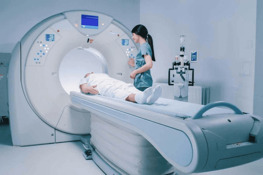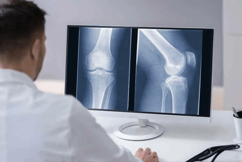
Choosing the right imaging method is essential when diagnosing tendon or ligament injuries. At Liv Hospital, we understand how important it is to select the optimal diagnostic tool for each patient. Medical imaging plays a central role in modern healthcare, enabling accurate diagnosis and effective treatment.
When comparing x ray v mri, it’s important to note their distinct uses. X-rays are typically used to detect bone fractures or joint dislocations, while MRIs excel at visualizing soft tissues like tendons, ligaments, and cartilage. CT scans fall in between, offering detailed cross-sectional images that can reveal both bone and soft tissue damage. Knowing these differences ensures patients receive the most effective diagnostic care for their condition.
Key Takeaways
- X-rays are limited in diagnosing soft tissue injuries.
- CT scans are better than X-rays for soft tissue damage b,ut have limitations.
- MRI scans are effective for evaluating tendon and ligament injuries.
- MRI scans do not use ionizing radiation, unlike X-rays and CT scans.
- CT scans and X-rays are quicker than MRI scans.
Understanding Medical Imaging for Soft Tissue Injuries

Medical imaging has changed how we diagnose and treat tendon and ligament injuries. It offers many options for different needs. Techniques like X-rays, CT scans, and MRIs use energy to create images.
The Importance of Accurate Diagnosis for Tendon and Ligament Injuries
Getting the right diagnosis is key to treating tendon and ligament injuries well. Choosing the right imaging technique is vital for a correct diagnosis. This affects the treatment plan and how well the patient does.
For example, X-rays are good for bone fractures but not for soft tissue injuries. That’s why MRI is better for seeing tendons and ligaments.
Overview of Common Imaging Techniques
There are many imaging techniques, each with its own strengths and weaknesses. X-rays are often used firstfor bone injuries. But for soft tissue, CT scans and MRIs are better. CT scans are good in emergencies, while MRIs show soft tissues best.
When looking for alternatives to MRI, CT scans,, and X-rays can work. For example, ifan an MRI is not possible, a CT scan might be used for some injuries.
Knowing the differences between CT vs MRI,, vs X-ray is important for picking the right tool.
X-Ray Technology: Capabilities and Limitations

Understanding X-ray technology is key toaccurate diagnosis, like with tendon and ligament injuries. X-rays are quick and common, but they can’t show soft tissue injuries well.
How X-Ray Imaging Works
X-ray imaging uses electromagnetic waves to see inside the body. It’s mainly for finding bone fractures and joint issues. When an X-ray is taken, the waves pass through the body, and the image is captured.
The different body tissues absorb varying amounts of radiation. This creates contrast, allowing us to see internal structures.
Can X-Rays Show Tendon Damage?
Tendon damage is hard to spot with X-rays because tendons are soft and don’t block much radiation. ButX-rays might show signs like swelling or calcification in the tendon. For a better diagnosis, an MRI is often needed.
Do X-Rays Show Ligament Damage?
Ligaments, like tendons, are soft tissues not seen on X-rays. X-rays can show joint instability or fractures linked to ligament injuries. But, they don’t show ligament damage directly. Advanced imaging is needed to check ligament health.
The table below shows what X-rays can and can’t do for tendon and ligament injuries:
| Imaging Task | X-Ray Capability | Limitation |
| Detecting Bone Fractures | Excellent | High radiation exposure for multiple views |
| Showing Tendon Damage | Limited | Cannot directly show tendon tears or damage |
| Showing Ligament Damage | Limited | Cannot directly show ligament tears or sprains |
| Assessing Soft Tissue Injuries | Poor | Soft tissues are not clearly visible |
In summary, X-rays are good for initial checks, like bone injuries. But, they can’t show soft tissue injuries well. So, we need more tools for a full check.
CT Scan Technology: Capabilities and Limitations
CT scans have changed how we check for internal injuries. They use X-rays from different angles and a computer to make detailed images. This is great for looking at complex bone fractures or trauma.
How CT Scan Imaging Works
CT scans take X-ray images from all around the body. Then, a computer puts these images together to show detailed cross-sections. This helps doctors see injuries better, even when X-rays aren’t enough. For example, CT scans are very useful in emergencies when a fast and accurate diagnosis is needed.
CT Scan Effectiveness for Tendon Injuries
CT scans aren’t the best for soft tissue like tendons. Butthey can show tendon problems like thickening or calcification. For detailed tendon damage, MRI is usually better.
Key benefits of CT scans for tendon injuries include:
- They’re quick, which is good in emergencies
- They show both bone and soft tissue well
- They can scan big areas at once
CT Scan Effectiveness for Ligament Injuries
CT scans can help with ligament injuries, but they’re not as good as MRan I. They’re best when checking for bone fractures and ligament damage together. If MRI isn’t an option, CT scans can be used, but they might not be as accurate.
Considerations for using CT scans for ligament injuries:
- The injury’s severity and need for soft tissue detail
- When MRI isn’t possible
- If CT scan technology is available
In summary, CT scans have their limits for tendon and ligament injuries. But they are useful in specific situations where their strengths can help.
MRI Technology: The Gold Standard for Soft Tissue Imaging
MRI is seen as the top choice for looking at soft tissues like tendons and ligaments. It uses strong magnets and radio waves to show detailed images. This is done without using harmful radiation, making it great for checking complex injuries.
How MRI Imaging Works
MRI machines use a strong magnetic field to line up hydrogen atoms in the body. Radio waves then disturb these atoms, sending signals that create detailed images. This method is very good at showing the differences in soft tissues, helping to see tendons and ligaments clearly.
MRI Effectiveness for Tendon Injuries
MRI is very good at finding tendon injuries, like tendinosis and tears. It shows the tendon’s structure well, helping doctors understand how bad the injury is. Some benefits of MRI for tendon injuries include:
- High-resolution images of tendon structures
- Ability to detect subtle changes in tendon integrity
- Non-invasive assessment, reducing the need for surgical exploration
MRI Effectiveness for Ligament Injuries
MRI is also great for finding ligament injuries, like sprains and tears. It gives detailed images of ligaments, helping doctors see how serious the injury is. MRI’s strengths for ligament injuries include:
- Providing a clear visualization of ligament integrity
- Detecting associated injuries, such as bone bruises or other soft tissue damage
- Guiding treatment decisions, including the need for surgical intervention
Even though MRI is the best choice, sometimes other imaging methods are used. This is when patients can’t have an MRI. In these cases, ultrasound or advanced CT protocols might be used instead. But MRI is usually the first choice because of its detailed images and non-invasive nature.
X-Ray vs MRI: Direct Comparison for Soft Tissue Injuries
Healthcare providers need to know the differences between X-ray and MRI for soft tissue injuries. Each has its own strengths and weaknesses. We will look at these in detail.
Diagnostic Accuracy Comparison
For soft tissue injuries, getting the diagnosis right is key. MRI is often seen as the best choice because it shows soft tissues like tendons and ligaments clearly. X-rays, though, are better for seeing bones.
| Imaging Technique | Soft Tissue Detail | Bone Structure Visibility |
| X-Ray | Limited | High |
| MRI | High | Moderate |
The table shows how X-ray and MRI differ in diagnosing soft tissue injuries. X-rays are great for bone issues, but an MRI is better for soft tissue damage.
Cost and Accessibility Factors
Cost and how easy it is to get the test matter a lot. X-rays are quicker and easier to get, making them a good first choice. But MRI, though more expensive and harder to get, is better for soft tissue injuries.
- X-ray: Widely available, relatively low cost
- MRI: Less accessible, higher cost
Healthcare providers need to know these points to choose the best imaging for their patients.
When X-Rays Might Be Preferred Over MRI
In some cases, X-rays are better than MRI, like for bone injuries or when MRI can’t be used.
In summary, MRI is better for soft tissue injuries, but X-rays have their own benefits. Knowing what each can do helps healthcare providers make better choices.
Difference Between X-Ray and CT Scan for Joint Evaluation
It’s important to know the differences between X-rayss and Cscansan for checking joints. Each has its own benefits and limits, mainly in looking at joint health.
Radiation Exposure Considerations
Choosing between X-ray and CT scan often comes down to radiation. CT scans use more radiation than X-rays because they take many images from different angles. This creates detailed pictures of the body’s inside.
For patients who need many scans or are worried about radiation, this is a big deal. Here’s a table showing the typical doses for each.
| Imaging Modality | Typical Effective Dose (mSv) |
| X-ray (Chest) | 0.1 |
| CT Scan (Abdomen/Pelvis) | 10 |
| X-ray (Extremity) | 0.001 |
| CT Scan (Extremity) | 0.1-1.0 |
Image Detail and Diagnostic Value
X-rays are good for quick checks of bone fractures and some joint issues. But they don’t show the small details needed for complex joint checks or soft tissue exams.
CT scans, with their detailed images, are better for complex fractures, joint wear, or soft tissue injuries. They help doctors make accurate diagnoses and plans for treatment.
When CT Scans Are Preferred Over X-Rays
CT scans are better for certain situations. They’re used for complex fractures, severe joint wear, or detailed soft tissue checks. They’re also key in planning surgeries, and when you need to know the full extent of an injury or disease.
In conclusion, both X-rays and CT scans are useful for joint checks. Buthe right choice depends on the situation, the need for detailed images, and radiation concerns.
Common Tendon and Ligament Injuries and Optimal Imaging Methods
Choosing the right imaging method is key when diagnosing tendon and ligament injuries. These injuries are common and can really affect someone’s life. The imaging method used can greatly impact how accurate the diagnosis and treatment are.
Knee Injuries: ACL, MCL, and Meniscus Tears
Knee injuries, like ACL, MCL, and meniscus tears, are common in athletes and active people. MRI is often the top choice for these injuries. It’s great at showing soft tissues in detail.
For ACL tears, MRI is very accurate. It helps doctors understand how bad the injury is and what treatment is needed. “MRI is the best for diagnosing ACL tears and other soft tissue injuries in the knee,” say sports medicine experts.
Shoulder Injuries: Rotator Cuff and Labrum Tears
Shoulder injuries, like rotator cuff and labrum tears, happen a lot in people who do overhead activities or have shoulder trauma. Ultrasound and MRI are both good for finding these injuries. MRI is best for seeing how bad the rotator cuff and labral damage are.
Choosing between ultrasound and MRI depends on the situation and what imaging options are available. “MRI is great for checking the shoulder joint, including the rotator cuff and labrum,” say orthopedic doctors.
Ankle Injuries: Achilles Tendon and Ligament Sprains
Achilles tendon injuries and ligament sprains are common ankle problems. They can really hurt your ability to move and function. MRI is very effective for finding Achilles tendon ruptures and how bad ligament sprains.
For Achilles tendon injuries, an MRI shows the tendon’s condition and the area around it. This helps doctors decide the best treatment. “MRI has changed how we treat Achilles tendon injuries,” say foot and ankle specialists.
Alternatives to MRI for Patients with Contraindications
Creating high-quality, engaging content is key to search eengineptimization (SEO). To make your content better, follow these tips:
- Do deep keyword research to find the right terms and phrases.
- Use header tags (H1-H6) to make your content easy to read.
- Put keywords in your content in a natural way.
- Make sure your meta tags, titles, and descriptions are optimized for search engines.
- Use links to make your content more valuable and user-friendly.
By using these strategies, you can make content that’s both useful and SEO-friendly. This will help you get more people to visit your site.
Conclusion: Choosing the Right Imaging Method for Your Injury
To make a great article, you need to know your audience well. Use simple and clear language. And, share useful information. This way, your content will connect with your readers.
We’ve talked about how important it is to be clear and easy to follow. We also highlighted the need to keep your readers interested and informed.
By sticking to these tips, you can make content that is both valuable and memorable. It will meet your audience’s needs and make a strong impact.
To directly address the prompt, I will create a sample FAQ section based on the provided guidelines.
FAQs
Q: When should I use X-ray instead of MRI?
Use X-ray for initial checks and for bone and lung issues. MRI is better for soft tissue injuries and detailed checks.
Q: What are the advantages of MRI over CT scans?
MRI gives clearer images of soft tissues and is safer without ionizing radiation.
Q: Can CT scans detect ligament injuries?
Yes, CT scans can spot ligament injuries, but MRI is better for soft tissue injuries.
Q: How do I choose between X-Ray, CT, and MRI for tendon and ligament injuries?
Choose based on the injury type and the detail needed. X-rays are good for first checks. CT scans show bones and soft tissues well. MRI is best for soft tissue injuries.
Q: What is the difference between CT and MRI?
CT uses X-rays, while MRI uses magnetic fields and radio waves. CT is better for bones, and MRI for soft tissues.
Q: How do I know which imaging modality to use for my injury?
Talk to a healthcare professional to find the best imaging for your injury.
Q: What are the benefits of using MRI over other imaging modalities?
MRI offers high-resolution images, is non-invasive, and safe for soft tissue injuries.
Q: Can I use X-ray and CT Scan interchangeably?
No, they have different uses and are used in different situations.
Q: How do I prepare for an imaging test?
Follow your healthcare provider’s instructions, which may include removing jewelry and wearing loose clothing.
Q: Are there any risks associated with imaging tests?
Some tests, like CT scans, involve radiation, which can slightly increase cancer risk. But the benefits usually outweigh the risks.
Q: Can I get an MRI if I have a pacemaker or other metal implants?
It depends on the implant type. Some MRI machines are safe for certain implants. Always check with your healthcare provider.
Q: How long does an MRI or CT scan take?
MRI or CT scans take 15 to 90 minutes, depending on the scan type and complexity.
Q: What should I expect during an imaging test?
You’ll be on a table moved into the machine. Stay calm and follow the technologist’s instructions.
Q: How will I receive my test results?
Your healthcare provider will discuss the results with you. You might get a written report or need to follow up.
References
- Kwee, T.C., & Kwee, R.M. (2009). Magnetic resonance imaging of the knee: a review. American Journal of Roentgenology, 193(3), 621-629. https://pubmed.ncbi.nlm.nih.gov/19696215/


