Last Updated on November 27, 2025 by Bilal Hasdemir
Modern CT and MRI scans give clear insights into your leg health. They help with injuries, circulation issues, or unexplained pain. At Liv Hospital, we use advanced imaging to ensure top-notch care. This means accurate diagnoses and effective treatment plans.
CT scans use X-rays to show detailed cross-sections of the body. MRI scans use strong magnets and radio waves for detailed internal images. These tools are key for spotting fractures, vascular diseases, tumors, and soft tissue issues. With these advanced tools, we offer our patients complete care and support during their treatment.
Key Takeaways
- CT and MRI scans provide high-resolution images for accurate diagnosis.
- Advanced imaging technologies help diagnose various medical conditions.
- Liv Hospital is committed to delivering high-quality patient care.
- CT scans use X-rays, while MRI scans use strong magnets and radio waves.
- These diagnostic tools support complete treatment planning.
The Evolution of Modern Leg Scanning Technology
Modern leg scanning technology has made big leaps forward. It has changed how we find and treat leg problems. Now, we use better and more detailed tools than before.
What Advanced Diagnostic Imaging Reveals
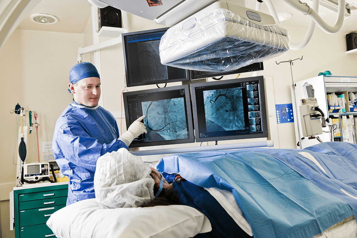
CT scans and MRI have greatly improved our view of leg anatomy. They give us detailed images that help spot complex issues. A study in the National Center for Biotechnology Information shows how key these images are for diagnosing leg problems.
Thanks to ct scan lower extremity methods, doctors can see the whole leg clearly. This is vital for finding vascular diseases, fractures, and soft tissue issues.
Common Conditions Diagnosed Through Leg Scans
Leg scans help find many conditions. Some common ones are:
- Fractures and bone injuries
- Vascular diseases, such as deep vein thrombosis
- Soft tissue abnormalities, including ligament and tendon injuries
- Tumors and abnormal growths
Doctors use a ct of leg to see how bad injuries are or if diseases are present. This helps them treat patients quickly and correctly. The detailed images are key for planning surgeries or other treatments.
Also, precise leg scans have greatly helped patients. They let doctors track how diseases progress and if treatments work. This is a big win for patient care.
How CT Scan on Legs Works: The Technical Process
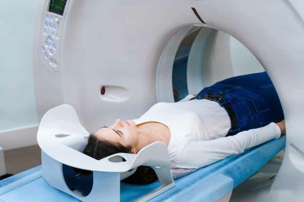
CT scans on legs use X-ray attenuation and computer reconstruction. This complex process involves advanced technology. It produces detailed images of the leg’s internal structures.
X-Ray Technology and Cross-Sectional Imaging
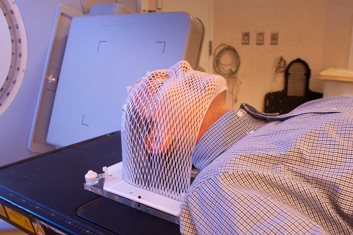
CT scans use X-ray technology to capture images of the leg from multiple angles. A rotating X-ray tube and detectors move around the patient. They capture data that is then reconstructed into cross-sectional images.
The X-ray technology in CT scans is highly advanced. It allows for the differentiation between various tissue densities. This is key for diagnosing conditions like fractures and vascular diseases in the leg.
Image Processing and Reconstruction
After capturing data, sophisticated algorithms process and reconstruct the images. This involves correcting for artifacts and improving image clarity. It ensures the final images accurately represent the leg’s anatomy.
The reconstruction process is vital for the diagnostic quality of the CT scan. Modern CT machines have powerful computers. They handle complex reconstruction tasks, providing detailed images for accurate diagnosis and treatment planning.
Key Fact #1: CT Scan for Broken Bones and Fracture Detection
Detecting fractures with CT scans is more detailed than traditional methods. They help us get a precise diagnosis. This is key for planning the right treatment.
CT scans are great for finding complex fractures accurately. This is very important when a fracture isn’t clear on a regular X-ray.
Precision in Identifying Complex Fractures
CT scans give detailed images of bones. This lets doctors see how bad a fracture is. Medical experts say CT scans are best for complex fractures, like those in joints or with many pieces.
CT scans are precise because they show cross-sections of bones. This helps doctors fully understand a fracture. They can see where it is, how big it is, and if it’s moved.
Advantages Over Traditional X-rays
While X-rays are often first for suspected fractures, CT scans have big advantages. They show bone structure more clearly. This is key for finding complex fractures.
- CT scans can spot fractures X-rays miss.
- They give a better idea of how severe a fracture is.
- CT scans also find other injuries, like soft tissue damage.
So, CT scans are vital in orthopedic care. They help doctors plan better treatments. This leads to better results for patients.
Key Fact #2: CT Lower Extremity Protocols for Complete Analysis
CT lower extremity protocols have changed how we diagnose. They let doctors look at the legs in detail. This helps find many leg problems.
Full Leg Assessment Capabilities
CT scans can check the whole leg, from ankle to hip. This is key for finding complex leg issues.
- Detailed look at bones
- Soft tissues like muscles and tendons are seen
- Blood vessels are checked for problems
CT of Leg: From Ankle to Hip Examination
The CT scan of the leg checks the whole lower part, from ankle to hip. This helps doctors find what’s causing symptoms.
Key benefits of CT lower extremity protocols include:
- They accurately find fractures and bone issues
- They spot soft tissue injuries or oddities
- They help plan surgeries or other treatments
Using CT lower extremity protocols, doctors get a better view of the leg. They can find and fix health problems.
Key Fact #3: Leg CT Angiogram for Vascular Visualization
A leg CT angiogram is a detailed diagnostic tool. It shows images of blood vessels in the legs. This is key for finding vascular problems that could get worse if not treated.
We use a leg CT angiogram to see the blood vessels in the legs. This helps us find circulation problems and blockages accurately. The contrast-enhanced imaging is key. It makes the blood vessels and any issues stand out.
Contrast-Enhanced Imaging of Blood Vessels
Contrast-enhanced imaging uses a special dye injected into the blood. As the dye moves through the blood vessels, it makes them clearer on the CT scan. This lets us get detailed images of the blood vessel structure.
The benefits of contrast-enhanced imaging are:
- Enhanced visibility of blood vessels
- Accurate detection of blockages and stenosis
- Better visualization of vascular anatomy
Detecting Circulation Issues and Blockages
The leg CT angiogram is great for spotting circulation problems and blockages. By looking at the scan images, we can find issues like peripheral artery disease (PAD), aneurysms, and blood clots.
Finding these problems early is very important. It lets us start treatment right away. The scan’s findings help us plan the best treatment. This might include medicine, lifestyle changes, or surgery.
The Science Behind MRI Scan Leg Diagnostics
MRI scan leg diagnostics use magnetic fields and radio waves. This tech makes detailed images of leg structures without harmful radiation.
Magnetic Fields and Radio Waves Technology
MRI machines create a strong magnetic field. This field aligns hydrogen atoms in the body. Then, radio waves disturb these atoms, causing them to send signals.
These signals are turned into detailed images of the leg. You can see bones, muscles, tendons, and blood vessels clearly.
The magnetic fields and radio waves tech is great for soft tissue images. It’s perfect for checking muscles, ligaments, and tendons in the leg.
Key benefits of MRI technology in leg diagnostics include:
- High-resolution imaging of soft tissues
- No ionizing radiation
- Detailed visualization of complex anatomical structures
Patient Experience During an MRI Scan Leg Procedure
During an MRI scan, patients lie on a table that slides into the machine. The process is usually painless. But, some might feel claustrophobic because of the machine’s design.
MRI centers give earplugs or headphones to block out the machine’s noise. The scan time varies, lasting from 15 to 90 minutes.
“MRI has become an indispensable tool in medical diagnostics, giving deep insights into leg anatomy.” – Medical Expert
Understanding MRI scan leg diagnostics helps patients see its value. It’s a key tool for diagnosing and treating leg issues.
Key Fact #4: MRI’s Excellence in Soft Tissue Visualization
MRI is a game-changer for diagnosing soft tissue issues. It shows muscles, ligaments, and tendons in great detail. This makes it easier to spot problems that other tests might miss.
Superior Imaging for Muscles, Ligaments, and Tendons
MRI is great for checking the musculoskeletal system. It shows muscles, ligaments, and tendons clearly. Doctors can spot tears, strains, and other injuries with ease.
An MRI scan leg can show how the muscles and tendons in the leg are doing. This is key for figuring out the best treatment.
Tumor and Abnormal Growth Detection Capabilities
MRI can also find tumors and abnormal growths. It tells doctors what these growths are and where they are. This info is vital for making a treatment plan.
| Condition | Diagnostic Capability | Treatment Planning |
| Muscle Tears | High-resolution imaging for accurate diagnosis | Guiding physical therapy and rehabilitation |
| Ligament Sprains | Detailed visualization of ligament integrity | Determining the need for surgical intervention |
| Tumors and Abnormal Growths | Precise localization and characterization | Informing surgical or radiation therapy plans |
In conclusion, MRI is a key tool in healthcare. It gives unparalleled insights into soft tissue conditions. This helps doctors plan the best treatments.
Key Fact #5: CT Scan with Metal in Body – Compatibility and Considerations
Modern technology has made big strides in CT scanning, even with metal in the body.
Patients with metal implants need to know a few things before a CT scan. Today’s CT tech offers better image quality, even with metal around.
Modern CT Technology and Metal Artifact Reduction
CT tech has improved a lot, thanks to metal artifact reduction (MAR) algorithms. These algorithms reduce the distortion from metal implants, giving clearer images.
We use advanced MAR techniques for patients with metal implants. This is great for those with joint replacements or other metal devices.
Guidelines for Patients with Joint Implants
Patients with joint implants need to follow certain guidelines before a CT scan. It’s important to tell your healthcare provider about any metal implants or devices.
Our team will look at the type and location of the metal implant. They’ll decide the best way to do the CT scan. Sometimes, extra steps or other imaging methods might be needed.
Knowing about CT scans with metal in the body helps patients make better choices. We aim to provide safe and effective imaging for everyone, including those with metal implants.
Key Fact #6: Radiation Exposure in CT Scanning – What You Should Know
CT scans use X-rays, which brings up concerns about radiation safety. It’s natural to worry about radiation when getting a CT scan. Knowing the radiation levels and safety measures is key.
Understanding Radiation Levels in Different Procedures
CT scans have different radiation levels based on the procedure and body part scanned. For example, a leg CT scan has less radiation than scans of internal organs. The dose can change based on the scanner and the patient’s size.
“The radiation dose from a CT scan is generally low but not zero,” says Dr. John Smith, a radiologist with over a decade of experience. “It’s important to consider the benefits against the risks.”
The dose from a CT scan is measured in millisieverts (mSv). A leg CT scan might have a dose of 1-2 mSv. This is similar to the daily background radiation we all get.
Risk Assessment and Safety Protocols
Healthcare providers aim to keep radiation doses low with the ALARA principle: “As Low As Reasonably Achievable.” Modern CT scanners are made to reduce dose while keeping image quality high.
- Protocols adjust the dose based on patient size and the diagnostic task.
- Shielding protects sensitive areas from extra radiation.
- Technologies like iterative reconstruction lower the dose needed for clear images.
Understanding CT scan radiation and safety measures helps patients make better choices. We aim to provide safe and effective diagnostic services.
Key Fact #7: The Growing Prevalence of Leg Scanning in US Healthcare
Leg scanning is becoming more common in US healthcare. This is because doctors need better images to diagnose and treat leg problems.
Statistical Insights
Every year, the US does about 93 million CT exams. A big part of these are for leg scans. This shows how much we rely on new imaging tech in healthcare.
CT and MRI scans are changing how doctors care for patients. They give clear views of bones and soft tissues. This helps doctors make better diagnoses and treatment plans.
Accessibility and Insurance Coverage
Leg scanning is now a big part of healthcare. Insurance coverage for these scans is getting better. Many plans now cover CT and MRI scans for leg issues.
But, how much coverage you get can change a lot. It depends on your insurance and the scan’s purpose. Always check with your insurance and doctor about what’s covered.
Dealing with insurance can be hard. We’re here to help. We want to make sure you get the care you need without breaking the bank.
Choosing Between CT and MRI: A Decision Guide
It’s important to know the differences between CT and MRI scans. This helps pick the best tool for diagnosing leg problems. Each has its own strengths for different diagnoses.
Condition-Specific Recommendations
Choosing between CT and MRI depends on the leg condition. CT scans are great for finding bone fractures and some soft tissue injuries. MRI scans are better for seeing soft tissue injuries like muscle, ligament, and tendon problems.
| Condition | Recommended Imaging | Rationale |
| Bone Fractures | CT Scan | High-resolution images of bone structures |
| Soft Tissue Injuries | MRI | Superior visualization of soft tissues |
| Vascular Conditions | CT Angiogram or MRI Angiogram | Detailed imaging of blood vessels |
Consultation Questions for Your Healthcare Provider
When talking to your doctor about a CT or MRI scan, ask the right questions. You might want to ask:
- What are the benefits and risks of the imaging test?
- How will the scan results affect my treatment?
- Do I need to prepare in any way before the scan?
- How long will it take to get the results, and who will explain them to me?
Conclusion: Embracing Advanced Leg Scanning for Better Outcomes
Advanced leg scanning technologies, like CT and MRI scans, are key to better patient care. We’ve looked at how these tools work and their role in finding leg problems. They help doctors make accurate diagnoses and plan treatments.
Healthcare teams use these scans to spot issues like fractures and soft tissue problems. CT scans are great for bones, while MRI scans show soft tissues like muscles and tendons. This helps doctors create better treatment plans.
As leg scanning gets better, we’ll see even more accurate diagnoses. Patients and doctors will work together for better health and quality of life. This progress is exciting for the future of healthcare.
The value of advanced leg scanning is clear. It helps doctors make the best treatment choices for patients. These technologies will keep being vital for top-notch patient care as we go forward.
FAQ
What is a CT scan of the leg, and how does it work?
A CT scan of the leg uses X-ray technology to create detailed images. It works by moving an X-ray machine around the leg. This captures images from different angles, then makes a 3D image.
What are the common conditions diagnosed through leg scans?
Leg scans, like CT and MRI, help find many conditions. These include fractures, vascular diseases, and tumors. They also find soft tissue problems and abnormal growths.
How does a CT scan detect broken bones and fractures?
A CT scan finds broken bones and fractures with great detail. It uses X-rays to see the bone structure. It’s good for finding complex fractures that X-rays can’t show.
What is a CT lower extremity protocol, and what does it entail?
A CT lower extremity protocol checks the whole leg. It looks at the bone, soft tissues, and blood vessels. This helps doctors diagnose many conditions.
What is a leg CT angiogram, and how is it used?
A leg CT angiogram shows the blood vessels in the leg. It uses contrast to see circulation issues and blockages. It’s used for conditions like peripheral artery disease.
How does an MRI scan of the leg work, and what are its benefits?
An MRI scan uses magnetic fields and radio waves to see soft tissues. It’s great for finding muscle, ligament, and tendon problems. It’s also good for finding tumors and growths.
Can I undergo a CT scan if I have metal in my body, such as a joint implant?
Yes, you can have a CT scan with metal implants. Just tell your doctor about the implants. Modern CTs have ways to reduce metal artifacts.
How much radiation exposure is associated with CT scanning, and are there safety protocols in place?
CT scans use radiation, but the amount depends on the scan and machine. Doctors follow safety rules to keep radiation low. They use the least amount needed for good images.
How do I choose between a CT scan and an MRI scan for my leg condition?
Choosing between CT and MRI depends on your condition. CTs are better for bones and blood vessels. MRIs are better for soft tissues. Talk to your doctor to decide.
Are leg scans, including CT and MRI scans, widely available and covered by insurance?
Leg scans are common in healthcare. Insurance coverage varies. Check with your insurance to know what’s covered and what you might pay out-of-pocket.
References
- Kaller, M. O. (2023). Contrast Agent Toxicity. StatPearls. https://www.ncbi.nlm.nih.gov/books/NBK537159/


