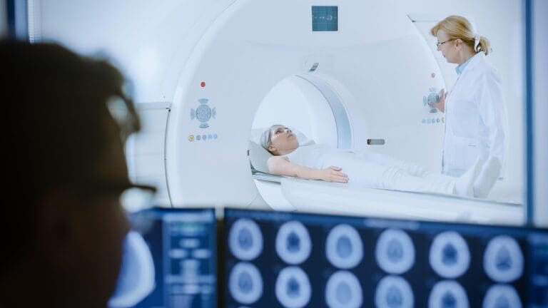
Choosing the right medical imaging test can be tough. With MRI, CT scans, and ultrasonography each offering unique advantages, it can be hard to know which one to choose. At Liv Hospital, we focus on what’s best for you, using the latest medical imaging technology.
Medical imaging plays a key role in diagnosing and treating many health conditions. But how do MRI and CT scan technologies compare to ultrasonography? The main difference lies in their imaging methods”MRI uses magnetic fields and radio waves, CT scans use X-rays, and ultrasound uses high-frequency sound waves.
Understanding these differences helps you make informed decisions about your health. In this article, we’ll explore the 7 main differences between these imaging techniques to help you choose the best option for your medical needs.
Key Takeaways
- Understanding the differences between MRI, CT scans, and ultrasonography is key to choosing the right test.
- Each imaging method has its own technology and use.
- MRI uses magnetic fields and radio waves, while CT scans use X-rays.
- Ultrasonography uses high-frequency sound waves.
- The right test depends on the health issue you’re dealing with.
The Evolution of Medical Imaging Technologies
Medical imaging started with X-rays and has grown into advanced digital technologies. We’ve seen big steps forward in non-invasive imaging techniques. These changes have changed how we diagnose and treat diseases.
From X-rays to Advanced Digital Imaging
Wilhelm Conrad Röntgen discovered X-rays in 1895, starting medical imaging. We’ve moved to better technologies like CT scans, MRI, and ultrasonography. These tools have made diagnostic accuracy of imaging better, helping doctors make better choices.
Digital imaging has made medical images better and easier to get. It lets us get images faster and store them better. For example, MRI and CT scans give detailed views of the body. They help find many conditions, from simple injuries to complex diseases.
The Role of Imaging in Modern Diagnostic Medicine
In today’s medicine, imaging is key for patient care. It helps us see inside the body to diagnose and plan treatments. The right imaging depends on the patient’s needs and condition.
Medical imaging will keep getting better, helping us diagnose and treat more effectively. The future of non-invasive imaging techniques looks bright for medicine.
Understanding the Fundamentals of MRI Technology

MRI technology uses magnetic fields and radio waves to create detailed images. It’s safe for patients and highly detailed. MRI machines make images of the body’s inside parts, helping doctors a lot.
How Magnetic Resonance Imaging Works
MRI aligns hydrogen atoms in the body with a strong magnetic field. Then, radio waves disturb these atoms. They send signals back to their aligned state.
These signals are caught by the MRI machine. It uses them to make detailed images of the body’s inside. This process is very advanced and makes sure the images are top-notch.
MRI is safe because it doesn’t use harmful radiation. This is great for patients who need many scans.
Key Components and Capabilities of MRI Machines
MRI machines have important parts like a superconducting magnet and gradient coils. The magnet makes the strong magnetic field. Gradient coils help make detailed images.
Radiofrequency coils send pulses and catch signals. MRI machines have gotten better over time. They can now take images faster and in higher detail.
But, MRI has some downsides. It’s expensive, needs special places, and some people get scared in the machine.
Compared to ultrasound, MRI is safer because it doesn’t use harmful radiation. But, the best choice depends on what the doctor needs to see, patient safety, and what’s available.
The Science Behind CT Scan Technology
CT scans use X-rays and computer algorithms to create detailed images of the body’s inside. This tech is key in medical diagnosis, giving quick and accurate views of bones and organs.
X-ray Based Imaging Principles
CT scans work by X-ray absorption, where tissues absorb X-rays differently. This lets us make detailed images based on how much X-rays are absorbed by different parts of the body.
A rotating X-ray tube and detectors gather data from many angles. Then, advanced computers turn this data into detailed cross-section images.
Modern CT Scanner Components and Operation
Today’s CT scanners have important parts like the gantry, X-ray tube, detectors, and a computer for image making. The gantry holds the X-ray tube and detectors, which move around the patient to get the needed data.
Key components include:
- X-ray tube: Makes the X-rays for imaging.
- Detectors: Catch the X-rays that go through the patient’s body.
- Computer system: Turns the data into images.
When we compare CT scans to MRI, we see big tech and use differences. MRI uses magnetic fields and radio waves, while CT scans use X-rays. This affects their uses, costs, and safety.
Knowing how CT scans work shows their importance in medical imaging. It also shows how they differ from MRI. The cost of MRI vs CT scan is a big factor, with CT scans being cheaper but involving radiation.
Ultrasonography: Principles and Technology
Ultrasonography is a safe and real-time imaging tool. It uses high-frequency sound waves to show internal body structures. This makes it key in modern medicine.
Sound Wave Imaging Fundamentals
Ultrasonography works by using sound waves to create detailed images. Sound waves go into the body and bounce back as echoes. These echoes are turned into images by the ultrasound device.
The sound waves used are between 2 and 15 MHz. The frequency needed depends on the body part being examined. Higher frequencies give better images but can’t go as deep. Lower frequencies go deeper but are less clear.
Key Advantages of Ultrasonography:
- Real-time imaging capabilities
- No ionizing radiation
- Non-invasive and painless
- Portable and relatively low-cost equipment
Ultrasound Equipment and Functionality
Today’s ultrasound machines have advanced technology. They improve image quality and help doctors diagnose better. The equipment includes a console, a transducer probe, and a control panel.
The transducer probe is essential. It sends and receives sound waves. Better transducers mean clearer ultrasound image quality.
A study in the Journal of Ultrasound in Medicine shows ultrasound’s growth. It’s now a top tool for diagnosing many conditions.
“The use of ultrasonography in clinical practice has become widespread due to its safety profile, real-time imaging capabilities, and cost-effectiveness.”
| Feature | Description | Benefit |
| Real-time Imaging | Captures movement and changes in real-time | Enhances diagnostic accuracy |
| No Ionizing Radiation | Safe for all patients, including pregnant women | Reduces risk of radiation exposure |
| Portability | Equipment is relatively portable | Increases accessibility in various clinical settings |
In conclusion, ultrasonography is safe, real-time, and cost-effective. Its ultrasonography advantages make it a top choice for many medical uses.
How Do MRI and CT Scan Technologies Compare to Ultrasonography? A Comprehensive Analysis
Medical imaging is key for accurate diagnoses and treatment plans. It’s important to know the differences between MRI, CT scans, and ultrasonography. We’ll look at their technology and how they work to compare them fully.
Technological Foundations: Magnetic Fields vs. X-rays vs. Sound Waves
MRI, CT scans, and ultrasonography work in different ways. MRI uses magnetic fields and radio waves to show body structures. CT scans create detailed images with X-rays. Ultrasonography uses high-frequency sound waves for internal organ images.
These technologies affect what each can do. MRI is great for soft tissue images without radiation. CT scans are fast and good for emergencies. Ultrasonography is perfect for live images and procedures like fetal monitoring.
Operational Differences in Clinical Settings
How MRI, CT scans, and ultrasonography work in hospitals is also key. MRI needs special rooms and is used for detailed exams. CT scanners are quicker and more common, great for emergency rooms. Ultrasound machines are easy to move and use at the bedside.
In hospitals, these differences help decide which imaging to use. MRI is best for detailed body scans. CT scans are fast for trauma cases. Ultrasonography is great for live monitoring and procedures.
Knowing these differences helps doctors choose the right imaging. This improves patient care and accuracy.
Difference #1: Image Resolution and Tissue Differentiation
It’s key to know how MRI, CT scans, and ultrasound differ in image quality and tissue detail. This knowledge helps pick the right imaging method for different health issues. The accuracy of these methods depends on their ability to show fine details and tell different tissues apart.
Spatial Resolution Comparison Across Modalities
Spatial resolution is about seeing two close objects as separate. MRI and CT scans usually do better than ultrasound. For example, MRI can show small body parts like knee menisci or brain details clearly. CT scans are great for seeing bones well and are often used for fracture or bone disease diagnosis.
Ultrasound has lower resolution but is useful for live imaging. It’s great for guiding biopsies or checking blood flow in vessels. Ultrasound’s quality can be improved with techniques like harmonic imaging and spatial compounding. But it’s not as sharp as MRI or CT.
Soft Tissue vs. Bone Visualization Capabilities
Seeing soft tissues versus bones is another area where these methods vary. MRI stands out for its clear soft tissue images. It’s best for checking complex soft tissues in the body, like muscles, brain, and spinal cord. It can spot different soft tissues by their magnetic properties, helping find tissue problems.
CT scans are better for bones and calcified things because they use X-rays. They’re good in emergencies to quickly find bleeding, fractures, or foreign objects. A study on Top Doctors says CT scans are fast and good for trauma cases because they show bones and bleeding well.
“The choice between MRI, CT, and ultrasound depends heavily on the specific clinical question being addressed, with each modality providing unique advantages in terms of image resolution and tissue differentiation.”
In summary, MRI, CT scans, and ultrasound have key differences in image quality and tissue detail. Knowing these differences helps doctors choose the best imaging method for each case. This improves patient care.
Difference #2: Radiation Exposure and Safety Profiles
It’s key for healthcare providers to know about MRI, CT scans, and ultrasonography. This knowledge helps them choose the best care for patients.
Radiation Risks in CT Scanning
CT scans use X-rays to make detailed images of the body. This means patients are exposed to ionizing radiation. While CT scans are often needed, we must think about the risk of cancer.
The risk comes from radiation damaging DNA and causing cancer. We need to balance the benefits of CT scans with the risks. Sometimes, other imaging methods might be safer.
Radiation Exposure Comparison
| Imaging Modality | Radiation Exposure | Typical Effective Dose (mSv) |
| CT Scan (Abdomen) | Yes | 10-20 |
| MRI | No | 0 |
| Ultrasonography | No | 0 |
MRI Safety Concerns and Contraindications
MRI doesn’t use ionizing radiation but has its own safety issues. The strong magnetic field can harm metal implants. It can also make patients with claustrophobia anxious.
We must check if patients can safely have an MRI. Their comfort is important too.
Ultrasonography’s Non-Ionizing Safety Advantage
Ultrasonography is safe because it uses sound waves, not radiation. This makes it great for patients who need many scans or are worried about radiation. It’s also safe for pregnant women and kids.
In summary, MRI, CT scans, and ultrasonography have different safety levels. Knowing these differences helps doctors choose the best imaging for each patient.
Difference #3: Cost, Accessibility, and Resource Requirements
The cost, accessibility, and resource needs for MRI, CT scans, and ultrasound vary. This affects their use in medical diagnostics. It’s important for healthcare providers and patients to understand these differences.
Equipment and Operational Expenses
The cost of MRI, CT scans, and ultrasound machines differs a lot. MRI machines are the priciest because of their complex tech and need for magnetic shielding. On the other hand, ultrasound machines are cheaper and more versatile, used in many ways.
Here are some key differences in equipment and operational expenses:
- Initial Cost: MRI machines are the most expensive, followed by CT scanners, with ultrasound machines being the least expensive.
- Maintenance and Upgrades: MRI and CT scanners need more frequent and costly maintenance compared to ultrasound machines.
- Operational Costs: The cost per scan varies, with MRI being the most expensive due to the high cost of cryogens and maintenance.
Patient Accessibility Factors
Accessibility to these imaging modalities is influenced by their availability, cost, and the need for specialized facilities. MRI accessibility is limited by the availability of MRI machines and the need for facilities with magnetic shielding. CT scans are more widely available, but they also need specialized equipment and trained personnel.
Key patient accessibility factors include:
- Geographic Availability: Urban areas tend to have better access to all three imaging modalities compared to rural areas.
- Insurance Coverage: The extent of insurance coverage can significantly impact patient access to these diagnostic tools.
- Waiting Times: The demand for MRI and CT scans can lead to longer waiting times compared to ultrasound.
Facility and Infrastructure Needs
The infrastructure needed for each imaging modality varies. MRI machines require specialized rooms with magnetic shielding, while CT scanners need rooms with radiation shielding. Ultrasound machines, being more portable, require less specialized infrastructure.
Key facility and infrastructure needs are:
- Space and Shielding: MRI and CT scanners require dedicated, shielded rooms.
- Power Requirements: MRI machines have specific power requirements due to their superconducting magnets.
- Technician Expertise: All three modalities require trained technicians, but MRI and CT scanning demand additional specialized training.
Difference #4: Clinical Applications and Diagnostic Specializations
MRI, CT scans, and ultrasonography each have unique uses in medicine. The right choice depends on the medical situation and what information is needed. Each modality offers special benefits for different conditions.
Optimal Uses for MRI Technology
MRI is great for seeing soft tissues like the brain and muscles. It’s safe for pregnant women and kids because it doesn’t use harmful radiation. MRI is key for diagnosing soft tissue problems and some vascular issues.
It’s also used for complex joint problems and soft tissue tumors. MRI’s clear images help doctors make precise diagnoses and plan treatments.
When CT Scans Provide Superior Diagnostic Value
CT scans are vital in emergencies for quick diagnosis. They’re good at finding internal injuries and spotting conditions like appendicitis or cancer.
CT scans are fast and accurate, making them essential in urgent care. They help doctors quickly spot internal injuries or bleeding, leading to better care.
Ultrasonography’s Unique Clinical Advantages
Ultrasonography is non-invasive and doesn’t use harmful radiation. It’s often used for pregnancy checks, gallbladder issues, and soft tissue exams.
It’s known for its ability to show things in real-time, like the heart and blood vessels. Plus, it’s portable, making it great for bedside checks in critical care.
Complementary Use of Multiple Imaging Modalities
While each modality has its strengths, sometimes combining them gives the best results. This approach helps doctors make more accurate diagnoses and tailor treatments.
Using MRI, CT scans, and ultrasonography together can improve patient care. For instance, CT scans for quick injury checks, then MRI for detailed soft tissue views, or ultrasonography for procedure guidance.
Difference #5: Real-time Imaging and Procedural Guidance
MRI, CT scans, and ultrasonography have different real-time imaging capabilities. This is important for choosing the right imaging method for various medical needs, like during procedures.
Dynamic Imaging Capabilities in Ultrasonography
Ultrasonography is great for showing real-time images. It helps doctors see moving parts and guide during procedures. This is useful for:
- Guiding needle placements for biopsies or aspirations
- Monitoring fetal movements during pregnancy
- Assessing blood flow in real-time
Real-time ultrasonography lets doctors make quick decisions. This improves the accuracy of many procedures.
Time Constraints and Processing in MRI and CT Scanning
MRI and CT scans don’t offer real-time images like ultrasonography. They take pictures and then process them for diagnosis.
MRI is getting faster, but it’s not yet good for real-time procedures. CT scans can take pictures quickly, but processing them takes time.
| Imaging Modality | Real-time Capability | Typical Use Cases |
| Ultrasonography | High | Procedural guidance, fetal monitoring, blood flow assessment |
| MRI | Low | Soft tissue imaging, neurological assessments |
| CT Scan | Moderate | Emergency trauma assessment, detailed organ imaging |
In summary, ultrasonography is best for real-time imaging and guiding procedures. MRI and CT scans are better for other medical uses. Knowing these differences helps choose the right imaging method for each case.
Difference #6: Operator Dependency and Technical Expertise Requirements
The quality of MRI, CT scans, and ultrasound depends a lot on the skills of the operators. The need for these skills varies among the imaging modalities. This shows how important technical skills are for each type of scan.
The Impact of Technician Skill on Diagnostic Quality
The technician’s skill level greatly affects the quality of the images. For example, ultrasound is more operator-dependent than MRI and CT scans. This is because ultrasound requires the technician to adjust the probe and interpret images live.
How well the operator does their job can really affect the accuracy of the diagnosis. With ultrasound, a skilled technician can get high-quality images. But, an inexperienced technician might miss important details.
Training and Certification Standards Across Imaging Modalities
The training and certification for MRI, CT scan, and ultrasound technicians differ a lot. MRI and CT scan operators need a lot of training because of the complex equipment. Ultrasound technicians, on the other hand, need special training to master the manual skills needed for quality images.
Here’s a comparison of the training and certification standards across the three imaging modalities:
| Imaging Modality | Typical Training Duration | Certification Requirements |
| MRI | 2-3 years | Certification in MRI technology |
| CT Scans | 2-3 years | Certification in CT scanning |
| Ultrasound | 2 years + Continuing Education | Certification in sonography (e.g., RDMS) |
Knowing these differences helps healthcare providers make better choices. It also helps patients understand the expertise behind their diagnostic care.
Difference #7: Patient Experience and Practical Considerations
Choosing between MRI, CT scans, and ultrasound goes beyond just finding out what’s wrong. How comfortable the patient feels is very important. We need to think about both physical and mental comfort during these tests.
Physical Comfort and Procedure Duration
Being comfortable during the test is a big deal. MRI scans can be tough for people who are scared of small spaces or have trouble moving. “The enclosed environment of an MRI machine can be daunting for many patients,” says a study on MRI comfort.
CT scans are quicker and don’t feel as tight, but patients must stay very quiet. Ultrasound is the most comfortable because it doesn’t need patients to stay in one place or be very quiet.
How long the test takes is also important. MRI scans can take a long time, sometimes over an hour. CT scans are much faster, done in just a few minutes. Ultrasound tests can be shorter than MRI but longer than CT scans.
Psychological Factors and Patient Preparation
How a patient feels mentally also matters a lot. MRI tests can make some people very anxious because of the small space. But, if patients know what to expect and are helped to relax, they can feel better.
CT scans and ultrasound tests usually make patients feel less stressed because they are quicker and don’t feel as tight. Getting ready for these tests is also easier, which helps patients feel more at ease.
In the end, when we look at how patients feel and what’s practical, we see big differences between MRI, CT scans, and ultrasound. Knowing these differences helps doctors make tests that are better for each patient. This makes patients more comfortable and willing to follow instructions.
Conclusion: Selecting the Optimal Imaging Technology for Patient-Centered Care
Choosing the right imaging technology is key for patient-centered care. MRI, CT scans, and ultrasound each have their own strengths and weaknesses. At Liv Hospital, we aim to offer top-notch medical care by using these imaging tools wisely.
It’s important to know the differences between MRI, CT scans, and ultrasound. For example, ultrasound gives real-time results and is safe. CT scans are quick but use radiation. MRI gives detailed images but can be risky for those with metal implants.
Healthcare providers should think about image quality, radiation, cost, and how the technology is used. At Liv Hospital, we’re dedicated to using these technologies to improve patient care and provide the best healthcare possible.
FAQ
What are the main differences between MRI, CT scans, and ultrasonography?
MRI, CT scans, and ultrasonography use different technologies to create images. MRI uses magnetic fields, CT scans use X-rays, and ultrasonography uses sound waves.
How do MRI and CT scans compare in terms of radiation exposure?
MRI doesn’t use ionizing radiation, but CT scans do. This makes MRI safer for patients who need many scans or are sensitive to radiation.
What are the advantages of ultrasonography over MRI and CT scans?
Ultrasonography is non-invasive and doesn’t use harmful radiation. It also shows images in real-time, making it great for guiding procedures or checking moving organs.
How do the costs of MRI, CT scans, and ultrasonography compare?
MRI is the most expensive, followed by CT scans, and then ultrasonography. Costs depend on equipment, operational costs, and facility needs.
What are the clinical applications where MRI is preferred over CT scans and ultrasonography?
MRI is best for soft tissues like the brain, spine, and joints. It offers high detail and can tell different tissues apart.
Can CT scans and MRI be used interchangeably?
No, CT scans and MRI are not interchangeable. CT scans are better for bones and acute hemorrhages. MRI is better for soft tissues.
How does the operator’s skill level impact the quality of ultrasound images?
A skilled technician greatly improves ultrasound image quality. They can get better images and make more accurate diagnoses.
What are the safety concerns associated with MRI?
MRI safety concerns include magnetic field effects on implants, claustrophobia, and the need for careful patient screening.
How do MRI, CT scans, and ultrasonography contribute to patient-centered care?
Choosing the right imaging modality improves diagnostic accuracy and patient safety. This aligns with patient-centered care, aiming for better outcomes.
References
- van Randen, A., et al. (2011). A comparison of the Accuracy of Ultrasound and Computed Tomography in common diagnoses causing acute abdominal pain. European Radiology, 21(7), 1535-1545. https://www.ncbi.nlm.nih.gov/pmc/articles/PMC3101356/


