Last Updated on November 27, 2025 by Bilal Hasdemir

When you get a CT scan of the neck with contrast, you might find surprises. These can include the uterus and cervix. At Liv Hospital, we make sure to check these findings carefully. We want to give you the best care possible.
CT scans with contrast can show more than expected. This includes the uterus and cervix. It shows how important it is to care for you fully. Our team works hard to give you the care you need with kindness.
Key Takeaways
- Incidental findings on CT scans can be significant for patient health.
- Thorough evaluation is key to unexpected discoveries.
- Liv Hospital focuses on patient-centered care and global best practices.
- Contrast-enhanced CT scans can reveal findings beyond the initial scan area.
- Comprehensive care includes follow-up for incidental findings.
The Unexpected Connection: Neck CT Scans and Pelvic Findings

Neck CT scans and pelvic findings might seem unrelated at first. Yet, they are key to full patient care. These scans focus on the neck but can also reveal insights into the pelvis.
How Contrast-Enhanced CT Scans Work
Contrast-enhanced CT scans use a special agent to highlight body structures. This agent is given through an IV and shows up bright on scans. It helps doctors see blood vessels, tumors, and other issues clearly.
These advanced scans can show more than expected. For example, a neck CT might find pelvic issues. This can be very important for health.
Incidental Discovery of Pelvic Abnormalities
CT scans can find things in the pelvis by accident. These can be simple or serious, like tumors. Doctors must check these findings carefully and decide what to do next.
“Incidental findings on female pelvic MRI present diagnostic challenges and may have significant clinical implications.” This shows why it’s important to thoroughly check and follow up on these findings.
Clinical Significance of Unexpected Findings
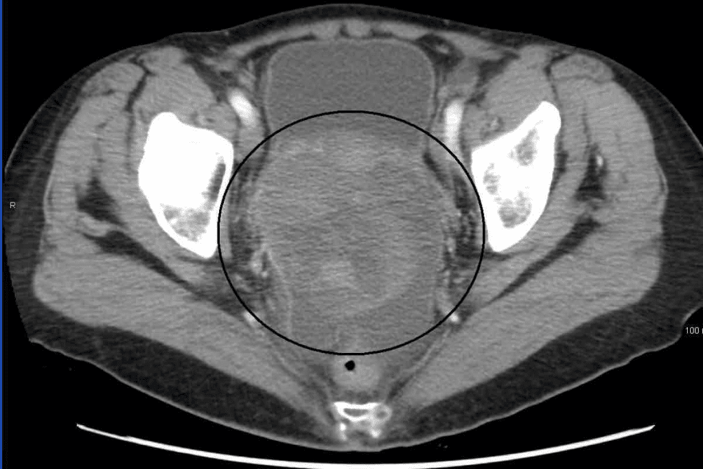
Unexpected CT scan findings are very important. For example, finding something unusual in the uterus or cervix can lead to early treatment. Below is a table showing what these findings might mean.
| Finding | Potential Implication | Recommended Action |
| Uterine Mass | Possible tumor or fibroid | Further evaluation with MRI or ultrasound |
| Cervical Abnormality | Potential cervical cancer or infection | Colposcopy or biopsy |
| Ovarian Cyst | Benign or malignant cyst | Ultrasound follow-up |
In conclusion, neck CT scans and pelvic findings are connected. This highlights the need for thorough patient care and attention to unexpected scan results.
Normal Uterine Appearance on CT Imaging

The normal uterus shows clear signs on CT scans. These signs help doctors spot any problems. It’s important to know what a normal uterus looks like on a CT scan.
Homogeneous Enhancement Patterns After Contrast
A normal uterus shows a homogeneous enhancement pattern after contrast. This means the uterus gets contrast evenly. This evenness shows the uterus is healthy and working properly.
Important points about homogeneous enhancement are:
- Uniform enhancement throughout the uterine tissue
- Absence of focal areas of hypo- or hyper-enhancement
- Symmetric enhancement patterns
Zonal Anatomy Visualization
A normal uterus also shows clear zonal anatomy on CT scans. The uterus has layers like the endometrium, myometrium, and serosa. These layers are visible after contrast, helping doctors check their thickness and health.
Key points about zonal anatomy are:
- Clear delineation between the endometrium and myometrium
- Normal thickness of the endometrium and myometrium
- Intact serosal layer
Baseline Measurements and Characteristics
It’s key to know the baseline measurements and characteristics of the uterus. This includes its size, shape, and where it sits. These details help doctors see if anything changes over time.
Important baseline characteristics are:
- Uterine size and volume
- Shape and orientation of the uterus
- Position relative to surrounding structures
Understanding these normal signs helps doctors spot problems early. This leads to better care for patients.
Key Factors Influencing Uterine Imaging Results
Understanding what affects uterine imaging results is key to accurate diagnosis and treatment. When looking at CT scans of the uterus, several important elements play a role.
Age-Related Changes in Uterine Appearance
As women get older, their uterus changes on CT scans. Age-related changes can alter the size, shape, and how it looks on scans.
A study in a top radiology journal showed that uterine volume decreases with age, more so after menopause. This change can affect how CT scans are read.
| Age Group | Average Uterine Volume | Typical CT Findings |
| Reproductive Age | 50-100 cm ³ | Homogeneous enhancement |
| Post-Menopausal | 20-50 cm ³ | Reduced enhancement, possible calcifications |
Impact of Menopausal Status on CT Findings
Menopausal status is a big factor in uterine imaging results. The shift from pre-menopause to post-menopause brings big hormonal changes that affect the uterus.
“The end of menstrual cycles marks a big change in a woman’s reproductive life. It affects hormonal balances and how the uterus looks on imaging studies.”
– Radiology Expert
After menopause, the uterus gets smaller. The CT scan enhancement pattern may also change due to less hormonal stimulation.
Hormonal Influences on Tissue Enhancement
Hormonal changes can greatly affect how tissues look on CT scans. Hormonal influences can change the blood flow and density of uterine tissue, altering its appearance on scans.
For example, during the menstrual cycle, changes in estrogen and progesterone levels can change uterine enhancement. Knowing these hormonal effects is key to accurate CT scan interpretation.
- Hormonal changes during the menstrual cycle can affect uterine enhancement patterns.
- Hormone replacement therapy (HRT) can also impact the appearance of the uterus on CT scans.
- Certain endocrine disorders can influence uterine imaging results.
By considering these factors, healthcare providers can improve the accuracy of uterine imaging interpretations. This leads to better patient outcomes.
Identifying Abnormal CT Scan of Neck with Contrast: Uterine Findings
Contrast CT scans of the neck can show unexpected uterine issues. It’s key to spot both normal and abnormal uterine findings. This helps in giving accurate diagnoses and care.
Congenital Uterine Anomalies
Congenital uterine anomalies happen during fetal development. They can be found on CT scans, even if not the main focus.
Some common anomalies include:
- Bicornuate uterus: A uterus with two separate horns.
- Uterus didelphys: A condition where there are two separate uteri.
- Septate uterus: A uterus divided by a septum.
These can affect reproductive health. Finding them on CT scans means more evaluation and care.
Uterine Masses and Their Distinguishing Features
Uterine masses on CT scans can differ in size and importance. Leiomyomas (fibroids) are benign tumors seen as defined masses in the uterine wall.
Features of uterine masses on CT scans include:
- Size and location: Size and location can affect their clinical meaning.
- Enhancement patterns: How a mass changes after contrast can hint at its type.
- Margins and borders: Clear margins often mean benign, while irregular ones might suggest cancer.
Correctly identifying uterine masses is vital. It helps decide the next steps, which might include an abnormal cervical MRI or other tests.
“The incidental detection of uterine abnormalities on CT scans performed for other indications highlights the importance of a thorough review of all imaged structures, not just those related to the primary clinical question.” –
Expert Radiologist
Understanding and spotting abnormal uterine findings on CT scans is key. It allows healthcare providers to give full care. They address both the main reason for the scan and any extra findings that need attention.
Normal vs. Abnormal Cervical Findings on CT
It’s important for radiologists to know the difference between normal and abnormal cervical findings on CT scans. They need to understand how the cervix looks after contrast is added.
Standard Cervical Appearance After Contrast
After contrast, the normal cervix looks uniform. This uniform look helps spot any issues. The different parts of the cervix show up differently after contrast.
The cervical canal is usually thin and less dense than the rest. Its size and shape can change due to age, hormones, and surgery.
Benign Cervical Conditions
Some benign conditions can make CT scans harder to read. Nabothian cysts are common and look like clear spots in the cervix.
Cervical leiomyomas are another benign condition. They show up as clear masses in the cervix. Knowing about these conditions helps avoid mistaking them for cancer.
By knowing what a normal cervix looks like after contrast and understanding benign conditions, radiologists can better spot problems on CT scans.
Critical Signs in Abnormal Cervical CT Scans
Cervical CT scans can show important signs of cervical problems. These signs help doctors diagnose and treat cervical issues well. We will look at the main signs that doctors check for in cervical CT scans.
Irregular Margins and Their Diagnostic Significance
Irregular margins on a cervical CT scan can mean there’s a problem. Irregularities in the cervical margins might show a tumor or other health issues. We closely check these irregularities because they could mean cervical cancer.
- Irregular margins may indicate tumor growth or invasion.
- The shape and extent of irregularities can provide insights into the stage of the disease.
- Careful evaluation of margin irregularities is critical for accurate diagnosis.
Parametrial Invasion Assessment
Parametrial invasion is when cervical cancer spreads to the tissue around the cervix. Checking for this spread is key to cancer staging and treatment planning. We use CT scans to see how far the cancer has spread.
Key factors to consider when assessing parametrial invasion include:
- The presence of soft tissue stranding or nodularity in the parametrium.
- The extent of parametrial involvement and its impact on surrounding structures.
- The relationship between the tumor and the parametrium.
Cervical Cancer Indicators and Staging Considerations
CT scans can show signs of cervical cancer, like changes in the cervix or masses. Knowing the stage of cervical cancer is important for choosing the right treatment.
| Indicator | Description | Staging Implication |
| Cervical Mass | A mass or tumor is visible on the CT scan. | May indicate local invasion or advanced disease. |
| Parametrial Invasion | Spread of cancer into the parametrium. | Affects staging and treatment planning. |
| Lymph Node Involvement | Enlarged lymph nodes indicate possible metastasis. | Impacts overall staging and prognosis. |
Understanding these signs helps us better diagnose and manage cervical issues. Accurate reading of cervical CT scans is key to giving patients the best care.
Advanced Imaging: From CT to MRI for Cervical Abnormalities
When looking at cervical problems, moving from CT scans to MRI can give a clearer picture. CT scans are often the first step to finding cervical issues. But MRI can show more details and is better for checking cervical problems.
When to Recommend Follow-up MRI Studies
We suggest getting an MRI if a CT scan shows possible cervical issues. This is key for those with suspected cervical cancer or complex lesions.
- Unclear or suspicious findings on the initial CT scan
- Patients with a history of cervical cancer or high-risk factors
- Complex cervical lesions that require detailed evaluation
Enhanced Tissue Characterization with MRI
MRI shows soft tissues better than CT scans. This means it can see cervical anatomy and problems more clearly. This is important for making accurate diagnoses and knowing how serious the problem is.
Key benefits of MRI in cervical imaging include:
- Improved detection of small lesions
- Better delineation of tumor extent and parametrial invasion
- Enhanced visualization of adjacent structures and possible involvement
Comparative Diagnostic Value in Cervical Pathology
Comparing CT and MRI for cervical issues, MRI usually gives a fuller view. Research shows MRI is better at measuring tumor size, how far it has spread, and if it’s touching nearby tissues.
In conclusion, MRI is essential for diagnosing cervical problems. It’s most useful when CT scans are not clear or when more detailed info is needed for treatment.
Clinical Management of Incidental Uterine and Cervical Findings
When CT scans show unexpected uterine and cervical issues, we need a clear plan. We must follow up and manage these findings carefully. This ensures patient safety and the best possible results.
Developing Follow-up Protocols
Creating good follow-up plans is key when dealing with unexpected uterine and cervical issues. We might need more tests, like an MRI or an ultrasound, to learn more. For example, an MRI can give more details about a uterine mass found on a CT scan.
We look at the patient’s health history, symptoms, and the first scan findings to plan the next steps. Good planning means setting clear follow-up times and telling the patient about them.
Patient Communication Strategies
Talking clearly and kindly to patients is very important. We explain the findings, the follow-up plan, and any treatments needed in simple terms. We avoid using hard medical words and listen to their worries.
Good communication also means giving patients written info and resources. We make sure they know who to contact if they have more questions or concerns.
Multidisciplinary Approach to Unexpected Findings
Working together is key when dealing with unexpected uterine and cervical issues. Radiologists, gynecologists, and others team up for better care. This teamwork helps us make a correct diagnosis and treatment plan.
Regular team meetings help us discuss tough cases. This way, we consider all viewpoints. This teamwork benefits the patient with a more effective care plan.
Conclusion: Integrating Imaging Insights into Comprehensive Patient Care
At Liv Hospital, we know how important it is to use imaging study results in patient care. An abnormal neck CT scan might show unexpected findings in the uterus or cervix. This shows why a detailed check is key.
Healthcare teams can make better plans when they understand what unexpected findings mean. Tools like MRI help figure out what’s found on a CT scan of the uterus.
We aim to give top-notch care by using the newest imaging tech and skills. Our team works together to support patients fully during their treatment.
Using imaging study results in care can lead to better health outcomes. Whether it’s a CT scan uterus or other tests, we’re all about giving the best healthcare. We focus on what each patient needs.
FAQ
What is the significance of incidental findings on a CT scan of the neck with contrast?
Incidental findings on a CT scan of the neck with contrast can be significant. They may show abnormalities in other areas, like the uterus and cervix. These need further evaluation and follow-up care.
How do contrast-enhanced CT scans work?
Contrast-enhanced CT scans use a contrast agent. This agent highlights different structures in the body. It helps to see organs and tissues better.
What does a normal uterus look like on CT imaging?
A normal uterus shows a uniform enhancement pattern after contrast. It has clearly visible zonal anatomy.
What factors can influence the appearance of the uterus on CT imaging?
Age, menopausal status, and hormonal influences can affect the uterus’s appearance on CT imaging.
What are some common abnormal uterine findings on CT scans?
Common abnormal uterine findings include congenital anomalies like a bicornuate or didelphys uterus. Uterine masses are also common.
How can abnormal cervical findings be identified on CT scans?
Abnormal cervical findings can be identified by looking for irregular margins and parametrial invasion. These signs may indicate cervical cancer.
When is a follow-up MRI recommended for cervical abnormalities?
A follow-up MRI is recommended for suspicious cervical abnormalities. It’s also needed when more detailed tissue characterization is required.
What is the role of MRI in evaluating cervical pathology?
MRI offers enhanced tissue characterization. It’s used to further evaluate cervical pathology, providing a more detailed assessment than CT scans alone.
How are incidental uterine and cervical findings managed clinically?
Incidental findings are managed through follow-up protocols and effective patient communication. A multidisciplinary approach is also used for unexpected findings.
What is the importance of understanding normal and abnormal uterine and cervical findings on CT scans?
Understanding these findings is key to accurate diagnosis and patient care. It ensures optimal outcomes.
How can CT scans of the neck with contrast be related to uterine and cervical health?
CT scans of the neck with contrast can incidentally reveal abnormalities in the uterus and cervix. This highlights the need for complete patient care and follow-up evaluation.
References
- Yitta, S., Hecht, E. M., Mausner, E. V., & Bennett, G. L. (2011). Normal or abnormal? Demystifying uterine and cervical contrast enhancement at multidetector CT. Radiographics, 31(3), 647-661. https://pubmed.ncbi.nlm.nih.gov/21571649/


