At Liv Hospital, we use top-notch diagnostic tools to give our patients quick and accurate diagnoses. A CT scan thorax with contrast shows detailed pictures of the chest. This includes the lungs, heart, blood vessels, and bones. It helps us spot problems like tumors, infections, and inflammatory diseases with great accuracy.
Using contrast material makes some areas easier to see. This helps us find issues in the chest and lungs more clearly. As RadiologyInfo.org explains, CT scans can show many lung problems. These include tumors, pneumonia, and cystic fibrosis. Knowing this helps us create better treatment plans.
Key Takeaways

The world of thoracic imaging has grown a lot. We now understand pulmonary diseases better than ever. Thanks to new chest imaging methods, diagnosing and treating respiratory issues has changed a lot.
X-rays were the first big step in chest imaging. Then, CT scans of the chest with contrast came along and changed everything. These scans give us clear pictures of the chest, showing lungs, airways, and more.
High-resolution CT scans have made things even better. They help spot small problems that regular CT scans might miss. This is key for finding and treating lung cancer, lung diseases, and blood clots in the lungs.
Advanced imaging, like CT with contrast chest and chest CT with contrast, is vital for lung health checks. They help in:
These advanced scans give doctors clear pictures of the chest. This helps them make accurate diagnoses and plan the best treatments. As technology gets better, we’ll see even more advanced imaging for lung health.

A CT scan thorax with contrast is a high-tech tool. It uses X-rays and computer technology to show detailed images of the chest. This method is great for spotting and tracking chest and lung problems.
This scan uses a contrast agent given through an IV. It makes blood vessels, organs, and other chest parts clearer. The tech works because different tissues absorb X-rays differently, and the contrast agent helps see these differences.
The CT scanner moves around the patient, taking X-ray images from many angles. A computer then makes detailed images of the chest. These images let doctors see the chest from different views, helping find problems.
Contrast agents are key to seeing chest structures clearly on a CT scan. They make blood vessels, and some lesions stand out.
They also help see how far tumors have spread. This info is important for figuring out cancer stages and treatment plans.
Iodine-based agents are the most common for CT scans. They’re safe for most people but can cause allergies in some. Barium-based agents are used less often for chest CTs.
Choosing the right contrast agent depends on the patient’s needs and health. Our team looks at kidney function and allergy history to pick the best one.
The CT scan procedure is straightforward but has several key steps. Knowing what to expect can help reduce anxiety and ensure the best results. It’s important to understand the process to have a better experience.
Before starting the CT scan thorax with contrast, there are a few things to do. Following your healthcare provider’s instructions is key to a successful scan.
You might also need to fast for a few hours before the scan or avoid certain medications. Following these instructions carefully is important to avoid complications.
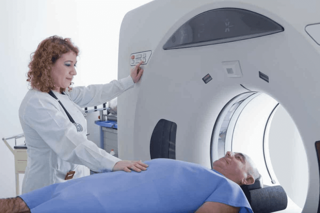
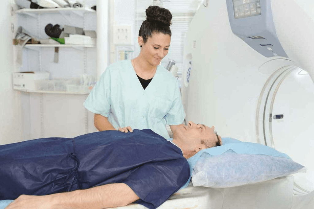
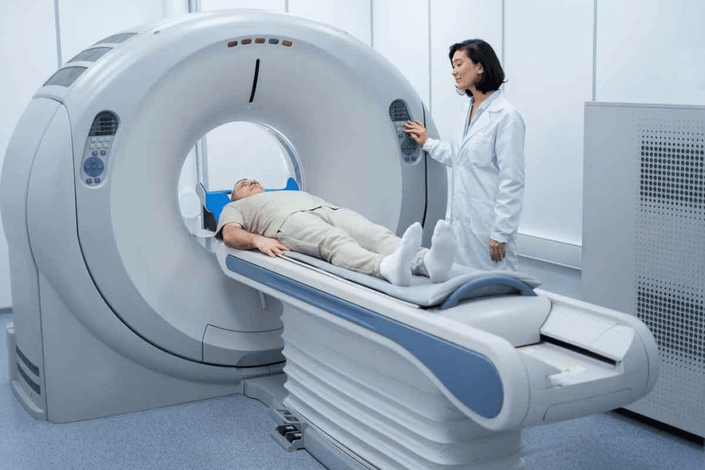
During the CT scan of the chest with contrast, you’ll lie on a motorized table that slides into the CT scanner. A trained radiologic technologist will perform the procedure.
The scan itself is quick, usually taking just a few minutes. You might be asked to hold your breath for short periods to get clear images.
After the CT chest with contrast scan, you can usually go back to your normal activities unless your healthcare provider says differently.
Post-scan care may include:
While complications from a CT scan thorax with contrast are rare, knowing the risks and taking precautions can make the procedure safer and more effective.
CT scans with contrast have changed how we see the thoracic cavity. They let us see the chest’s inner parts in detail. This is thanks to contrast enhancement.
Thoracic CT imaging is great for seeing the lungs and airways clearly. We can spot problems like nodules or bronchiectasis. Contrast agents help us tell different lung issues apart.
CT scans also help us look at the heart and the big blood vessels. We can see the heart’s chambers and the aorta. Contrast helps find issues like aneurysms.
The mediastinum, with its important structures, is well seen with CT scans. We can find problems in the lymph nodes, like swelling. This could be from infection or cancer.
Thoracic CT imaging also checks the chest wall and pleural space. We can see if there are effusions or thickening in the pleura. It also helps find masses on the chest wall.
CT scans of the chest with contrast give us a full view of these structures. They are key in diagnosing and treating chest problems.
Contrast in CT thorax scans makes it easier to spot lung problems. These scans are key to finding many lung diseases.
Lung cancer is a big worry, and CT scans help find it early. The contrast agent makes tumors and nodules stand out. This helps doctors tell if they are cancerous or not.
A study in the Journal of Thoracic Imaging found that contrast CT scans are better at spotting lung nodules.
| Nodule Characteristics | Benign | Malignant |
| Size | Typically small (<1 cm) | Variable, often larger |
| Margins | Smooth | Irregular or spiculated |
| Contrast Enhancement | Minimal | Significant |
CT scans with contrast are great for spotting pneumonia and other infections. They show how far the infection has spread. They also find complications like abscesses.
ILDs are a group of lung diseases. CT scans with contrast help see how bad the disease is. They check for inflammation or scarring.
COPD makes it hard to breathe and gets worse over time. CT scans help see how bad COPD is. They look at emphysema and other changes.
We use CT scans with contrast to check for lung diseases. This helps doctors make the right diagnosis and treatment plan.
Contrast in CT chest scans helps a lot in finding heart diseases. It makes the heart and blood vessels stand out. This lets doctors see more clearly.
CT chest with contrast is key for spotting pulmonary embolism (PE). PE is when a blood clot blocks a lung artery. The contrast makes it easier to see these clots. Early detection is key to saving lives.
It’s also great for finding aortic aneurysms and dissections. An aneurysm occurs when the aorta gets too big. A dissection is when the aorta tears. The contrast helps doctors measure the aorta and spot dissections.
CT chest with contrast also checks the coronary arteries. It looks for plaque buildup that can cause heart problems. The contrast makes it easier to see the arteries.
It also looks at the heart’s chambers. Doctors can see how big and how well the chambers are. They can also find problems like blood clots or tumors.
Thanks to CT chest with contrast, doctors can make better choices for patients. This might mean surgery, medicine, or more tests.
A CT scan thorax with contrast is key for checking pleural and mediastinal issues. It gives us detailed views of the thoracic area. This helps us spot and treat problems in the pleura and mediastinum.
This imaging method is great for spotting pleural effusions and thickening. It shows us how big and what kind of issues these are. Knowing this helps us figure out what’s causing them and how to treat them.
The mediastinum is a complex area with important structures. A CT scan thorax with contrast is vital for checking masses and abnormalities here. It helps us see the size, location, and type of masses, which is important for planning treatment.
Lymphadenopathy means enlarged lymph nodes, which can signal infections, inflammation, or cancer. A CT scan thorax with contrast lets us check the lymph nodes in the thorax. We can see their size, number, and how they react to contrast, which is key for diagnosing and treating diseases like lymphoma.
The esophagus and trachea are vital in the thorax, and problems here can affect health a lot. A CT scan thorax with contrast gives us clear images of these structures. This helps us diagnose issues like esophageal tumors, tracheal stenosis, and fistulas.
Dual-phase scanning in CT chest imaging is a big step forward in medical imaging. It takes pictures both with and without contrast. This gives a full view of the chest’s structure and any problems.
Dual-phase CT scanning is very useful in many situations. Patients with complex chest conditions need both types of images to get a clear diagnosis. For example, in suspected pulmonary embolism, a non-contrast scan can spot other causes, while a contrast scan shows the embolism.
Also, some lung nodules or tumors are clearer without contrast, but their position is better seen with it. This way, no important details are left out.
Dual-phase CT scanning shines in complex cases. It combines non-contrast and contrast images for a deeper understanding. For instance, in trauma, it can show bleeding (with contrast) and bone breaks (without contrast).
Many clinical scenarios benefit from dual-phase CT chest imaging. These include:
In conclusion, dual-phase CT chest imaging with and without contrast is very helpful. It gives a detailed look at the chest’s anatomy and problems. This helps doctors diagnose and treat many chest conditions better.
Advanced CT imaging techniques are changing how we diagnose and treat lung and chest problems. They combine new technology with medical skills. This improves patient care and makes clinical work easier.
Three-dimensional (3D) CT images give a detailed look at complex body parts. This is great for planning surgeries and seeing how different parts fit together. Virtual bronchoscopy lets doctors look inside the airways without surgery. It helps find problems in the airways.
CT imaging studies blood flow in the lungs and chest. This is key for spotting issues like blood clots and checking lung health. It also helps see how well lungs work and how severe lung diseases are.
CT-guided procedures are key for diagnosing and treating chest problems. They let doctors precisely target areas for biopsies or treatments. This reduces risks and makes procedures safer and more effective.
Artificial intelligence (AI) is changing radiology with CT scans. AI quickly goes through lots of data to help doctors spot issues. It’s making diagnoses faster and more accurate, improving patient care.
As we explore more with CT imaging, these advanced methods will be vital. They help us make better diagnoses and treatments. This leads to better patient outcomes.
CT scans of the thorax with contrast are very useful for diagnosing chest and lung issues. But they also have some limitations and risks. It’s important to weigh their benefits against possible drawbacks.
One big worry with CT scans is radiation exposure. These scans use X-rays to show detailed images of the body. This exposure can slightly increase the risk of cancer, mainly for younger people and those needing many scans.
To lower this risk, we follow the ALARA principle. This means we use the least amount of radiation needed for clear images. New CT technology helps by reducing doses and improving image quality.
| Radiation Dose Reduction Strategies | Description | Benefits |
| Low-Dose Protocols | Adjusting scanner settings to reduce dose | Significant dose reduction with maintained image quality |
| Iterative Reconstruction | Advanced image reconstruction techniques | Improved image quality at lower doses |
| Tube Current Modulation | Adjusting X-ray tube current based on patient size and anatomy | Optimized dose for different patient types |
Contrast agents make certain body areas more visible in CT scans. But they can cause reactions in some patients. These reactions can range from mild to severe.
Patients with severe kidney disease are at risk of kidney damage from contrast agents. We check each patient’s health and kidney function before using these agents to lower risks.
Choosing the right patients for CT scans is key to reducing risks. We look at each patient’s condition, medical history, and the scan’s benefits to decide if it’s the best choice.
For those at higher risk, we explore other imaging options or adjust our scans to reduce exposure. This might mean using non-contrast CT scans, MRI, ultrasound, or other strategies.
For patients at high risk from CT scans with contrast, there are other options. These include:
By carefully choosing the right imaging modality for each patient, we ensure safe and effective diagnosis.
Thoracic CT with contrast has changed chest imaging a lot. It gives us clear pictures of the lungs, heart, and nearby areas. This tool is key for spotting and figuring out many health issues, like lung cancer and heart problems.
Using a CT scan thorax with contrast lets doctors see the chest and lungs very well. The contrast makes things clearer, helping doctors make better diagnoses. This is really helpful when a CT chest with contrast is needed to see how far a disease has spread or to plan treatments.
The CT scan of the lungs with contrast is great because it shows everything in the chest area. Doctors can spot problems, track how diseases change, and plan the best treatments. As medical imaging gets better, thoracic CT with contrast will keep being a big help in caring for patients.
A CT scan thorax with contrast is a test that uses X-rays and a special dye. It shows detailed pictures of the chest and lungs. This helps doctors find and diagnose many health issues.
The contrast agent makes blood vessels, organs, and other parts stand out. This makes it easier to spot problems like lung cancer or heart disease.
Iodinated contrast is the most common dye used in CT scans. It’s given through an IV. Gadolinium-based agents are less common but uare sed in some cases.
You’ll lie on a table that slides into a CT scanner. A contrast agent will be given through an IV. You might need to hold your breath for a few seconds for clear images.
CT scans can expose you to radiation. You might also have an allergic reaction to the dye. People with kidney disease could face kidney damage.
If you have kidney disease, there’s a risk of kidney damage from the dye. Your doctor will weigh the risks and benefits. They might suggest other tests or take steps to protect your kidneys.
Dual-phase scanning takes images with and without contrast. It’s used for complex cases or when both blood vessels and other structures need to be checked.
CT imaging has many advanced uses. These include 3D models, virtual bronchoscopy, and studies of blood flow. They help doctors diagnose and treat many conditions.
Artificial intelligence can analyze CT images. It spots problems and provides detailed data. This makes diagnosis more accurate and efficient.
Yes, there are other tests like MRI, ultrasound, and PET scans. These might be suggested for those at risk of dye reactions or with other reasons to avoid CT scans.
A CT scan thorax with contrast shows detailed images of the chest and lungs. It helps find issues like lung cancer, blood clots, and heart problems.
It’s used to check for problems like fluid in the lungs, growths in the chest, and swollen lymph nodes. It helps diagnose conditions affecting the pleura and mediastinum.
It’s great for spotting heart and lung problems. It can find blood clots, aortic issues, and check the heart’s chambers and arteries.
Subscribe to our e-newsletter to stay informed about the latest innovations in the world of health and exclusive offers!