Dealing with back pain means knowing about the tools doctors use to diagnose. A CT scan is a special X-ray that shows the body’s inside clearly. It’s great for finding out what’s causing back pain.
At Liv Hospital, we aim for top-notch care and results. Our team is here to help you grasp your spinal health. We use tools like CT scans to tackle back pain.
Key Takeaways
- CT scans provide detailed images of the spine, helping diagnose the cause of back pain.
- Understanding CT scan results is key to planning treatment.
- Liv Hospital has advanced CT scan technology for accurate diagnoses.
- Our team focuses on patient care and support.
- CT scans are a key tool in modern medicine for spinal conditions.
What Is a CT Scan Back Examination and How Does It Work?
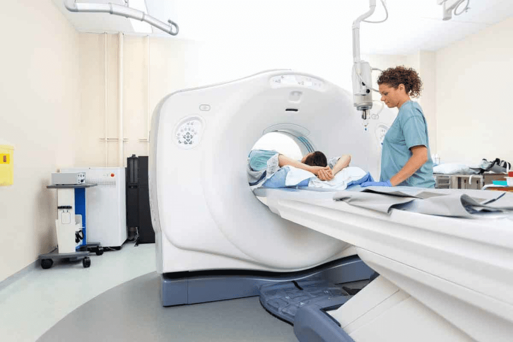
CT scans are key in modern medicine for checking the spine and back. They create detailed images of the body. This helps us find and diagnose back pain causes.
The Science Behind CT Imaging Technology
CT scans use X-rays to make detailed images. The scanner moves around the patient, taking X-ray shots from many angles. A computer then turns these shots into detailed images of the spine.
Cross-Sectional Imaging and 3D Reconstruction
CT scans are great for cross-sectional imaging of the spine. They let us see the spine from different angles and layers. This gives us a full view of the spinal anatomy.
Also, CT scans can make 3D reconstructions of the spine. These are very helpful for planning surgeries and checking complex spinal issues.
A study in Frontiers in Radiology shows 3D reconstructions in CT scans improve the diagnosis of spinal problems.
Difference Between CT Scans and Traditional X-rays
While X-rays are used, CT scans are better for diagnosing back pain. CT scans show more detail, like soft tissues and complex bones. They give a three-dimensional view of the spine, leading to more accurate diagnoses.
CT scans are also better at finding fractures, tumors, and degenerative diseases. These are often missed on X-rays. So, CT scans are a vital tool for diagnosing back pain.
Key Fact #1: When CT Scans Are Recommended for Back Pain
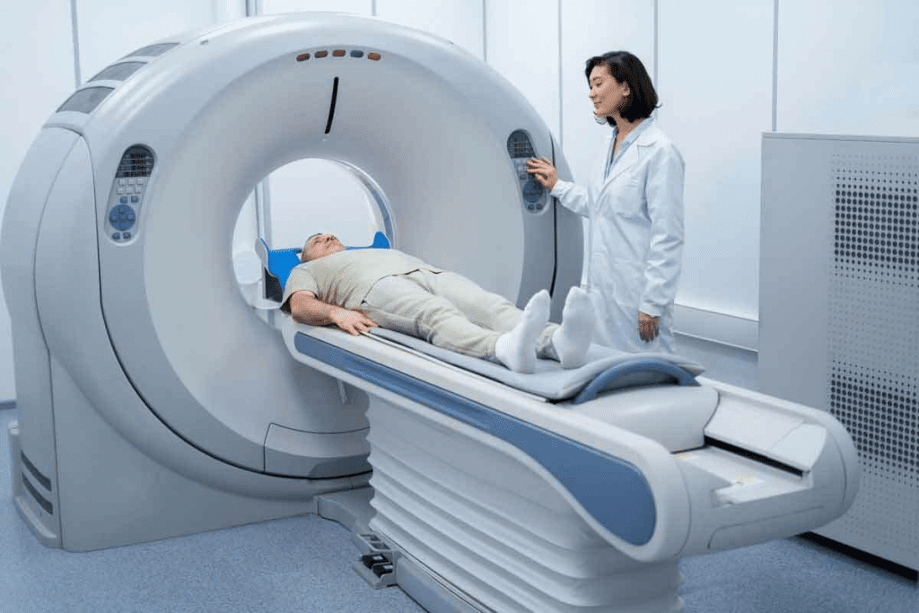
Back pain diagnosis often starts with simpler tests. But CT scans are key in certain cases. They are not the first choice, but are vital for detailed imaging to guide treatment.
Not a First-Line Diagnostic Tool
CT scans are not the first test for back pain. Doctors start with a detailed medical history and physical exam. If imaging is needed, X-rays or MRI scans are usually the first choice.
But, if these tests are unclear or more detailed imaging is needed, a CT scan might be suggested.
Specific Conditions That Warrant CT Imaging
There are specific conditions where a CT scan is very useful for diagnosing back pain:
- Severe injuries, such as those from a car accident or a fall
- Suspected tumors or infections in the spine
- Osteoporotic fractures, mainly in older patients
- Complex spinal deformities or scoliosis
- Post-surgical complications or hardware failure
In these cases, CT scans give detailed images of bone structures. This helps doctors accurately diagnose and plan treatment.
Emergency Situations Requiring Immediate Spine CT
In emergencies, an immediate CT scan of the spine is critical. These include:
- Trauma cases with suspected spinal fractures or dislocations
- Patients with neurological deficits, such as sudden numbness or weakness
- Severe back pain following a recent injury
In urgent cases, CT scans provide quick, high-resolution images. This helps emergency doctors make fast decisions about patient care.
Knowing when CT scans are recommended for back pain helps both patients and healthcare providers. It ensures this valuable tool is used effectively and safely.
Key Fact #2: Different Types of Back CT Scans and Their Applications
CT scans for back pain come in different types, each for a specific spinal area. This makes it easier for doctors to choose the right scan for each patient.
CT Thoracic Spine: Mid-Back Assessment
A CT thoracic spine scan looks at the middle back. It shows detailed images of the vertebrae, discs, and surrounding areas between the neck and lower back. This scan is great for finding issues like:
- Fractures or osteoporosis in the thoracic spine
- Tumors or infections affecting the mid-back
- Degenerative changes or scoliosis
Doctors can spot the cause of mid-back pain with this scan. Then, they can plan the best treatment.
Lumbar Spine CT Scan: Lower Back Evaluation
The lumbar spine CT scan checks the lower back. This area carries a lot of the body’s weight and stress. It’s key for spotting problems like:
- Herniated discs or spinal stenosis
- Spondylolisthesis or degenerative disc disease
- Fractures or osteoporosis in the lumbar vertebrae
This scan gives doctors clear images of the lower back. They can then find the cause of pain and suggest treatments.
Cervical Spine CT: Upper Back and Neck Imaging
A cervical spine CT scan looks at the neck and upper back. It shows detailed views of the vertebrae, discs, and soft tissues. It’s useful for finding issues like:
- Neck injuries or trauma
- Degenerative conditions such as cervical spondylosis
- Tumors or infections in the cervical spine
Doctors can use this scan to find neck pain. They can then guide the right treatment.
Full Spine CT: Comprehensive Evaluation
Sometimes, a full spine CT scan is used to check the whole spine. This scan gives a complete view of the spine, from top to bottom.
This scan is great for complex spinal issues. It helps plan surgeries for conditions like scoliosis or kyphosis.
Knowing about the different back CT scans helps both patients and doctors. They can make better choices about imaging and treatment.
Key Fact #3: Contrast vs. Non-Contrast CT Lumbar Spine Scans
CT lumbar spine scans can be done with or without contrast. Each method has its own use. The choice depends on the condition being checked and the patient’s health history.
CT Lumbar Spine Without Contrast: When It’s Preferred
Non-contrast CT scans are best for bone issues like fractures or bone spurs. They show bone structures clearly without contrast. They’re great for emergencies when a fast diagnosis is needed.
They’re also good for patients with kidney problems or allergies to contrast dyes. This reduces the risk of bad reactions and keeps patients safe.
Situations Requiring Contrast Enhancement
Contrast-enhanced CT scans are better for soft tissue problems like tumors or infections. The contrast makes these areas stand out. This is key for spotting issues not seen in non-contrast scans.
Contrast is also used for checking after surgery or for spine inflammation. It helps see different soft tissue structures and problems, leading to a better diagnosis.
In summary, whether to use contrast or not depends on the situation and what’s being looked for. Knowing the pros and cons of each helps ensure the best care for patients.
Key Fact #4: What Conditions a Spine CT Scan Can Accurately Detect
Spine CT scans are key in finding many spinal problems. They show the vertebrae, discs, and tissues in detail. This helps doctors spot different issues.
Fractures and Bone Abnormalities
Spine CT scans are great for finding fractures and bone problems. They are very useful in emergencies when quick checks are needed. They can spot tiny fractures that X-rays miss.
Tumors and Growths
CT scans can also find tumors and growths in the spine or nearby tissues. This info is key for cancer staging and treatment planning. The scans give doctors clear images of the tumors’ size and where they are.
Post-Surgical Complications
After spinal surgery, a CT scan can spot complications like hardware failure or bone fusion issues. This is important for checking surgery success and planning more treatment if needed.
Can CT Scans Show Herniated Discs?
CT scans can find herniated discs, but they’re not the first choice. Yet, a herniated disc CT scan can offer useful insights, like when an MRI isn’t an option. Whether a CT scan shows a herniated disc depends on its size and where it is.
In summary, spine CT scans are very useful. They can find many spinal issues, from fractures and tumors to problems after surgery and herniated discs.
Key Fact #5: CT Scan vs. MRI for Back Pain Diagnosis
It’s important to know the differences between CT scans and MRI for back pain diagnosis. Each has its own strengths and is better for different needs.
Strengths of Spinal CT Scans
CT scans are great at showing bone structures. They give clear pictures of bones, discs, and joints. This makes them perfect for finding fractures, bone spurs, and other bone issues. CT scans are also quicker than MRI, taking just a few minutes. This is good for patients who can’t stay long or are scared of tight spaces.
When MRI Is the Preferred Imaging Method
MRI is better for soft tissues like discs, nerves, and the spinal cord. It’s the top choice for diagnosing herniated discs, spinal stenosis, or nerve compression. MRI can also spot infections, tumors, and soft tissue problems around the spine.
Situations Where CT Is the Better Option
While MRI is great for soft tissues, CT scans are better for certain issues. For example, if a patient might have a bone injury, a CT scan is a better choice. CT scans are also used in emergencies when quick answers are needed.
Complementary Use of Both Imaging Techniques
Sometimes, both CT scans and MRI are used together for a full diagnosis. For example, a CT scan might check bones, while an MRI looks at soft tissues. This way, doctors get a detailed view of the patient’s health, even in complex cases.
The Complete Back Pain CT Scan Procedure: What to Expect
Understanding the CT scan process for back pain can reduce anxiety. We’ll walk you through everything, from getting ready to after the scan.
Preparation Before Your Spine CT Scan
Before your spine CT scan, there are a few steps to take:
- Tell your doctor about any allergies to contrast dye.
- Take off any metal items, like jewelry or glasses.
- Wear loose, comfy clothes.
- If you think you might be pregnant, tell your doctor.
Contrast dye might be used to make things clearer. If it is, you’ll get it through an IV. Our team will explain everything and answer your questions.
During the Procedure: Positioning and Scanning
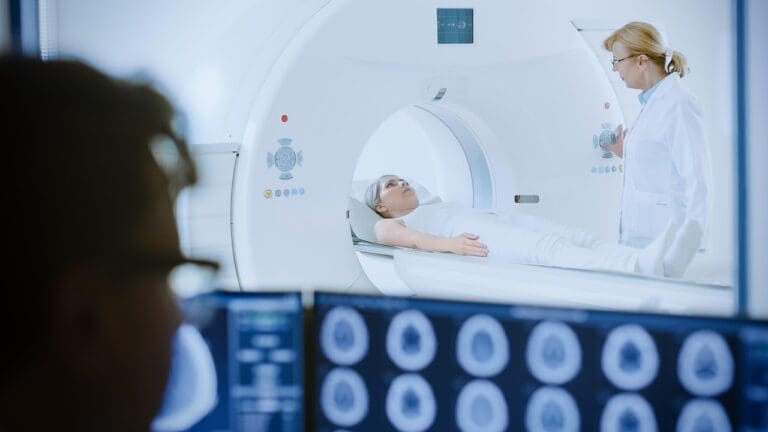
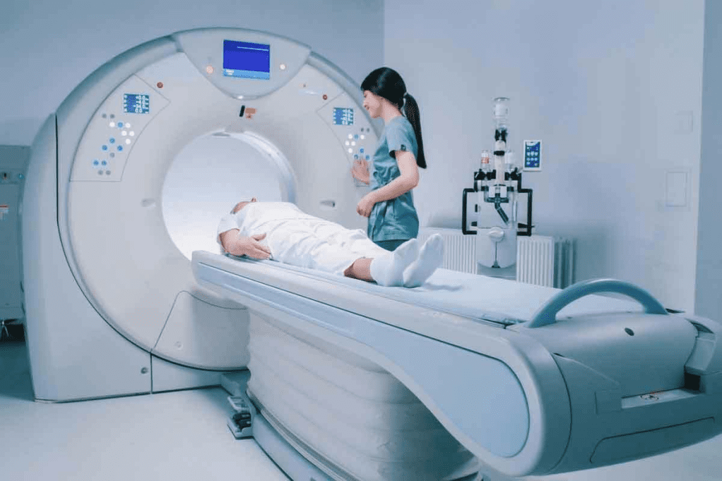
During the CT scan procedure, you’ll lie on a table that slides into a big machine. The technologist will help you get into the right spot and give you instructions through a speaker.
The scanning process includes:
- Getting into position and a quick scan at the start.
- The actual scan, which might ask you to hold your breath briefly.
- Maybe moving you for more scans.
The whole spine CT scan is fast, usually taking just a few minutes.
After the Scan: Results and Follow-up
After the scan, the images are checked by a radiologist. Your doctor will then talk to you about the findings and what to do next.
It’s important to follow up with your doctor. They’ll explain what your back pain CT scan results mean and any treatment plans.
Typical Duration and Comfort Considerations
The CT scan procedure is short, lasting 10 to 30 minutes. The scan itself is painless, but some might feel a bit uncomfortable. This could be from staying very quiet or the contrast dye.
Our facilities are made to keep you comfortable. We aim to reduce your anxiety and make your experience better.
Key Fact #6: Advanced Applications:
SPECT/CT for Precise Diagnosis
SPECT/CT technology has changed how we diagnose back pain. It combines SPECT’s functional info with CT’s detailed images. This gives us a full view of spinal issues.
How SPECT/CT Enhances Diagnostic Accuracy
SPECT/CT gives us both function and anatomy in one scan. This lets doctors pinpoint problems and match them to body parts. So, we can find the real cause of back pain and plan better treatments.
Together, SPECT and CT fill in each other’s gaps. SPECT might miss body details, while CT might miss functional issues. SPECT/CT gives a complete spine picture, leading to precise diagnosis and targeted treatment planning.
Identifying Active Degeneration and Pain Sources
SPECT/CT is great at finding where pain comes from in the spine. It shows where the body is working hard, helping us tell chronic problems from new ones.
This info is key when there are many issues. It helps us focus on what’s really hurting the patient. This way, we can plan treatments that are more likely to work, avoiding big surgeries.
Benefits for Targeted Treatment Planning
SPECT/CT gives us detailed information for targeted treatment planning. It helps us pinpoint where to focus treatments like injections or surgery. This makes treatments more effective and personal.
It also lets us check how treatments are working. This is super helpful for long-term conditions, where we need to keep adjusting plans.
Recent Research and Clinical Outcomes
New studies show SPECT/CT is really useful for spinal problems. It helps doctors make better decisions and can lead to better patient results.
As SPECT/CT gets better, we’ll see even more accurate diagnoses and treatments. By keeping up with these advances, we can give our patients the best care and improve their lives.
Key Fact #7: Radiation Exposure and Safety Considerations
It’s important to know the risks and benefits of CT scans. They help us understand back pain, but also expose us to radiation. We need to think about how to reduce these risks.
Understanding Radiation Doses in Spinal CT
CT scans use X-rays to see the spine clearly. The radiation dose depends on the scanner, the scan type, and the patient’s size. New CT scanners aim to use less radiation while keeping images sharp.
Typical Effective Doses for Spinal CT Scans:
| Spine Region | Typical Effective Dose (mSv) |
| Cervical Spine | 2-3 |
| Thoracic Spine | 4-6 |
| Lumbar Spine | 5-8 |
Risk vs. Benefit Assessment
Doctors consider the benefits and risks of CT scans. For many, the scan’s benefits are worth the radiation risk. This is true when it helps plan better treatment.
The risk of radiation-induced cancer is a primary concern. But this risk is usually low. It’s less than the benefits of accurate diagnosis and treatment.
Minimizing Exposure During CT of the Back
To cut down radiation, several steps are taken:
- Using the lowest dose needed for good images.
- Using new CT scanner technologies to reduce dose.
- Choosing the right scan range to cover only the needed areas.
- Shielding organs outside the scan area when it’s possible.
Special Considerations for Different Patient Groups
Some groups need extra care with radiation:
- Pediatric Patients: Kids are more sensitive. Lower doses are used.
- Pregnant Women: The risks to the fetus are carefully weighed.
- Young Adults: The long-term risks of radiation are considered for younger patients.
By understanding these points and taking steps to reduce exposure, doctors can use CT scans safely. This helps in diagnosing and treating back pain effectively.
Insurance Coverage and Cost Factors for Spine CT Scans
Insurance and cost are key when deciding on a spine CT scan. Knowing these can help patients manage their healthcare costs better.
Typical Costs of Different Back CT Scans
The price of a spine CT scan varies a lot. It depends on the scan type, where it’s done, and where you live. Without insurance, a spine CT scan can cost between $500 and $1,500 or more. For example, a CT scan of the lumbar spine might cost differently from a full spine scan.
Cost factors include:
- The specific type of CT scan required (e.g., cervical, thoracic, or lumbar spine)
- The use of contrast material
- The healthcare provider’s fees
- Facility fees (hospital vs. outpatient imaging center)
- Geographic location
Questions to Ask Your Provider
To understand costs, patients should ask their healthcare provider or the imaging facility these questions:
- What is the total cost of the CT scan?
- Does the cost include the radiologist’s interpretation?
- Are there additional fees for contrast material?
- What are the payment options available?
- Does the facility offer any financial assistance programs?
Appealing Insurance Denials
If insurance denies a spine CT scan claim, patients can appeal. Steps to appeal include:
- Review the denial reason with your insurance provider
- Gathering additional information or documentation from your healthcare provider to support the necessity of the CT scan
- Submitting a formal appeal to the insurance company
- Following up on the appeal status
Patients need to be proactive in understanding their insurance coverage and the costs associated with diagnostic imaging, like spine CT scans.
Limitations of CT Scan Vertebrae Imaging
It’s important to know the limits of CT scans for vertebrae imaging. They give valuable bone structure info, but have some drawbacks. Healthcare pros need to keep these in mind.
Soft Tissue Visualization Challenges
CT scans struggle to see soft tissues well. This is unlike MRI, which is better at spotting soft tissue issues. This can make it hard to find problems with muscles, ligaments, and discs.
For example, a CT scan might miss a herniated disc or nerve pressure. This could lead to more tests. To get a full picture, doctors might use CT scans and MRIs togetherCarelon Medical Benefits Management says combining scans can help get a clearer diagnosis.
When Additional Testing May Be Needed
CT scans might not always give a clear diagnosis. This is why more tests like MRI or PET scans might be needed. The choice for more tests depends on the patient’s symptoms and medical history.
Interpreting Incidental Findings
CT scans often find things not related to the main reason for the scan. These findings can add to the challenge of making a diagnosis. Doctors must carefully look at these findings to avoid extra tests or worry.
Emerging Technologies Addressing Current Limitations
New technologies are improving CT scans for vertebral imaging. Better CT scanners and AI in image analysis are helping spot problems better. This means CT scans will soon be even more useful for spine health.
As these new tools get better, doctors will be able to give more accurate diagnoses. This will help patients get the best care possible.
Conclusion: Making Informed Decisions About CT Scans for Back Pain
Understanding CT scans’ role in diagnosing back pain is key. We’ve looked at when they’re used, the types of scans, and their uses in medical conditions. This helps in making smart choices about their use.
Deciding on CT scans for back pain diagnosis means weighing their pros and cons. Knowing how they work and what they can find helps patients. This knowledge lets them ask the right questions and work with doctors to find the best diagnostic path.
CT scans are vital in modern medicine, mainly for back pain diagnosis. They help doctors give accurate diagnoses and treatment plans. This teamwork is vital for the best back pain management results.
FAQ
What is a CT scan, and how does it work?
A CT scan is a medical test that uses X-rays and computers to show body parts inside. It works by moving an X-ray machine around you. Then, a computer makes images from the data it gets.
What is the difference between a CT scan and an X-ray?
CT scans and X-rays both use X-rays. But CT scans give detailed, cross-sectional images. X-rays show two-dimensional pictures.
When are CT scans recommended for back pain?
Doctors suggest CT scans for back pain if it’s severe or if other tests didn’t help. They’re good for checking injuries, infections, or tumors.
What types of back CT scans are available?
There are many back CT scans, like the CT thoracic spine and lumbar spine CT scan. Each type looks at different parts of the spine.
What is the difference between a CT lumbar spine scan with and without contrast?
A CT scan without contrast shows bones and some soft tissues. In contrast, it highlights blood vessels or tumors, helping diagnose conditions.
Can a CT scan show a herniated disc?
Yes, CT scans can spot herniated discs. They’re good for large or pressing discs.
How does a CT scan compare to an MRI for back pain diagnosis?
CT scans are better for bones and fractures. MRI is better for soft tissues like discs and nerves. The choice depends on the cause of pain.
What should I expect during a CT scan for back pain?
During a CT scan, you’ll lie on a table that moves into a scanner. You might need to stay very quiet and hold your breath. The scan is fast, and you can go back to normal activities soon.
Are there any risks associated with CT scans?
CT scans use radiation, which is a risk. But the benefits often outweigh the risks. Steps are taken to reduce radiation, mainly for sensitive groups.
How much does a spine CT scan cost?
The cost of a spine CT scan varies. It depends on where you are, the facility, and whether contrast is used. Check with your healthcare provider or insurance for costs.
Will my insurance cover a CT scan for back pain?
Insurance coverage for CT scans varies. Many plans cover them when they’re medically necessary. It’s important to check with your insurance and understand any costs you might have.
What are the limitations of CT scan vertebrae imaging?
CT scans are great for bones, but not as good as MRI for soft tissues. They might find things that need more checking.
Reference
- Villagrán, J. M., et al. (2023). Diagnostic accuracy of lumbar CT and MRI in the evaluation of chronic low back pain. European Journal of Radiology, 162, 110847. https://pubmed.ncbi.nlm.nih.gov/37858354/
–


