Last Updated on November 27, 2025 by Bilal Hasdemir

What is the difference between mri and x ray? Medical imaging is key in diagnosing many health issues, and understanding what is the difference between MRI and X-ray is important for everyone to make informed health decisions.
At Liv Hospital, we focus on your health and the latest medical care. We use state-of-the-art imaging technologies for accurate and effective treatment. Two common imaging methods are magnetic resonance imaging (MRI) and X-ray scans.
Both scans show detailed images of the inside of the body, but they work in different ways. Knowing how they differ helps get the right diagnosis and treatment.
Key Takeaways
- Medical imaging is key to diagnosing many health issues.
- MRI and X-ray are two common imaging methods.
- Understanding the difference between MRI and X-ray is essential for accurate diagnosis.
- Liv Hospital uses advanced medical imaging technologies for precise care.
- State-of-the-art imaging helps provide effective treatment plans.
The Role of Medical Imaging in Modern Healthcare

Diagnostic imaging is key in today’s medicine. It helps us see inside the body, leading to better health checks and treatments.
Evolution of Diagnostic Imaging Technologies
Medical imaging has changed a lot, starting with Wilhelm Conrad Röntgen’s X-rays in 1895. Now, we have CT scans, MRI, and more. These tools give us clearer images faster and make scans more comfortable for patients.
New tech in physics, computer science, and engineering has driven these changes. For example, digital X-ray detectors have replaced old film systems. This makes getting and processing images quicker.
“The development of new imaging technologies has revolutionized the field of medicine, enabling healthcare professionals to diagnose and treat diseases more effectively.”
A Senior Radiologist
Impact on Patient Diagnosis and Treatment
Medical imaging is very important for finding and treating health problems. Tools like MRI and X-ray show what’s inside the body. This helps doctors spot issues like bone breaks and soft tissue injuries.
| Imaging Modality | Diagnostic Capability | Treatment Planning |
| X-ray | Excellent for bone fractures and lung conditions | Limited soft tissue detail |
| MRI | Superior soft tissue contrast and detail | Essential for neurological and musculoskeletal assessments |
What we learn from imaging helps decide how to treat patients. Knowing what each imaging tool can do helps doctors plan better treatments.
In short, medical imaging is vital in today’s healthcare. It helps us make better diagnoses and treatments. As tech keeps improving, we’ll see even better care for everyone.
What Is the Difference Between an MRI and an X-Ray?

It’s important to know how MRI and X-ray differ. This knowledge helps doctors and patients understand their options. We’ll cover the main differences between these two imaging methods.
Fundamental Technology Differences
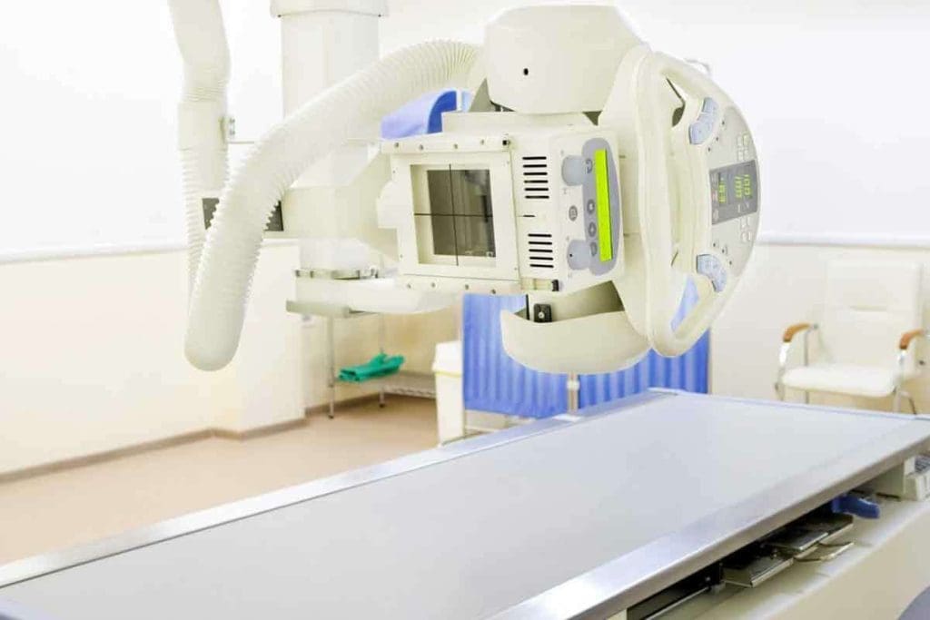
X-rays and MRIs work in different ways. X-rays use electromagnetic waves to see inside the body, focusing on bones. MRIs, on the other hand, use magnets and radio waves to show both bones and soft tissues.
Comparing Image Production Methods
X-rays create images by how much radiation is absorbed by tissues. Bone absorbs more, showing up white, while softer tissues appear gray or black.
MRIs, though, show soft tissues in detail. They do this by detecting signals from hydrogen atoms in the body. This is something X-rays can’t do.
| Characteristics | X-Ray | MRI |
| Primary Use | Bone imaging, detecting fractures and lung conditions | Soft tissue imaging, neurological , and musculoskeletal assessments |
| Technology | Electromagnetic radiation | Powerful magnets and radio waves |
| Image Detail | Excellent for bone structures, limited soft tissue detail | Excellent for soft tissues, detailed neurological and musculoskeletal imaging |
Radiation vs. Magnetic Fields
One big difference is radiation. X-rays use ionizing radiation, which is a worry for some, like kids and pregnant women. MRIs, though, use magnetic fields and radio waves without ionizing radiation. This makes MRIs safer for some patients.
We’ll explore more about these differences later. But for now, choosing between MRI and X-ray depends on what’s needed for diagnosis and the patient’s situation.
How X-Ray Technology Works
X-ray technology uses the fact that different tissues absorb radiation at different rates. This allows X-rays to show detailed images of what’s inside the body.
The Physics of X-Ray Radiation
X-ray radiation is a type of electromagnetic wave with a shorter wavelength than visible light. It can pass through soft tissues but gets blocked by denser materials like bone. When an X-ray beam hits the body, different tissues absorb it at different rates. This creates a contrast that shows up on the X-ray image.
Key factors influencing X-ray absorption include:
- The density of the material
- The atomic number of the material
- The energy level of the X-ray beam
Image Formation and Density Differentiation
How X-rays form images is based on how different tissues absorb them. Denser materials, like bone, block more X-rays and show up white. Softer tissues block less and appear gray or black. This helps us see inside the body.
The quality of the X-ray image depends on several factors, including:
- The quality of the X-ray beam
- The sensitivity of the detector
- The patient’s positioning
Types of X-Ray Procedures
There are many types of X-ray procedures, each for different body parts. Some common ones are:
- Chest X-rays for lung conditions
- Orthopedic X-rays for bone fractures
- Dental X-rays for tooth and jaw assessments
Knowing about these different uses shows how useful X-ray technology is in medical care.
How MRI Technology Works
Magnetic Resonance Imaging (MRI) uses strong magnetic fields and radio waves. It creates detailed images of the body’s inside without surgery. This technology has changed how doctors see inside the body.
Magnetic Fields and Radio Frequency Pulses
At the heart of MRI is the magnetic field and radio waves. The MRI machine makes a strong magnetic field. This field lines up the hydrogen atoms in the body.
Hydrogen is everywhere in the body, mostly in water. When the magnetic field hits, these atoms line up. Then, radio waves disturb them, making them send signals.
These signals are caught by the MRI machine. It uses them to make detailed pictures of the body’s inside.
Hydrogen Atom Alignment and Signal Generation
Aligning hydrogen atoms is key to MRI. When radio waves hit, the atoms move. As they go back to their place, they send out signals.
The strength and how long these signals last tell MRI what tissue it’s looking at. This lets MRI see soft tissues like organs and tendons. X-rays can’t do this as well.
Computer Processing and Image Creation
The MRI machine catches these signals and sends them to computers. The computers make detailed images from this data. They use the signal strength and how fast the atoms relax to create clear images.
Today’s MRI machines can show images in many ways. They help doctors see the body’s inside in detail. This is great for diagnosing many health issues, from brain problems to muscle injuries.
| Aspect | X-Ray | MRI |
| Primary Use | Bone fractures, lung conditions | Soft tissue injuries, neurological disorders |
| Imaging Principle | X-Ray absorption | Magnetic resonance |
| Radiation Exposure | Yes | No |
What X-Rays Can and Cannot Visualize
X-rays are great at showing what’s inside our bodies, but they have their limits. They work best for bones and some lung issues. This makes them a key tool in many medical situations.
Excellent Visualization of Bone Structures
X-rays are top-notch at showing bones. They help find fractures, check bone health, and spot osteoporosis. The difference between bones and soft tissue makes bones stand out clearly.
In emergencies, X-rays are often the first choice. They’re quick and easy to get, making them essential in urgent care.
Detection of Certain Lung Conditions
X-rays also do well with lung problems like pneumonia and tumors. The lungs’ airiness contrasts with soft tissue, making it easier to spot issues.
“Chest X-rays remain a key tool for lung health checks. They’re fast and don’t hurt.” – A Senior Radiologist
Limitations in Soft Tissue Imaging
But X-rays struggle with soft tissues like muscles and organs. These are hard to see because they’re similar in density. This makes X-rays less good for soft tissue injuries or diseases.
| Imaging Modality | Bone Visualization | Lung Condition Detection | Soft Tissue Imaging |
| X-Ray | Excellent | Good | Limited |
| MRI | Good | Limited | Excellent |
In short, X-rays are great for bones and some lung issues. But they can’t see soft tissues well. This is why we need other tools like MRI for a full picture.
What MRIs Can and Cannot Visualize
Magnetic Resonance Imaging (MRI) has changed how we diagnose diseases. It shows soft tissues in great detail. This makes MRI key in healthcare for diagnosing and tracking many conditions.
Superior Soft Tissue Contrast
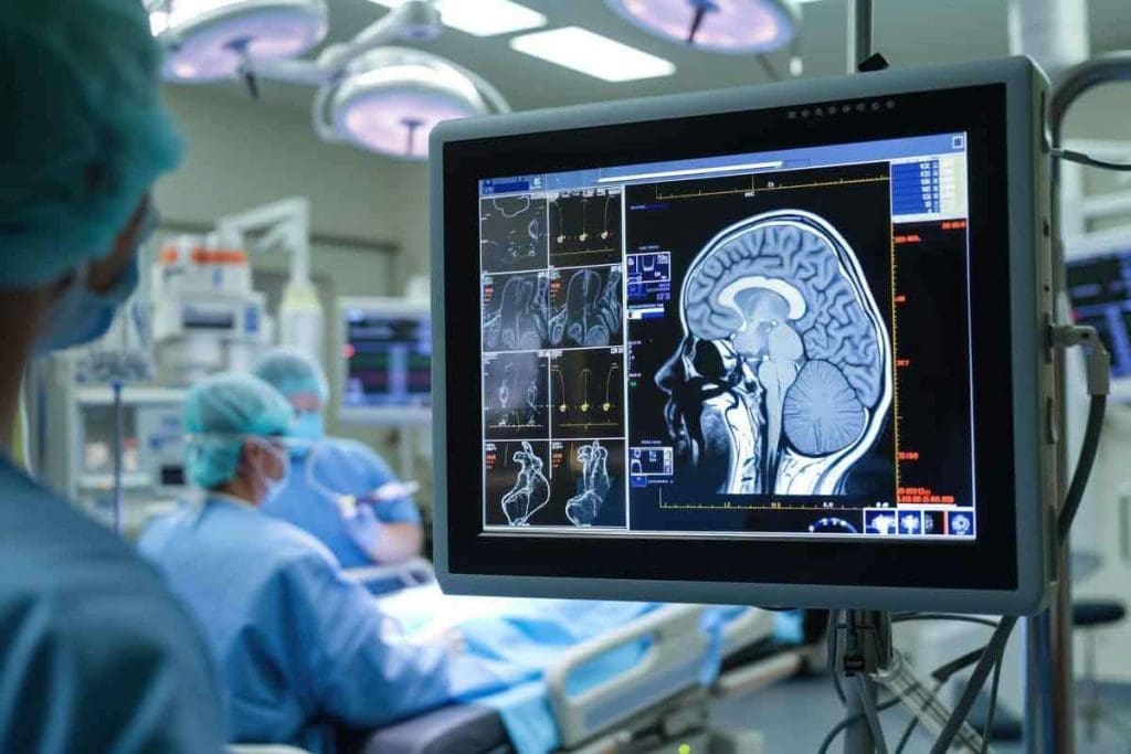
MRI stands out because it shows soft tissues better than X-rays. X-rays are good for bones, but MRI is better for muscles, tendons, and ligaments. This helps doctors spot problems in these areas.
For example, MRI can spot tendon and ligament injuries around joints. It gives clear images for treatment plans. MRI also finds tumors and checks how big they are.
Detailed Visualization of Neural Structures
MRIs are great at showing the brain and spinal cord. They are key to diagnosing and tracking neurological issues. This includes conditions like multiple sclerosis and spinal cord injuries.
These detailed images help doctors find damage or disease in the brain and spinal cord. They help plan treatments. Functional MRI (fMRI) also looks at brain activity, helping with neurological disorder diagnosis and study.
Key benefits of MRI in neural structure visualization include:
- High-resolution imaging of the brain and spinal cord
- Detection of lesions and abnormalities
- Assessment of neurological conditions
Joint and Cartilage Assessment
MRI is also great for checking joint health and cartilage. It shows the cartilage, synovium, and soft tissues around joints. This is key for diagnosing and tracking osteoarthritis.
MRI also checks how bad joint injuries are. It helps plan treatments and rehabilitation. It’s a top choice for many musculoskeletal issues because it doesn’t use harmful radiation.
Important aspects of MRI in joint assessment include:
- Detailed visualization of cartilage and joint spaces
- Evaluation of soft tissue injuries around joints
- Monitoring of degenerative joint diseases
Even though MRI is very useful, it has some limits. It’s not safe for people with metal implants or pacemakers. Some might feel claustrophobic during the test. Also, contrast dye can cause allergic reactions.
Knowing what MRI can and can’t do is important. It helps doctors make better diagnoses and treatment plans. This improves patient care and outcomes.
Clinical Applications: When to Use X-Rays
X-ray technology has many uses in healthcare. It helps find bone fractures and lung problems. X-rays give doctors a clear look at what’s inside our bodies.
Fracture Detection and Bone Disease
X-rays are key spotting bone fractures and diseases. They show bone details well, making them a first choice for fracture checks. They also help find osteoporosis, a condition that weakens bones.
Doctors use X-rays to see if a bone is healing riproperlyThey show bone density and structure clearly. This makes X-rays very useful in orthopedic care.
Pneumonia and Lung Disorders
X-rays are also vital for lung issues like pneumonia and tumors. They let doctors see lung problems. This is very important in emergencies when a quick diagnosis is needed.
For pneumonia, X-rays show lung inflammation. This helps doctors decide how to treat it. While X-rays can’t solve all lung problems, they’re a good start for further tests.
Foreign Object Detection
X-rays are also great for finding objects inside the body. This is helpful for accidental ingestions or inserted objects. They quickly find where these objects are, helping doctors remove them safely.
| Application | Description | Benefits |
| Fracture Detection | X-rays visualize bone structures to detect fractures. | Quick and effective for assessing bone injuries. |
| Lung Disorders | X-rays help diagnose conditions like pneumonia and COPD. | Provides initial assessment for lung abnormalities. |
| Foreign Object Detection | X-rays identify ingested or inserted foreign objects. | Aids in the safe removal of foreign objects. |
| Dental Diagnostics | X-rays visualize dental structures to detect problems. | Essential for diagnosing issues not visible to the naked eye. |
Dental Diagnostics
In dentistry, X-rays are very important. They help find cavities, tooth decay, and other dental issues. Dental X-rays give a detailed look at teeth and bones, helping catch problems early.
Dentists use X-rays to find problems not seen during regular checks. This helps prevent bigger issues and avoid more complex treatments.
Clinical Applications: When to Use MRIs
MRI technology helps us see complex body structures clearly. This has greatly improved patient care. It’s a key tool in today’s healthcare for detailed body checks.
Brain and Spinal Cord Assessment
MRI is great for checking the brain and spinal cord. It shows soft tissues in high detail. This helps find tumors, infections, and diseases like multiple sclerosis.
Sports Injuries and Joint Problems
In sports medicine, MRI checks for injuries in ligaments, tendons, and cartilage. It’s essential for spotting issues like torn ACLs and meniscal tears.
Organ and Tissue Abnormalities
MRI also looks at organs and soft tissues all over the body. It spots problems like liver disease and heart issues, and checks the stomach area too.
Cancer Detection and Staging
In cancer care, MRI is key to finding and checking cancer types. It shows how far tumors have spread and if treatments are working.
| Clinical Application | MRI Capability |
| Brain and Spinal Cord | High-resolution imaging of soft tissues, ideal for diagnosing tumors, infections, and degenerative diseases. |
| Sports Injuries | Detailed evaluation of ligament, tendon, and cartilage injuries. |
| Organ Abnormalities | Assessment of liver disease, cardiovascular conditions, and abdominal organ abnormalities. |
| Cancer Detection | Detection and staging of various cancers, evaluation of tumor spread, and treatment assessment. |
Patient Experience and Preparation
Diagnostic imaging, like X-rays and MR, needs special preparation. This ensures accurate results and a better experience for patients. Knowing what to expect can help reduce anxiety and make patients more cooperative.
What to Expect During an X-Ray
Patients stand in front of an X-ray machine for this test. It’s quick, lasting just a few minutes. The X-ray technologist will guide you and help you get into the right position.
Preparation for an X-ray is simple. You might need to take off jewelry or clothes that could mess up the image. For some tests, like those of the stomach, you might need to drink a special liquid or wear a certain gown.
What to Expect During an MRI
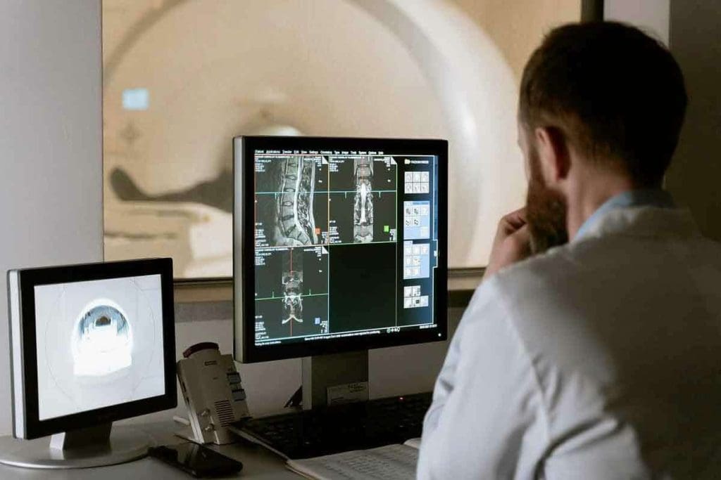
MRI scans involve lying on a table that moves into a big, round magnet. The machine makes loud noises during the scan. We give out earplugs or headphones to make it less uncomfortable.
Preparation for an MRI means removing all metal. This includes jewelry, glasses, and clothes with metal parts. We also check for metal implants, like pacemakers, as they can’t be scanned.
Clothing and Accessory Requirements
For both X-rays and MRIs, you’ll need to change into a hospital gown. This is to make sure your clothes don’t get in the way of the images. We also ask you to remove any accessories that could affect the quality of the images or be dangerous.
Knowing what to expect beforehand makes the process smoother. It helps patients feel more at ease and prepared for their diagnostic imaging tests.
Safety Considerations and Contraindications
Knowing about X-rays and MRIs is key to making smart health choices. Each has its own safety rules that doctors must think about before tests.
X-Ray Radiation Exposure Risks
X-rays use radiation that can raise cancer risk, more so in kids. The danger grows with the dose. It’s important to think hard about the need for X-rays, like for pregnant women or young kids.
Reducing radiation is a big goal. This means using the least amount of radiation needed for clear images. New X-ray tech has made it safer.
MRI Safety Concerns
MRIs don’t use harmful radiation, making them safer. But they have risks too. The strong magnetic field can harm metal implants or fragments in the body.
Some people, like those with pacemakers, might not be able to have an MRI. Claustrophobia is also a worry, as the MRI machine’s tight space can scare some.
Pregnancy Considerations for Both Modalities
Pregnancy makes choosing between X-rays and MRIs tricky. X-rays have radiation, which is a no-go for pregnant women. But MRIs are safer in this area. Yet, MRI contrast agents are used with caution during pregnancy.
Doctors must decide carefully for pregnant patients. They look at how urgent the test is and weigh the risks and benefits. Ultrasound is often the first choice when it can be used.
Understanding these safety points helps doctors make choices that protect patients while getting the needed information.
Accessibility, Cost, and Insurance Factors
It’s important to know the cost, insurance, and availability differences between MRI and X-ray. These factors help patients choose the right imaging test.
Comparing Procedure Costs
MRIs are usually more expensive than X-rays. They cost between $800 and $2,500 or more. This depends on the body part scanned and whether contrast dye is used.
X-rays, on the other hand, are cheaper. They cost between $100 and $500. This makes X-rays a more affordable first choice for diagnosis.
Insurance Coverage Differences
Insurance for MRI and X-ray varies. Most plans cover X-rays with little cost to patients. But insurance for MRIs can be tricky. It might need pre-approval or have higher copays because of its cost.
- Check if your insurance covers the MRI or X-ray your doctor recommends.
- Know the costs, including deductibles and copays, for both tests.
- See if MRI procedures need pre-authorization.
Availability and Wait Times
X-rays are easier to find and have shorter wait times. MRIs are less common and may have longer waits. This is because MRIs are more expensive and complex.
- X-rays are widely available and have quick wait times.
- MRIs have longer waits because they’re less common.
- Some centers offer MRI services quickly but at a higher cost.
In summary, MRI and X-ray are both key for diagnosis. But their costs, insurance, and availability are different. Knowing these details helps patients make better healthcare choices.
Conclusion
Knowing the difference between MRI and X-ray imaging is key. It helps choose the right tool for different medical issues. We’ve looked at how these two imaging methods work, what they’re used for, and their limits.
Whether to use an MRI or an X-ray depends on the health issue. X-rays are great for seeing bones and some lung problems. MRIs, on the other hand, show soft tissues, like the brain and joints, in more detail.
When comparing MRI and X-ray, it’s important to know their strengths and weaknesses. MRIs are best for checking the brain, spinal cord, sports injuries, and joint issues. X-rays are good for finding bone fractures, pneumonia, and foreign objects in the body.
In summary, the choice between MRI and X-ray is critical for accurate diagnosis and treatment. Understanding the differences helps healthcare professionals make better decisions. This leads to better care for patients.
FAQ
What’s the difference between an MRI and an X-ray?
MRI and X-ray are two different ways to see inside the body. X-rays use radiation to show bones. MRIs use magnetic fields and radio waves to show soft tissues.
Does an MRI use X-rays?
No, MRI doesn’t use X-rays. It uses a strong magnetic field and radio waves to create images.
What does an MRI show that an X-ray does not?
MRI shows soft tissues like organs and tendons. It also finds cancer and checks the brain and spinal cord. X-rays are better for bones and lungs.
Is an MRI an X-ray?
No, MRI and X-ray are different. They work on different principles and are used for different reasons.
What is the difference between an MRI and Xan -ray in terms of radiation exposure?
X-rays use ionizing radiation. MRIs don’t. MRIs are safer, which is why they’re often chosen for pregnant women.
How are MRIs different from X-rays in terms of image production?
MRIs use hydrogen atoms in the body to make images. X-rays use the density of tissues to make images.
What are the clinical applications where MRI is preferred over X-ray?
MRI is better for soft tissue injuries, cancer, and brain and spinal cord issues. It’s great for detailed soft tissue images.
When is X-ray preferred over MRI?
X-rays ares better for bone fractures, lung problems, and dental issues. It’s also used for finding foreign objects and guiding procedures.
Are there any safety concerns specific to MRI or X-ray?
Yes, X-rays have radiation risks. MRI has a strong magnetic field and risks and can cause claustrophobia.
How do the costs and insurance coverage compare between MRI and X-ray?
MRI is more expensive because of its technology. Insurance varies, but both are covered when needed.
What should patients expect during an MRI or X-ray procedure?
For X-rays, remove metal and get in different positions. For MRIs, remove metal and wear a special gown. Stay very quiet during the scan.
References:
- Xiong, C. (2021). An analysis of clinical values of MRI, CT and X-ray in differential diagnosis of benign and malignant bone metastases. Medical Science Monitor, 27, e930180. https://www.ncbi.nlm.nih.gov/pmc/articles/PMC8290716/


