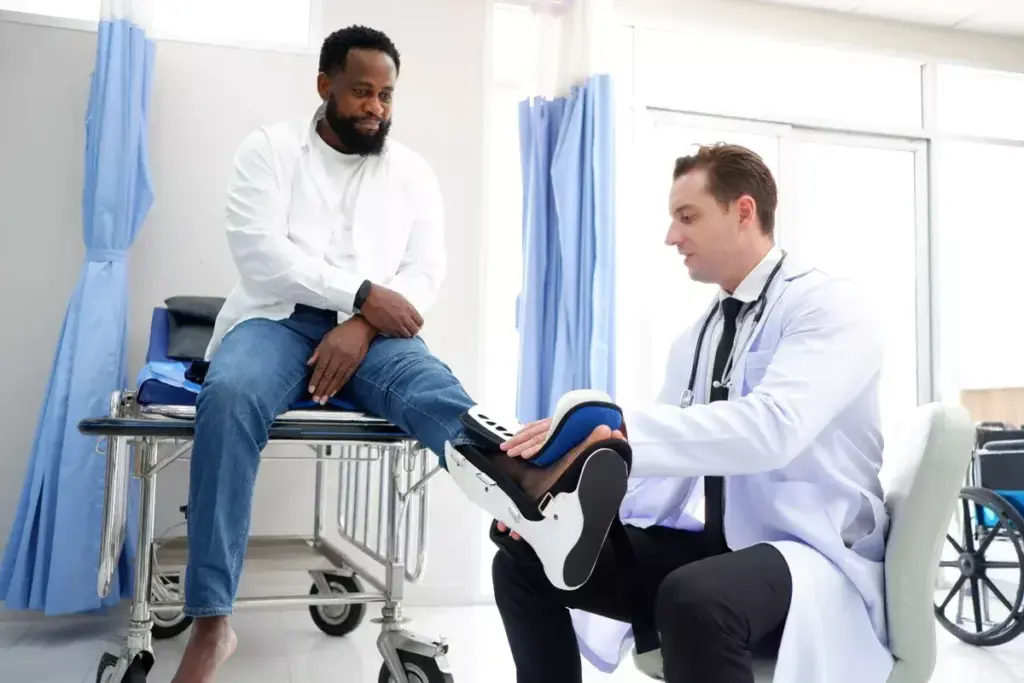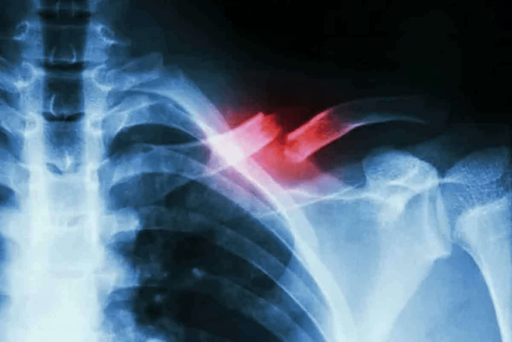
Accurate diagnosis of musculoskeletal injuries is key for good treatment. When it comes to finding broken bones, we use CT scans, X-rays, and MRIs. Each tool has its own strengths and is best for different injuries.
Liv Hospital shows us the key differences between these imaging methods. CT scans give detailed views of inside structures, like bones, soft tissues, and blood vessels. They’re great for complex bone injuries and severe trauma.
Key Takeaways
- CT scans offer high-resolution images and 3D reconstructions, valuable for complex fractures.
- X-rays are typically used for initial assessments of bone injuries.
- MRIs are ideal for soft tissue injuries, including ligament and tendon tears.
- Choosing the right imaging modality depends on the nature of the injury.
- Accurate diagnosis is critical for effective treatment planning.
The Critical Role of Diagnostic Imaging in Bone Injuries
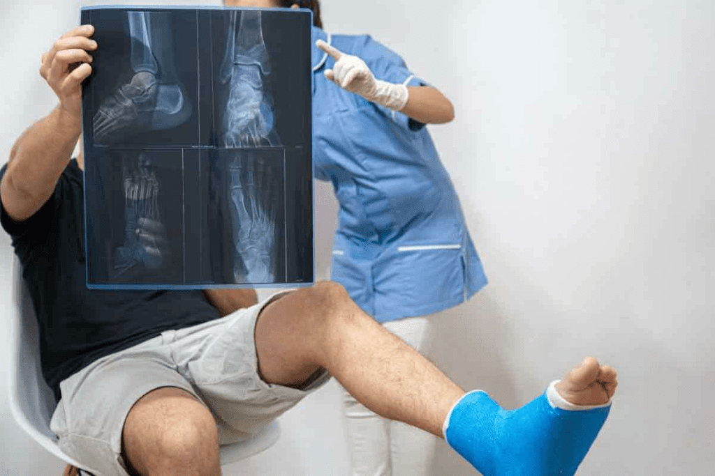
Diagnostic imaging is key in handling bone injuries. It helps doctors figure out the extent of the damage. This information guides how to treat the injury.
Modern Approaches to Fracture Detection
Finding fractures has gotten better with new imaging tech. X-rays are often the first choice because they’re easy to get. But CT scans show more detail with 3D images. This makes diagnosis more accurate.
A study in Signa Vitae shows how better imaging helps find fractures more accurately.
| Imaging Modality | Fracture Detection Rate | Advantages |
| X-ray | Initial assessment | Quick, widely available |
| CT Scan | 88-98% | High-resolution 3D images |
| MRI | Soft tissue evaluation | Excellent soft tissue detail |
Impact of Accurate Diagnosis on Treatment Outcomes
Getting the diagnosis right is very important. It helps doctors plan the best treatment. This leads to better recovery and fewer problems for the patient.
Can CT Scan Show Broken Bones? Effectiveness and Accuracy
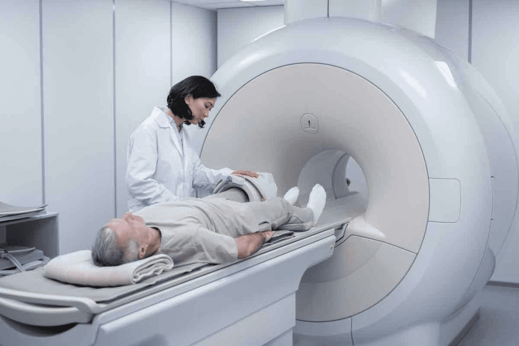
CT scans are very good at finding broken bones. They are a key tool in medical care, very important in emergencies. Quick and accurate checks are vital.
Detection Rates: 88-98% of Fractures Identified
Research shows CT scans can spot 88% to 98% of fractures missed by X-rays. This is very important in trauma care. Quick and right diagnosis can greatly help treatment.
A study by NYU Langone Medical Center found ultra-low-dose CT scans work well. They can find fractures without using too much radiation. This shows how good CT scans are for keeping patients safe.
- High sensitivity: CT scans are very good at finding bone injuries, perfect for tough cases.
- Rapid assessment: They help doctors quickly check trauma patients, which is key in emergencies.
- Comprehensive imaging: CT scans show clear images of bones and soft tissues around them.
High-Resolution Imaging Capabilities for Complex Fractures
CT scans are great at showing high-resolution images of hard-to-read fractures. This is very helpful for planning surgeries. Detailed pictures are needed for good surgery plans.
With CT scans, doctors can:
- See how bad and complex fractures are with great detail.
- Find any extra problems, like bone pieces or soft tissue damage.
- Make specific treatment plans based on the detailed images.
In short, CT scans are a top choice for finding broken bones. They have high success rates and give detailed images. These are key for managing hard fractures well.
Difference #1: Image Resolution and Detail
When diagnosing bone injuries, the detail in images matters a lot. Seeing the full extent of a fracture helps doctors make better treatment plans. This affects how well a patient will recover.
X-Ray Resolution vs. CT Scan Clarity
X-rays are often used first because they’re quick and cheap. But, they don’t show as much detail, which can be a problem in complex cases.
CT scans, on the other hand, show much clearer images. They catch details that X-rays miss. This is really helpful for complex fractures or those near joints.
- Key advantages of CT scans over X-rays:
- Higher resolution images
- Better visualization of complex fractures
- Ability to detect smaller fractures or fragments
3D Reconstruction Advantages of CT Technology
CT technology’s biggest leap is making 3D images. This lets doctors:
- See the fracture from different views
- Understand how the pieces fit together
- Plan surgeries more accurately
The 3D feature of CT scans is a game-changer for tough cases. It gives a full view that 2D images can’t match. This is key for fractures in the spine, pelvis, or joints.
Difference #2: Fracture Complexity Assessment
The complexity of a fracture is key in choosing between X-rays and CT scans. It’s important to assess fracture complexity to decide the best diagnostic approach.
Simple Fractures: When X-Rays Are Sufficient
For simple fractures, X-rays are usually the first choice. They are quick and cost-effective for checking bone alignment and finding obvious fractures. Simple fractures have a single break and usually don’t have complicating factors.
Characteristics of simple fractures that make X-rays sufficient include:
- Single, clean break in the bone
- Minimal displacement of bone fragments
- No significant soft tissue damage
Complex Fractures: When CT Scans Become Essential
Complex fractures need more detailed imaging. CT scans are essential for fractures with multiple fragments, significant displacement, or complex anatomy.
CT scans offer advantages in complex fractures by:
- Providing detailed 3D reconstructions of the fracture
- Allowing for better assessment of fragment displacement
- Enabling evaluation of surrounding soft tissue damage
A comparison of imaging modalities for simple and complex fractures is shown in the table below:
| Fracture Type | Imaging Modality | Key Features |
| Simple | X-ray | Quick, cost-effective, sufficient for single breaks |
| Complex | CT Scan | Detailed 3D imaging, better for multiple fragments and soft tissue assessment |
In conclusion, the choice between X-rays and CT scans for fracture assessment depends on fracture complexity. Understanding this difference is key for healthcare providers to make the right diagnostic imaging choices.
Difference #3: Soft Tissue Visualization Capabilities
Choosing the right imaging modality is key for soft tissue injury diagnosis. Soft tissue injuries, like tendon and ligament damage, are often linked to complex fractures. It’s important to see these injuries clearly.
X-rays and CT scans can’t compare to MRI in soft tissue imaging. Knowing their limits helps doctors choose the best imaging for patients.
Can X-Rays Show Tendon or Ligament Damage?
X-rays aren’t good at showing tendon or ligament damage. They work better for bones. Doctors often use other methods to check for these injuries.
Limitations of X-rays in soft tissue visualization:
- Poor soft tissue contrast
- Inability to directly visualize tendons and ligaments
- Reliance on indirect signs for soft tissue injury diagnosis
Limitations of CT Scans in Soft Tissue Evaluation
CT scans are better than X-rays for bones, but they’re not perfect for soft tissues. They can show some soft tissue details, but not as well as MRI.
Key limitations of CT scans in soft tissue evaluation include:
- Lower soft tissue contrast compared to MRI
- Difficulty in detecting subtle soft tissue injuries
- Limited ability to assess tendon and ligament integrity
In summary, X-rays and CT scans are great for bones but not for soft tissues. Knowing their limits is vital for accurate diagnosis and treatment.
Difference #4: Radiation Exposure Considerations
Radiation exposure is a big deal when choosing between X-rays and CT scans for bone injuries. It’s key to know the risks of radiation for better patient care. This is important for both doctors and patients.
Comparing Radiation Doses: X-Ray vs. CT Scan
CT scans give off much more radiation than X-rays. For example, a head CT scan has about 20 times more radiation than a chest X-ray. This is important when thinking about the risks and benefits for patients, like when they need imaging again.
“CT scans are used more in emergency rooms for checking injuries,” a study on radiation in medical imaging says. “But because CT scans have more radiation, we need to think about other imaging options when we can.”
Risk-Benefit Analysis in Different Clinical Scenarios
Choosing between X-rays and CT scans depends on weighing the risks and benefits. For simple fractures or first checks, X-rays are often better because they have less radiation. But for complex cases or when detailed images are needed, CT scans might be better, even with more radiation.
In trauma cases with possible complex fractures or internal injuries, CT scans can be life-saving. Here, the benefits of CT scans usually outweigh the higher radiation risks.
New CT technology has made CT scans safer. “Today’s CT scanners use advanced dose reduction methods,” medical papers say. “These methods cut down radiation while keeping image quality high.”
In short, CT scans have more radiation than X-rays, but careful analysis is needed for each case. By considering each patient’s needs and using new imaging tech, doctors can improve care and lower radiation risks.
Difference #5: Time and Accessibility Factors
When diagnosing bone injuries, how fast we get and understand imaging results matters a lot. X-rays and CT scans affect how quickly we can treat patients and their outcomes.
Speed of Obtaining and Interpreting Results
X-rays are fast to do and understand, making them great for emergencies. CT scans are quicker than MRI but take longer to set up and scan. But, new CT tech has made scanning times much shorter.
Comparison of Imaging Modalities: Here’s a table showing the time differences between X-rays and CT scans.
| Imaging Modality | Procedure Time | Interpretation Time |
| X-ray | 5-10 minutes | Immediate to 30 minutes |
| CT Scan | 15-30 minutes | 30 minutes to several hours |
Availability and Cost Implications
X-rays are easy to find and cheap, fitting into most healthcare budgets. CT scans are pricier but give detailed images, essential for complex cases.
Cost Comparison: X-rays cost between $100 and $500. CT scans can be $500 to $3,000 or more, based on the scan’s complexity and where it’s done.
Choosing between X-rays and CT scans depends on the patient’s needs and the cost. Knowing these factors helps doctors make the best choices for both diagnosis and budget.
Difference #6: MRI’s Superior Soft Tissue Evaluation
MRI is the top choice for soft tissue injuries. It shows tendons, ligaments, and cartilage very well. This makes MRI key in today’s medical world.
How MRI Visualizes Tendons, Ligaments, and Cartilage
MRI uses advanced tech to see soft tissues clearly. It combines magnetic fields and radio waves for detailed images. This is great for checking tendons, ligaments, and cartilage.
MRI’s key advantages in soft tissue evaluation include:
- High contrast resolution, enabling clear differentiation between various soft tissue types
- Ability to detect subtle changes in soft tissue structures, aiding in early diagnosis
- Multi-planar imaging capabilities, allowing for a full view from different angles
When MRI Is the Preferred Choice Over CT and X-Ray
MRI is best when soft tissue details matter. For example, it’s great for spotting ligament sprains or tears. It’s also top for checking cartilage, like in osteoarthritis.
| Imaging Modality | Soft Tissue Contrast | Usefulness in Tendon/Ligament Evaluation |
| MRI | High | Excellent |
| CT Scan | Moderate | Fair |
| X-Ray | Low | Poor |
In short, MRI is essential for soft tissue checks. It gives clear images and detailed views. This makes it the go-to for many medical needs.
Difference #7: Emergency and Trauma Assessment Protocols
In emergency medicine, the right imaging choice can greatly affect patient outcomes in trauma cases. The protocols for emergency and trauma assessment aim to quickly and accurately diagnose injuries. This ensures timely and proper treatment.
First-Line Imaging Choices in Emergency Settings
In emergency settings, a CT scan is often the first choice for trauma patients. This is because CT scans are fast and can show detailed images of internal injuries. CT scans are very valuable in polytrauma cases, where many injuries are suspected.
The benefits of using CT scans first include:
- They allow for quick image acquisition, speeding up assessment and decision-making.
- They are highly sensitive for detecting a wide range of injuries, including internal organs and bones.
- They can image multiple body regions at once, which is great for polytrauma patients.
Sequential Imaging Approaches for Complete Diagnosis
While CT scans are often the first choice in trauma cases, other imaging methods might be used for a more complete diagnosis. This could include X-rays or MRI, depending on the patient’s condition and the suspected injuries.
| Imaging Modality | Primary Use in Trauma | Advantages |
| CT Scan | Initial assessment of polytrauma patients | Rapid, detailed imaging of internal injuries |
| X-ray | Initial assessment of bone fractures | Quick, low-cost, and widely available |
| MRI | Detailed assessment of soft tissue injuries | High sensitivity for soft tissue damage |
The choice of imaging modality and the decision to use sequential imaging depend on several factors. These include the patient’s condition, the injury mechanism, and the suspected injuries. By choosing the right imaging protocols, healthcare providers can ensure trauma patients get accurate and timely diagnoses. This improves patient outcomes.
Alternatives to MRI for Soft Tissue Evaluation
While MRI is highly effective, alternatives like ultrasound offer distinct advantages for soft tissue evaluation. These alternatives are key for patients who can’t have MRI due to claustrophobia, metal implants, or other reasons.
Ultrasound as a Diagnostic Option
Ultrasound is a safe and real-time imaging modality for soft tissue evaluation. It’s great for checking superficial structures and guiding interventions. Ultrasound’s portability and relatively low cost make it an attractive option for bedside assessments and in resource-limited settings.
But ultrasound has its limits. It depends a lot on the operator, and image quality can be affected by patient body habitus and the presence of gas or bone. Despite these, ultrasound is a valuable tool for evaluating soft tissue injuries, like tendons and ligaments.
Emerging Technologies in Soft Tissue Imaging
Beyond ultrasound, new technologies are improving soft tissue imaging. One is Contrast-Enhanced Ultrasound (CEUS), which uses ultrasound contrast agents to better see tissue vascularity and perfusion.
Another area is the use of Artificial Intelligence (AI) in medical imaging. AI algorithms can improve image analysis, catching subtle abnormalities that might be missed. These technologies are set to change soft tissue imaging by making diagnoses more accurate and efficient.
| Imaging Modality | Advantages | Limitations |
| Ultrasound | Real-time imaging, portable, relatively low cost | Operator-dependent, limited by patient factors |
| CEUS | Enhanced vascularity and perfusion assessment | Requires contrast agents, limited availability |
| AI-enhanced Imaging | Improved diagnostic accuracy, efficient analysis | Dependent on algorithm quality, data privacy concerns |
Clinical Decision Making: Selecting the Optimal Imaging Modality
Choosing the right imaging technique is key in today’s healthcare. It depends on the patient’s condition, the question being asked, and what each imaging modality can do.
Patient-Specific Considerations
When picking an imaging method, the patient’s needs are top priority. Age, medical history, and any issues with certain scans are important. For example, people with metal implants or pacemakers can’t have MRI scans. Studies show that these factors greatly affect the choice of imaging and how accurate it is.
Key patient-specific factors include:
- Age and medical history
- Presence of metal implants or pacemakers
- Allergies to contrast agents
- Claustrophobia or other anxiety disorders
Modern Diagnostic Protocols at Leading Medical Centers
Top hospitals, like Liv Hospital, follow modern diagnostic plans. They pick the best imaging for each patient. This ensures quality care and accurate diagnoses. New technologies, like 3D reconstruction and high-resolution CT scans, have boosted diagnostic skills.
| Imaging Modality | Key Advantages | Common Applications |
| CT Scan | High-resolution images, quick scanning time | Trauma, complex fractures, emergency diagnoses |
| MRI | Excellent soft tissue visualization, no radiation | Soft tissue injuries, neurological disorders, certain cancers |
| X-Ray | Quick, low cost, low radiation dose | Simple fractures, initial assessments, routine checks |
Healthcare providers make smart choices by considering patient needs and following modern protocols. This way, patients get the best care and results.
Conclusion: Making Informed Decisions About Diagnostic Imaging
Diagnostic imaging is key in today’s healthcare. It helps doctors make accurate diagnoses and plan effective treatments. The choice between CT scans, X-rays, and MRI depends on the patient’s condition and the issue’s complexity.
It’s important for patients to make informed decisions about their imaging tests. This ensures they get the best care possible. Healthcare professionals can pick the right imaging test for each case by knowing the differences.
Choosing the right imaging test is about finding a balance. It involves considering image quality, radiation exposure, and how easy it is to get the test. CT scans are great for complex fractures because they provide detailed images. X-rays are quick and affordable for a first look. MRI is best for soft tissues, like tendons and ligaments.
In the end, making smart choices about imaging tests leads to better health outcomes. Healthcare professionals can offer top-notch care by considering each patient’s needs and the strengths of each imaging method. This approach improves treatment results.
FAQ
Can a CT scan show broken bones more effectively than an X-ray?
Yes, CT scans are better at showing broken bones, like complex fractures. They offer high-resolution images and 3D views.
Can X-rays show tendon or ligament damage?
No, X-rays can’t show tendon or ligament damage. They mainly show bone structures. MRI is used for soft tissue injuries.
What’s the difference between a CT scan and an MRI in diagnosing musculoskeletal injuries?
CT scans are great for bone fractures and complex bone injuries. MRI is better for soft tissues like tendons, ligaments, and cartilage.
How does radiation exposure compare between X-rays and CT scans?
CT scans have more radiation than X-rays. But, they provide detailed images of complex fractures. This makes them worth the risk.
Are there alternatives to MRI for soft tissue evaluation?
Yes, ultrasound is a good alternative for soft tissue checks. It’s cheaper and shows movement. New technologies are also being looked into.
Can X-rays detect ligament damage?
No, X-rays can’t directly show ligament damage. They might show signs of injury, but MRI is more accurate.
What’s the preferred imaging modality for emergency and trauma assessment?
CT scans are often first choice in emergencies. They’re fast, provide clear images, and quickly check for complex injuries.
How do CT scans compare to X-rays in terms of time and accessibility?
CT scans take longer than X-rays. But, new tech has made them faster. X-rays are quicker and cheaper.
Can CT scans visualize tendons and ligaments?
CT scans can give some info on soft tissues. But, MRI is better for seeing tendons and ligaments because of its soft tissue contrast.
What factors influence the choice of imaging modality for diagnosing bone injuries?
Many things decide which imaging to use. These include the fracture’s complexity, soft tissue needs, radiation risks, and patient factors like age and health history.
Reference
- Aldhyani, T. H. H., et al. (2025). Diagnosis and detection of bone fracture in radiographic images using deep learning techniques. Computational and Mathematical Methods in Medicine. https://www.ncbi.nlm.nih.gov/pmc/articles/PMC11803505/
- François, M. A., et al. (2025). Diagnostic performance of spectral CT in detecting bone marrow edema in vertebral fractures. Journal of Radiology. https://www.sciencedirect.com/science/article/pii/S0720048X24005734


