At Liv Hospital, we know how important an accurate diagnosis is for your health. A CT scan of the thyroid gland is key. It helps us see how the gland is doing and find any problems.
A thyroid CT scan gives us important information about your thyroid. It helps us spot issues like nodules, cysts, and enlargement. Knowing what a CT of thyroid shows is key to finding the right treatment.
Key Takeaways
- CT scans help diagnose thyroid abnormalities.
- Understanding CT findings is vital for treatment.
- Liv Hospital offers expert care for thyroid conditions.
- A thyroid CT scan evaluates gland structure.
- Accurate diagnosis ensures overall well-being.
Understanding CT of Thyroid: Basic Principles and Applications
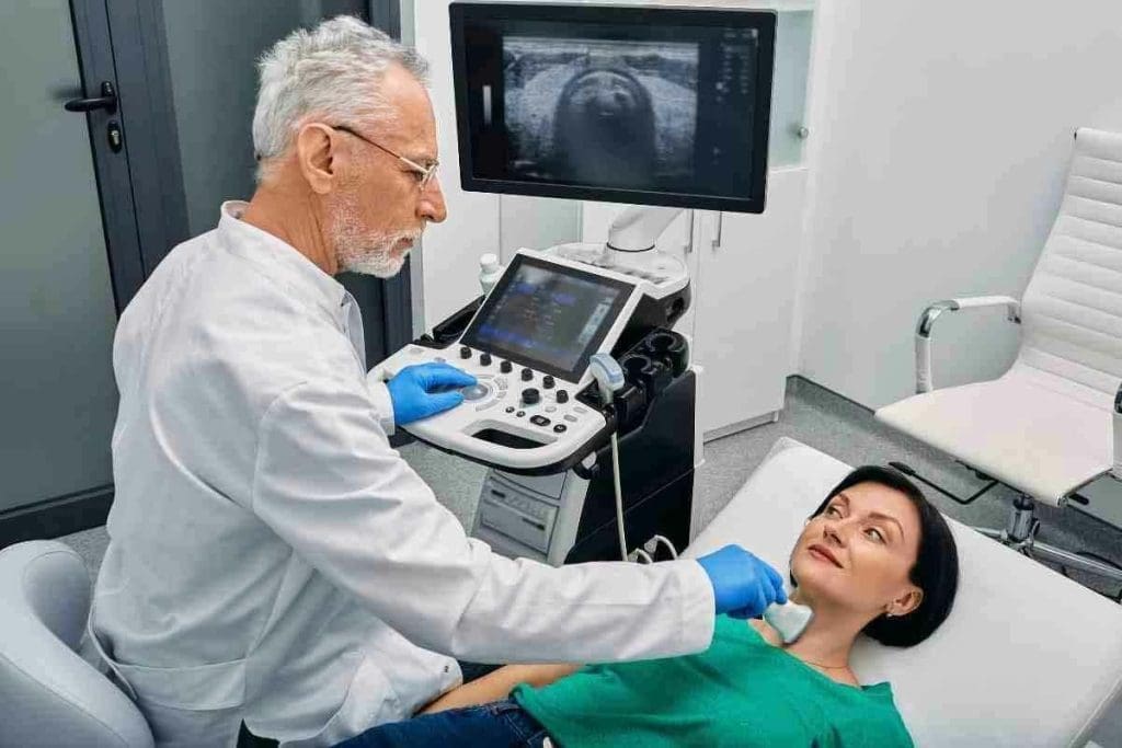
The thyroid gland is key to our endocrine system. CT scans give us a clear view of it. This helps us understand thyroid disorders better and manage them well.
What Is a Thyroid CT Scan?
A thyroid CT scan is a non-invasive test that uses X-rays. It creates detailed images of the thyroid gland. This tool is vital for checking the thyroid’s size, shape, and any issues like nodules or cancer.
CT scans give us a detailed look at the thyroid. This helps us see how far thyroid disease has spread. It also helps us plan the right treatment.
How CT Imaging Works for Thyroid Evaluation
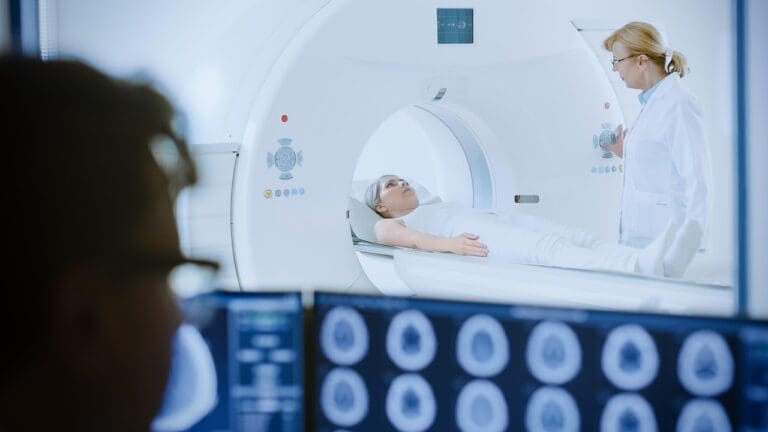
CT imaging combines X-rays and computer technology to show the thyroid gland. During a CT scan and thyroid check, the patient lies on a table. The table moves into a CT scanner, which takes X-ray images from all sides.
These images are then put together to show detailed pictures of the thyroid. This lets us check its structure and find any problems.
Some benefits of CT imaging for thyroid checks include:
- High-resolution images that show the thyroid gland’s details.
- The ability to spot small nodules and other issues not seen by other methods.
- Helpful for planning surgery and understanding the extent of thyroid disease.
For more on CT scans, check out this article on the National Center for Biotechnology Information’s website.
Differences Between CT and Other Thyroid Imaging Methods
While ultrasound is often used, a thyroid gland CT scan has its own benefits. CT scans show how the thyroid relates to nearby structures like the trachea and esophagus. This is important for surgery planning. Also, CT scans can find thyroid issues that are in the chest, known as substernal or retrosternal goiters.
Here are some key differences between CT and other imaging methods:
| Imaging Modality | Key Features | Clinical Use |
| CT Scan | High-resolution images, detailed anatomy | Pre-surgical planning, detecting substernal goiters |
| Ultrasound | Non-invasive, real-time imaging | Initial thyroid evaluation, guiding fine-needle aspiration |
When Is a CT Scan of the Thyroid Recommended?

We use CT scans of the thyroid gland when a detailed look is needed. This is often when thyroid cancer is suspected or when goiters are large. This tool gives us important information that helps in planning care and treatment.
Clinical Indications for Thyroid CT
CT scans are recommended for several reasons. They help when thyroid cancer is suspected, when large goiters cause problems, or when ultrasound results are unclear. They are great for seeing how far the disease has spread and if it’s touching nearby tissues.
The table below summarizes the common clinical indications for thyroid CT scans:
| Clinical Indication | Description |
| Suspected Thyroid Malignancy | CT scans help assess the extent of disease and possible lymph node involvement. |
| Large Goiters | CT is useful for checking the substernal extension and how it affects nearby structures. |
| Inconclusive Ultrasound Findings | CT provides more information when an ultrasound is hard to read due to calcifications or deep tissue. |
Limitations of Thyroid Ultrasound
Thyroid ultrasound is great for starting, but it has its limits. It can’t always see big or deep thyroid masses well. A CT scan can give a clearer picture, helping doctors decide what to do next.
Pre-operative Planning and Staging
Before surgery for thyroid disease, a CT scan is very helpful. It shows how far the disease has spread, if it’s in the chest, and if lymph nodes are affected. This info is key for surgeons to plan the best surgery.
For thyroid cancer patients, CT scans are key for staging. Accurate staging helps predict outcomes and decide on treatments like radioactive iodine or radiation therapy.
The Normal Thyroid CT Scan: What to Expect
When you get a CT scan of your thyroid, knowing what a normal result looks like is key. It helps doctors and patients understand thyroid health. A normal scan is the first step in diagnosing and treating thyroid issues.
Typical Appearance of a Healthy Thyroid Gland
A healthy thyroid gland looks homogeneous and dense on CT scans. It’s usually denser than the muscles around it. This helps doctors tell a normal gland from one that’s not.
Normal Anatomical Variations
Thyroid glands can vary in size, shape, and position. For example, they might be uneven or have a pyramidal lobe. Knowing these variations is key to reading CT scans right.
Density and Enhancement Patterns
The density and how the gland appears on CT scans tell a lot about its health. A normal gland shows uniform enhancement after contrast. Its density and how it enhances can change, like with iodine.
| Characteristics | Normal Thyroid Gland |
| Density | Higher than the surrounding muscles |
| Homogeneity | Homogeneous |
| Enhancement Pattern | Uniform after contrast |
Experts say knowing the normal thyroid gland on CT scans is vital. It helps spot problems and guide treatment.
This knowledge is key to telling normal variations from serious issues. It ensures patients get the right care.
Thyroid CT With Contrast: Enhanced Visualization
Contrast-enhanced CT scans offer better views of complex thyroid issues. They help doctors see the thyroid gland more clearly. This makes CT scans more accurate for diagnosing thyroid problems.
Benefits of Contrast Enhancement
Contrast material makes thyroid CT scans better. It shows the thyroid’s anatomy and any problems more clearly. This is key for spotting and understanding thyroid nodules, tumors, and other issues.
One big plus of contrast is seeing the thyroid’s blood flow. This is vital for spotting and planning treatment for thyroid cancers. It helps doctors know how to best approach surgery.
Vascular Assessment and Perfusion Patterns
Contrast CT scans show the thyroid’s blood structure in detail. They help spot the blood flow to thyroid nodules or tumors. This can tell doctors if a growth is likely to be cancerous.
| Characteristics | Benign Lesions | Malignant Lesions |
| Vascularity | Typically less vascular | Often highly vascular |
| Perfusion Patterns | Homogeneous enhancement | Heterogeneous enhancement |
| Contrast Uptake | Slow and gradual uptake | Rapid uptake and washout |
Potential Risks and Contraindications
While contrast CT scans are helpful, there are risks. Some people might have allergic reactions to the contrast. This can be mild or severe, depending on the person.
Another risk is how contrast affects the kidneys. People with kidney problems might face issues. So, doctors check kidney health before using contrast.
In summary, contrast CT scans are very useful for thyroid exams. But it’s important to be careful and choose the right patients. This way, the benefits of contrast CT scans can be enjoyed without too many risks.
Key Finding #1: Thyroid Nodules on CT
Thyroid nodules found on CT scans are common and need careful handling. We will look into their importance, risks, and the best follow-up steps.
Incidental Nodules: Significance and Management
Thyroid nodules are quite common and often found on CT scans. Most are harmless, but a few might be cancerous. The key is to figure out which ones need more testing.
We suggest that people with thyroid nodules on CT scans get more tests. This includes ultrasound and sometimes a biopsy, based on the nodule’s look and the person’s risk factors.
Risk Factors for Malignancy in CT-Detected Nodules
Some features make thyroid nodules on CT scans more likely to be cancerous. These include:
- Nodule size greater than 1 cm
- Microcalcifications within the nodule
- Irregular margins or invasion into surrounding structures
- PPatient’shistory of radiation exposure
- Family history of thyroid cancer
People with these risk factors need a closer look. Studies have found that microcalcifications are a big sign of cancer.
Follow-up Recommendations for Incidental Findings
Handling thyroid nodules found by chance involves imaging and a doctor’s advice. Here’s what we recommend:
| Nodule Size | Risk Factors | Recommended Follow-up |
| No | Clinical monitoring, no immediate further imaging | |
| >1 cm | No | Ultrasound and clinical follow-up |
| Any size | Yes | Ultrasound, potentially fine-needle aspiration biopsy |
By focusing on risk levels, we can manage thyroid nodules on CT scans well. This ensures patients get the right care without too many tests.
Key Finding #2: Hypodense Thyroid Nodules
CT scans help check thyroid health. They find hypodense thyroid nodules, which are less dense than the rest of the thyroid. These nodules can mean different things.
Characteristics and Appearance
Hypodense thyroid nodules show up as less dense on CT scans. This makes them stand out. Knowing how they look is key to spotting them.
- Density: Hypodense nodules are less dense than the surrounding thyroid tissue.
- Appearance: They may appear as well-defined or ill-defined lesions.
- Size: The size of hypodense nodules can vary significantly.
Understanding these traits is vital for correct diagnosis and treatment.
Differential Diagnosis of Hypodense Lesions
Hypodense thyroid nodules can be many things. They can be benign or even cancerous.
- Thyroid Cysts: Simple or complex cysts can appear hypodense.
- Neoplastic Lesions: Certain tumors may present as hypodense nodules.
- Inflammatory Lesions: In some cases, inflammatory processes can result in hypodense areas.
It’s important to figure out what a hypodense nodule really is.
Clinical Significance and Further Evaluation
Hypodense thyroid nodules can be serious. They might be harmless or could be cancer. It’s key to check them out more.
We suggest a detailed check-up. This includes looking at the patient’s history, more scans, and maybe a biopsy. This helps figure out what the nodules are.
Understanding hypodense thyroid nodules helps doctors give better care. They can make more accurate diagnoses and plans for treatment.
Key Finding #3: Thyroid Calcifications on CT
Seeing calcifications in the thyroid gland on CT scans is a big deal. These are calcium deposits in the thyroid tissue. They show up clearly on CT scans.
Types of Calcifications and Their Appearance
Thyroid calcifications come in different shapes and sizes. The main types are:
- Microcalcifications: Tiny, point-like calcifications often linked to papillary thyroid carcinoma.
- Macrocalcifications: Larger, rough calcifications are found in both good and bad conditions.
- Egg-shell calcifications: A ring of calcification around a nodule, usually a sign of something benign.
Knowing the type and where the calcifications are is key to understanding their importance.
Benign vs. Malignant Calcification Patterns
The way calcifications show up can tell us if a thyroid lesion is bad or not. For example:
- Microcalcifications point strongly to papillary thyroid cancer.
- Macrocalcifications can be in both good and bad conditions, so they need more checking.
A study on thyroid calcifications found that microcalcifications in nodules are a big sign of cancer, mainly papillary thyroid carcinoma. This shows how important it is to look closely at calcification patterns on CT scans.
Correlation With Pathology
Diagnosing thyroid calcifications needs to match what the CT scan shows with what the pathology says. CT scans give us clues about calcifications, but we need to look at tissue samples to know for sure.
| Calcification Type | Common Associations |
| Microcalcifications | Papillary thyroid carcinoma |
| Macrocalcifications | Benign and malignant conditions |
| Egg-shell calcifications | Benign thyroid nodules |
We stress the need for a team effort. We should use what the CT scan shows, along with clinical and pathological data, to correctly diagnose and treat thyroid calcifications found on CT scans.
Key Finding #4: Abnormal Thyroid CT Scan Patterns
When we look at thyroid health through CT scans, spotting abnormal patterns is key. These patterns help us figure out what’s going on with the thyroid gland. They give us important clues about its condition.
Heterogeneity and Nodularity
One common finding on thyroid CT scans is heterogeneity. This means the thyroid gland looks uneven or non-uniform. It can hint at underlying problems.
Nodularity, or nodules in the thyroid gland, is another important sign. These nodules can be harmless or cancerous. Their look on CT scans helps us tell them apart.
Seeing many nodules or a gland that looks mixed up might mean conditions like multinodular goiter or thyroiditis. These can lead to thyroid problems or even cancer risk.
Mass Effect on Surrounding Structures
An abnormal thyroid CT scan might also show a mass effect. This happens when the thyroid gland or a mass presses on nearby structures. This can cause symptoms like trouble swallowing or breathing problems.
Checking for a mass effect is key to figuring out how serious the issue is. It helps doctors understand how bad the thyroid problem is and how it affects nearby tissues.
Signs of Local Invasion
In serious cases, a thyroid CT scan might show local invasion. This means cancer has spread into nearby structures like the trachea or major blood vessels. Finding this is very important because it changes how we treat thyroid cancer.
Spotting these abnormal patterns on a thyroid CT scan is vital. It lets doctors give a full picture of thyroid health. This helps them create a treatment plan that fits the patient’s needs.
Key Finding #5: Thyroid Cysts on CT Scan
CT scans often show thyroid cysts, which need careful attention. These cysts can be simple or complex. They must be distinguished from solid lesions.
Differentiating Cystic from Solid Lesions
It’s important to tell cystic from solid lesions on a CT scan. Cystic lesions show up as clear, less dense areas in the thyroid. They are usually less dense than the thyroid tissue around them.
Characteristics of Cystic Lesions:
- Well-defined borders
- Hypodense appearance compared to thyroid tissue
- Possible presence of septations or solid components
Simple vs. Complex Cysts
Simple cysts are usually harmless and look the same on CT scans. They are filled with fluid and don’t have solid parts. Complex cysts, though, might have solid parts or septations. This makes them harder to diagnose and could mean a higher risk of cancer.
| Cyst Type | Characteristics | Clinical Implication |
| Simple Cyst | Uniform, fluid-filled, no solid components | Typically benign, may not require intervention |
| Complex Cyst | Contains solid components or septations | May require further evaluation, potentially including fine-needle aspiration |
Management Approaches Based on CT Findings
The treatment of thyroid cysts depends on what the CT scan shows. Simple cysts might not need any action. But complex cysts might need more checks. Doctors consider the patient’s overall health and symptoms when deciding what to do.
“The accurate characterization of thyroid cysts on CT scans is vital for the right treatment plan. It helps avoid unnecessary tests.”
Explains a radiology specialist from a leading medical center.
Understanding thyroid cysts on CT scans helps doctors make better choices. This leads to better care for patients.
Conclusion: Interpreting Your Thyroid CT Results
Understanding your thyroid CT scan results is key to the right diagnosis and treatment. We’ve looked at what a CT scan can show, like nodules and calcifications. These signs can point to different thyroid diseases.
To make sense of your scan, you need to know what the images mean. We talked about how a CT scan can spot nodules and decide if they’re important. This helps doctors plan your care and make sure you get the right treatment.
A thyroid CT scan gives important details about your thyroid gland and nearby areas. Knowing your scan results helps doctors create a treatment plan just for you. If you’ve had a thyroid CT scan, talking to your doctor about it can help you understand your diagnosis and treatment choices.
FAQ
What is a thyroid CT scan, and how does it work?
A thyroid CT scan is a test that uses X-rays and computer technology to show detailed images of the thyroid gland. It works by moving an X-ray machine around the body. This captures data to make detailed images.
When is a CT scan of the thyroid recommended?
A CT scan of the thyroid is suggested in many cases. This includes when there’s a chance of thyroid cancer. It’s also used to check how far thyroid disease has spread or when ultrasound results are unclear.
What does a normal thyroid CT scan look like?
A normal thyroid CT scan shows a gland that’s uniform in density and smooth. The gland’s size and shape can vary. But it should be symmetrical and without any nodules or masses.
What are the benefits of using contrast enhancement in thyroid CT scans?
Using contrast in thyroid CT scans helps see thyroid lesions better. It also lets doctors check vascular structures and how blood flows in the gland.
How are thyroid nodules detected and managed on CT scans?
Thyroid nodules found on CT scans are checked for size, density, and shape. How they’re managed depends on these features. Suspicious nodules might need a biopsy.
What does a hypodense thyroid nodule on a CT scan indicate?
A hypodense thyroid nodule on a CT scan is less dense than the gland itself. This can mean different things, like cysts, benign nodules, or even cancer. More tests are needed to find out.
How are thyroid calcifications identified on CT scans, and what do they signify?
Thyroid calcifications on CT scans are high-density spots in the gland. They can mean benign or malignant conditions. Some patterns suggest cancer more than others.
What are the signs of an abnormal thyroid CT scan?
Abnormal thyroid CT scans show heterogeneity, nodularity, or mass effect. They can also show local invasion. These signs point to possible thyroid problems that need more investigation.
How are thyroid cysts differentiated from solid lesions on CT scans?
Thyroid cysts on CT scans are fluid-density and well-defined. They’re different from solid lesions because of their density and how they look after contrast is added.
What is the significance of a thyroid CT scan with contrast in evaluating thyroid disease?
A thyroid CT scan with contrast helps detect and describe thyroid lesions better. It shows their vascularity and disease extent. This helps in diagnosis and treatment planning.
References:
- Sohn, S. Y., et al. (2025). Thyroid dysfunction risk after iodinated contrast media exposure: A longitudinal cohort study. Journal of Clinical Endocrinology and Metabolism, 110(4), e1204. https://academic.oup.com/jcem/article/110/4/e1204/7663938


