Last Updated on November 27, 2025 by Bilal Hasdemir
Ever wondered about the difference between a CT scan and an X-ray? Knowing about these imaging methods is key for accurate diagnosis. At Liv Hospital, we offer exceptional medical services with the latest technology and caring service.
CT scans and X-rays are used in medical imaging, but they’re not the same. CT scans give detailed 3D images, while X-rays show 2D views. This difference makes CT scans better for complex cases, and X-rays good for first checks.What is the difference between a CT scan and X ray? Learn 7 essential facts about image detail, technology, and clinical uses.
We’ll look at the main differences between these imaging tools. We’ll talk about their uses, benefits, and downsides. Knowing these differences helps patients understand their diagnostic path better.
Key Takeaways
- CT scans provide detailed 3D images, while X-rays offer 2D views.
- CT scans are often used for complex diagnoses, while X-rays are ideal for initial screenings.
- X-rays are generally more affordable and deliver less radiation compared to CT scans.
- CT scans are preferred for emergency situations and detecting subtle findings.
- Low-Dose Computed Tomography (LDCT) significantly reduces radiation exposure.
The Crucial Role of Advanced Medical Imaging in Modern Healthcare
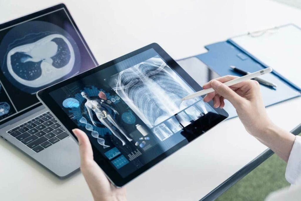
Accurate diagnostic imaging is key in today’s healthcare. Techniques like CT scans and X-rays are vital. They help doctors diagnose and treat diseases well.
Why Accurate Diagnostic Imaging Matters
Accurate imaging is important for many reasons:
- It leads to precise diagnoses, which are vital for good treatment plans.
- It helps track disease progress and treatment success.
- In emergency care, quick and correct diagnoses can save lives.
Our trained personnel knows how critical accurate imaging is. Our goal is to give the best care, and we invest in cutting-edge imaging technology.
Liv Hospital’s Commitment to Cutting-Edge Imaging Technology
We’re proud of our top-notch imaging facilities. They have the latest in medical imaging tech. Our radiology team is skilled and focused on quality service.
Our technology includes:
- High-resolution images for clear visuals.
- Advanced software for better image analysis.
- A focus on patient comfort and safety during scans.
As medical tech advances, Liv Hospital stays at the top. We use the latest advanced medical imaging to improve patient care.
What Is an X-Ray? Understanding the Basics
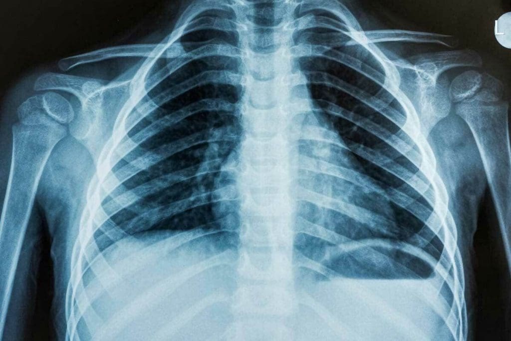
The X-ray is a key tool in healthcare today. It helps us see inside the body. This is important for diagnosing and treating many health issues.
How X-Ray Technology Works
X-rays use electromagnetic waves to show us what’s inside the body. When an X-ray beam hits the body, different parts absorb it differently. For example, bones absorb more than soft tissues. This makes it possible to see detailed images of the body’s inside.
Common Applications in Medical Diagnosis
X-rays are used a lot for checking bones, lungs, and teeth. They are great for:
- Spotting bone problems like osteoporosis and fractures.
- Finding lung infections like pneumonia.
- Looking at dental issues, like cavities and abscesses.
We also use X-rays to help with some medical procedures. This includes putting in catheters or fixing broken bones.
Here are some important points about X-rays:
- They give quick and affordable diagnostic info.
- Most healthcare places have X-rays.
- New digital X-rays improve image quality and cut down on radiation.
Understanding X-rays and their uses shows their importance in healthcare. They help us check bone health and lung conditions. X-rays are a basic but essential tool in medicine.
What Is a CT Scan? Comprehensive 3D Imaging
Computed Tomography (CT) scans have changed medical imaging. They give detailed 3D views of the body’s inside.
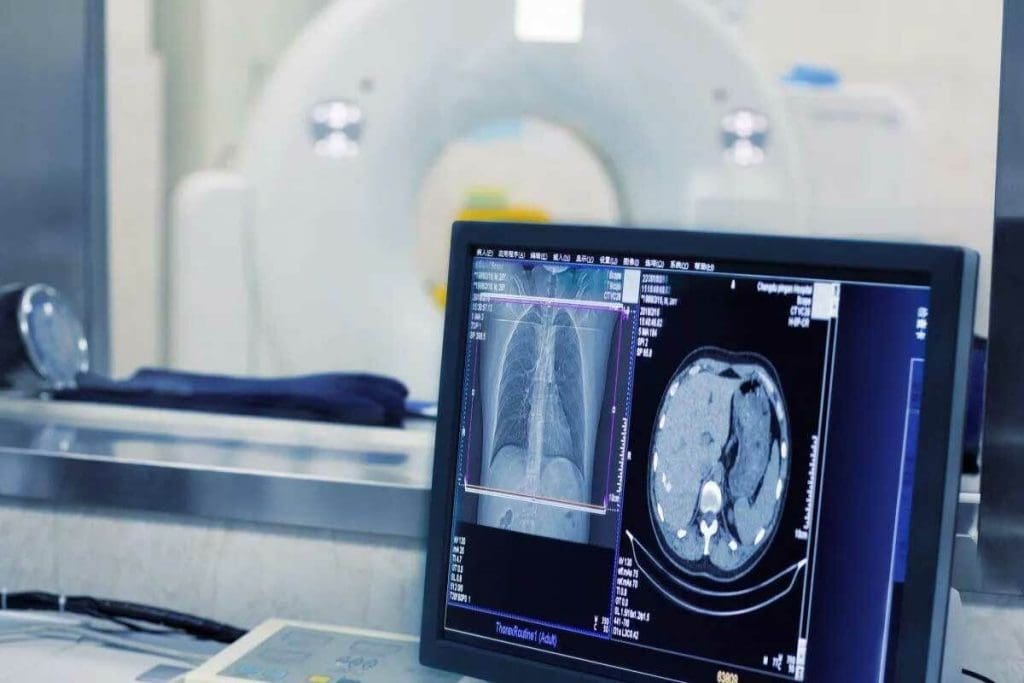
CT scans combine X-rays with computer technology. This makes cross-sectional images of the body. These images can be turned into detailed 3D models.
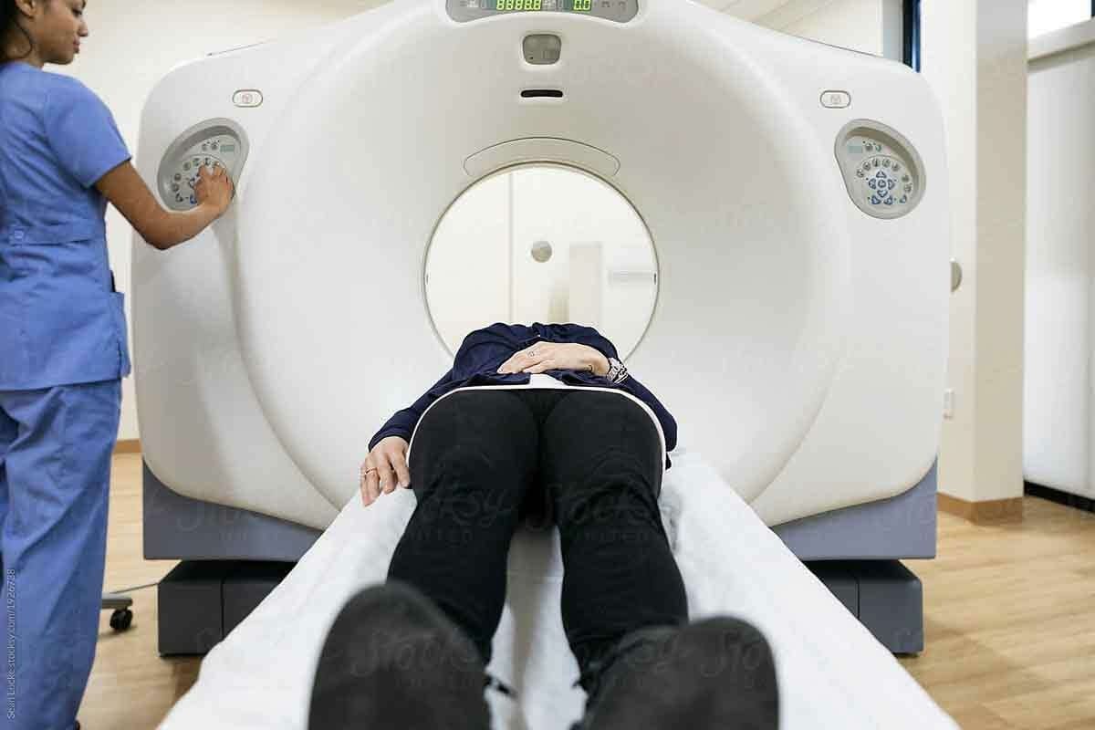
The process involves the patient on a table moving through a doughnut-shaped machine. The machine takes X-ray images from many angles.
The Science Behind Computed Tomography
CT scans work by using X-rays differently in various body tissues. They can tell apart tissues like bone, muscle, and fat. This helps diagnose many conditions, from injuries and cancers to vascular diseases.
When Physicians Recommend CT Scans
Doctors suggest CT scans for many reasons. They help check injuries, find cancers, and guide treatments. CT scans are key when detailed views of internal structures are needed.
In short, CT scans are a key diagnostic tool. They offer detailed 3D imaging of the body’s inside. Understanding CT scans helps patients see their value in healthcare.
The Fundamental Difference Between a CT Scan and X-Ray
CT scans and X-rays differ mainly in their ability to show internal structures. We’ll look at how these differences affect diagnosis and care.
Dimensional Capabilities: 2D vs. 3D Visualization
CT scans and X-rays differ in their imaging depth. X-rays give 2D views of inside structures. This can make it hard to see details clearly. On the other hand, CT scans offer 3D imaging, giving a better look at complex structures.
For example, CT scans are better for spine injuries. Their 3D view helps doctors make accurate diagnoses and plans.
Tissue Differentiation Abilities
CT scans are great at tissue differentiation. They help doctors tell different soft tissues apart. This is key for finding tumors or abscesses.
X-rays, though, are better for bones. They help spot fractures or osteoporosis. But they can’t tell soft tissues apart as well as CT scans.
Image Resolution and Detail Comparison
CT scans have better image resolution than X-rays. They take many images from different angles. Then, they make a detailed 3D picture.
| Imaging Modality | Dimensionality | Tissue Differentiation | Image Resolution |
| X-Ray | 2D | Limited | Lower |
| CT Scan | 3D | High | Higher |
Knowing these differences is key for doctors and patients. The right imaging choice helps get accurate diagnoses and treatments.
Fact 1: Radiation Exposure Levels Vary Significantly
Radiation exposure changes a lot between different imaging tests. It’s key to know the differences for safety and making smart choices.
X-Ray Radiation Dosage
X-rays have low radiation levels. They use millisieverts (mSv) to measure. A chest X-ray is about 0.1 mSv. This makes X-rays safe and good for first checks and regular visits.
CT Scan Radiation Considerations
CT scans have more radiation than X-rays. They can be 2 to 10 mSv, based on the scan area and type. Though more than X-rays, CT scans give detailed images for complex diagnoses.
Safety Protocols at Liv Hospital
Our team at Liv Hospital focuses on keeping patients safe. Our radiology team follows global guidelines to keep doses low. We use the latest tech and tailor scans for each patient.
Here’s a comparison of radiation doses:
| Imaging Modality | Typical Radiation Dose (mSv) |
| Chest X-Ray | 0.1 |
| CT Scan (Abdomen) | 8 |
| CT Scan (Head) | 2 |
Knowing about radiation helps patients and doctors choose better tests.
Fact 2: Soft Tissue Visibility Differs Dramatically
Soft tissue visibility is key in medical imaging. X-rays and CT scans show soft tissues differently. This affects how well they can diagnose injuries or conditions in tendons, ligaments, or other soft tissues.
Can X-Rays Show Tendon or Ligament Damage?
X-rays mainly show bones and struggle with soft tissues like tendons and ligaments. They might hint at soft tissue injury through swelling or calcification. But, they can’t directly show tendon or ligament damage.
An X-ray might reveal a fracture or dislocation. This could suggest soft tissue injury indirectly. Yet, it won’t clearly show the tendons or ligaments.
How CT Scans Visualize Soft Tissues
CT scans, though, offer a clearer view of soft tissues. They can spot injuries to tendons and ligaments, and other soft tissue issues. CT scans are great in emergency situations for quick, accurate diagnoses.
A study found CT scans are very good at diagnosing complex injuries, including soft tissue ones. For more on X-ray and CT scan differences, check out this article.
Limitations of Both Technologies
Though CT scans outdo X-rays for soft tissue, both have limits. CT scans might find it hard to tell apart some soft tissues. They’re not as good as MRI scans in all cases.
X-rays, not great for soft tissue, are useful for quick bone imaging. They’re also widely available.
| Imaging Modality | Soft Tissue Visibility | Bone Imaging Capability |
| X-Ray | Limited | Excellent |
| CT Scan | Good to Excellent | Excellent |
In conclusion, X-rays and CT scans each have their roles in medical imaging. The right choice depends on the clinical situation and the need for soft tissue visibility. Knowing these differences helps healthcare providers make better imaging decisions.
Fact 3: Cost, Time, and Accessibility Considerations
It’s important to know the differences in cost, time, and access between CT scans and X-rays. These factors help decide the best diagnostic tool for a patient’s needs.
Procedure Duration and Patient Convenience
The time it takes for a procedure affects how comfortable and convenient it is for patients. X-rays are generally quicker, taking just a few minutes. CT scans, while quicker during scanning, may take longer due to preparation and waiting.
We aim to make our facilities comfortable and quick for patients. This is why we try to reduce waiting times and make our facilities welcoming.
- Quick Procedure: X-rays are faster, making them ideal for initial assessments.
- Detailed Imaging: CT scans, while potentially longer, provide more detailed information.
Insurance Coverage and Out-of-Pocket Expenses
The cost of imaging is a big factor. Insurance coverage can vary a lot. X-rays are usually cheaper than CT scans because of their technology and complexity.
We help patients understand their insurance and any costs they might face. This way, they can make informed decisions about their tests.
- Check your insurance policy to understand what is covered.
- Discuss any out-of-pocket expenses with your healthcare provider.
- Think about the test’s value when looking at costs.
Availability in Different Healthcare Settings
Where you can get CT scans and X-rays differs a lot. X-ray technology is ubiquitous and found in almost all medical places. CT scanners are more common in hospitals or specialized centers because of their cost and complexity.
We make sure patients can get both X-ray and CT scan services in a place that’s easy and comfortable for them.
Fact 4: Diagnostic Applications and Limitations
It’s important to know how X-rays and CT scans work for your health. Each imaging method has its own use. Choosing the right one depends on what you need to check.
When X-Rays Are the Preferred Choice
X-rays are great for checking bones. They help find fractures, osteoporosis, and bone shape problems. They’re fast, easy to find, and show bone health well.
For example, X-rays are perfect for seeing if bones are broken or if they’re in the right place after being fixed.
Conditions Best Diagnosed with CT Scans
CT scans are best for complex problems. They give detailed pictures of inside the body. They’re great for finding internal injuries, cancers, and blood vessel diseases.
CT scans show more than X-rays. They help see how big an injury or disease is.
| Imaging Modality | Diagnostic Strengths | Common Applications |
| X-Ray | Bone imaging, quick and widely available | Fractures, osteoporosis, bone deformities |
| CT Scan | Detailed cross-sectional images, soft tissue visualization | Internal injuries, cancers, vascular diseases |
| MRI | Soft tissue imaging, detailed views of tendons and ligaments | Soft tissue injuries, neurological conditions |
X-Ray vs. CT Scan vs. MRI: Choosing the Right Tool
Choosing between X-rays, CT scans, and MRI depends on what you need to see. X-rays are good for bones. CT scans show bones and soft tissues. MRI is best for soft tissues like tendons and ligaments.
At our place, we pick the best imaging for each patient. Our radiologists work with doctors to make sure we get the right info. We also try to use less radiation and avoid risks.
Fact 5: Patient Experience and Preparation Requirements
Knowing what to expect during a CT scan or X-ray can make your experience better. At our institution, we aim to make the process easy and comfortable for our patients.
What to Expect During Each Procedure
A CT scan involves lying on a table that slides into a large, doughnut-shaped machine. The whole process usually takes 10-30 minutes, depending on the area scanned. You might need to hold your breath or stay very quiet to get clear images.
An X-ray is much quicker, taking just a few minutes. You’ll be positioned by the radiographer, who then steps away to operate the machine from another room.
Preparation Guidelines and Restrictions
For a CT scan, you might need to remove jewelry, glasses, or metal objects. You could also be asked to wear a hospital gown. Sometimes, a contrast dye is used, which requires fasting for a few hours beforehand.
Preparation for an X-ray is simple, usually just removing clothes or jewelry that could get in the way. No special diet is needed.
Accommodations for Special Populations
We know some patients need special care. Pregnant women or those who might be pregnant should tell their doctor, as it might change the scan choice. Children and those with claustrophobia might need sedation or a parent present.
Our team works hard to create a safe and welcoming space for everyone. We tailor our care to meet each patient’s unique needs.
Fact 6: Alternative Imaging Options When CT or X-Ray Isn’t Ideal
When CT scans or X-rays aren’t the best choice, doctors use other imaging methods. These options are key for patients who can’t have CT scans or X-rays. Or when these methods don’t give enough information.
Alternatives to MRI for Claustrophobic Patients
For those who feel anxious or claustrophobic during MRI scans, there are other choices. The open MRI offers a bigger space, making patients feel less trapped. Doctors might also suggest ultrasound or nuclear medicine scans. These can give important details without needing a closed space.
Ultrasound and Nuclear Medicine Options
Ultrasound uses sound waves to see inside the body. It’s great for checking organs like the liver and thyroid. It’s also used to watch how a baby grows in the womb.
Nuclear medicine scans use tiny amounts of radioactive materials. They help doctors see how different parts of the body work. This can give detailed info on body functions and structures.
Emerging Imaging Technologies
Medical imaging is always getting better, with new tech coming out. New tools like AI-enhanced imaging can make pictures clearer and faster. Other new tech includes better contrast agents and scanner designs. These aim to make patients more comfortable and help doctors diagnose better.
Fact 7: How Physicians Determine the Most Appropriate Imaging Method
Physicians look at many things to choose the best imaging method. This choice is key to making sure patients get the right diagnosis and stay safe.
Clinical Decision-Making Process
The process starts with looking at the patient’s medical history and current symptoms. Healthcare professionals also check the results of initial tests. They decide which imaging method, like X-rays or CT scans, will give the most helpful information.
We follow evidence-based guidelines for choosing imaging methods. These guidelines help us find the right balance between getting clear diagnoses and avoiding risks.
Balancing Diagnostic Needs with Patient Safety
It’s important to balance the need for accurate diagnoses with keeping patients safe. Physicians must compare the benefits of different imaging methods with their risks, like radiation or contrast agents.
At our institution, we focus on keeping patients safe. We follow strict protocols for imaging, using the least amount of radiation and choosing methods that are safe yet effective.
The Role of Radiologists in Image Interpretation
Radiologists are key in understanding imaging results. Their knowledge is vital for making accurate diagnoses and spotting issues that might not be obvious.
We work with radiologists to make sure imaging results fit the patient’s overall health picture. This teamwork helps us give the most accurate diagnoses and plan the best treatments.
Conclusion: Making Informed Decisions About Your Medical Imaging Needs
Knowing the difference between CT scans and X-rays is key. It helps you make smart choices about your medical imaging. By looking at the 7 key facts in this article, you can understand your options better.
At Liv Hospital, we aim to provide top-notch healthcare. We use the latest technology and care for our patients with compassion. Our team makes sure you get the right imaging tests, whether it’s a CT scan or an X-ray.
Choosing the right place for medical imaging is important. It helps you get accurate diagnoses and effective treatments. We suggest talking to your healthcare provider about your imaging needs. This way, you can decide the best action for you.
FAQ
Can X-rays show tendon or ligament damage?
X-rays can’t clearly see soft tissues like tendons and ligaments. They might show signs of damage indirectly. But, they’re not the best for diagnosing these injuries.
What’s the difference between an X-ray and a CT scan?
X-rays and CT scans differ in what they show. X-rays give 2D views, while CT scans offer 3D images. This makes CT scans better for detailed looks inside the body.
Can a CT scan show soft tissue injuries?
Yes, CT scans can show soft tissues more clearly. This helps doctors make more accurate diagnoses of soft tissue injuries.
Is there an alternative to an MRI scan?
Yes, there are other options like ultrasound and nuclear medicine. New technologies are also coming. The right choice depends on the situation and what the doctor needs to know.
How do CT scans and X-rays compare in terms of radiation exposure?
X-rays use less radiation than CT scans. But, CT scans give more detailed images. This is important for making accurate diagnoses.
Can X-rays diagnose complex conditions like internal injuries or cancers?
No, X-rays aren’t used for complex conditions like internal injuries or cancers. CT scans are better for these diagnoses.
What’s the difference between a CT scan, X-ray, and MRI?
CT scans show 3D images and are good for complex diagnoses. X-rays are for bone-related issues and show 2D views. MRI is best for soft tissues, like tendons and ligaments, and gives detailed images.
Are CT scans or X-rays more accessible?
X-rays are quicker and easier to get than CT scans. They’re a good choice for first checks.
How do physicians determine the most appropriate imaging method?
Doctors decide based on what they need to diagnose and the patient’s safety. They use a careful process to choose the best imaging method.
References
- Avci, M., et al. (2019). Comparison of X-Ray Imaging and Computed Tomography. PMC. https://pmc.ncbi.nlm.nih.gov/articles/PMC6843286/


