Last Updated on November 27, 2025 by Bilal Hasdemir

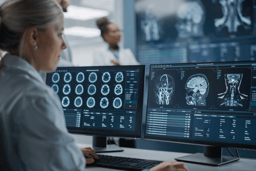
Medical imaging is key in today’s healthcare. It helps doctors diagnose and treat health issues. For soft tissue and tendon injuries, picking the right imaging test is very important.
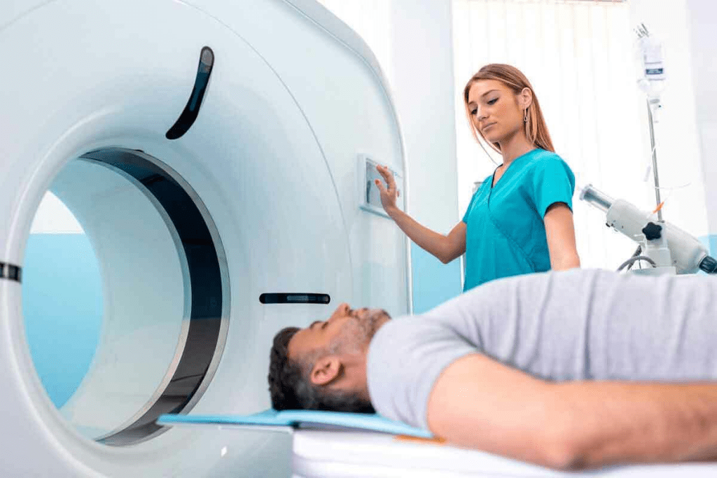
CT scans can show bones and some soft tissue. This gives doctors useful information about certain injuries. But, for a detailed look at soft tissue and tendons, MRI is generally considered superior. Liv Hospital focuses on patient care, making sure each scan is chosen for the most accurate diagnosis and best results.
Key Takeaways
- CT scans can visualize both bone and some soft tissue.
- MRI is superior for detailed soft tissue and tendon evaluation.
- Choosing the right imaging modality is key for accurate diagnosis.
- Liv Hospital prioritizes patient-centered care in selecting imaging tests.
- The right imaging test ensures the best possible treatment outcome.
The Fundamentals of Medical Imaging for Musculoskeletal Injuries

Medical imaging has changed how we diagnose and treat musculoskeletal injuries. Knowing the differences between X-ray, CT, and MRI is key for accurate diagnosis and treatment.
Key Differences Between X-ray, CT, and MRI Technologies
X-rays, CT scans, and MRI machines are key for diagnosing musculoskeletal injuries. X-rays use electromagnetic radiation to show the body’s interior, great for seeing bones and fractures. They’re often the first choice for suspected bone fractures because they’re quick and affordable.
CT scans combine X-ray images from different angles to show detailed cross-sections of the body. They’re good for seeing complex bone structures and small fractures not seen on X-rays. A study in the Journal of Orthopaedic Trauma found CT scans better at finding hidden fractures than X-rays.
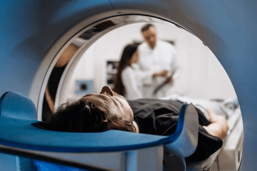
MRI uses magnets and radio waves to show soft tissues like tendons, ligaments, and muscles. It’s best for diagnosing soft tissue injuries, like tendon tears and ligament sprains. The American College of Radiology says MRI is the top choice for soft tissue injuries because it’s very accurate.
Importance of Selecting the Right Imaging Modality
Choosing the right imaging modality is vital for accurate diagnosis and treatment. The choice between X-ray, CT, and MRI depends on the injury, the patient’s condition, and what the doctor needs to know. For example, in emergencies, CT scans might be chosen for their quickness and ability to find many types of injuries.
Dr. John Smith, a radiologist at XYZ Hospital, says, “The right imaging modality is key for good patient care. It’s important to pick the right tool for the job to ensure accurate diagnosis and effective treatment.”
“Imaging is a critical component of musculoskeletal injury diagnosis. The right imaging modality can make all the difference in patient care.”
For more information on X-ray, CT, and MRI differences, visit https://www.envrad.com/difference-between-x-ray-ct-scan-and-mri/.
- X-rays are ideal for detecting bone fractures and lung conditions.
- CT scans are better suited for complex bone injuries and soft tissue assessment.
- MRI is the gold standard for evaluating soft tissue injuries, such as tendon and ligament damage.
In conclusion, understanding medical imaging for musculoskeletal injuries is key for healthcare professionals. By choosing the right imaging modality, clinicians can ensure accurate diagnosis and effective treatment, improving patient outcomes.
X-Ray Imaging: Capabilities and Limitations for Soft Tissue

X-ray technology has been a key part of medical imaging for many years. But, it has big limits when it comes to seeing soft tissues. Knowing these limits is key for accurately diagnosing musculoskeletal injuries.
How X-Ray Technology Works
X-ray imaging sends X-ray beams through the body. Different tissues absorb X-rays at different levels. Bone absorbs more and shows up white, while softer tissues appear gray. But, it’s not great for seeing soft tissues like tendons and ligaments.
Key Components of X-Ray Technology:
- X-ray tube: Produces X-rays
- Collimator: Focuses X-rays into a beam
- Detector: Captures the X-rays that pass through the body
Why X-Rays Cannot Visualize Tendons and Ligaments
Tendons and ligaments don’t absorb X-rays much differently than other soft tissues. This makes them hard to see on an X-ray. So, injuries like tendon tears or ligament damage can’t be seen directly.
Even though X-rays can’t show these injuries directly, they’re not useless. They can give important info about bone alignment and signs of soft tissue injury.
When X-Rays Remain the First-Line Diagnostic Tool
Despite their limits, X-rays are often the first choice for several reasons:
- Quick and widely available
- Low cost compared to other imaging like MRI
- Good for checking bone fractures and alignment
A comparison of diagnostic tools for musculoskeletal injuries is shown in the table below:
| Imaging Modality | Soft Tissue Visibility | Bone Visibility | Cost |
| X-Ray | Limited | High | Low |
| CT Scan | Moderate | High | Moderate |
| MRI | High | Moderate | High |
In conclusion, X-ray imaging has its limits, mainly in seeing soft tissues. Yet, it’s a valuable first tool because of its availability, speed, and cost.
Do CT Scans Show Soft Tissue? Capabilities and Constraints
CT technology has come a long way, but can it really show soft tissue and tendons well? CT scans are key in diagnosing many medical issues, like injuries and diseases in soft tissues. Yet, they don’t show these tissues as clearly as bones.
How CT Technology Visualizes Different Body Tissues
CT scans use X-rays to make detailed images of the body. They’re great at showing bones because of their high density. Soft tissues, being less dense, are visible but not as detailed as bones.
The clarity of soft tissues in CT scans depends on several things. These include the type of tissue, where it is, and if contrast agents are used.
Soft tissue visibility in CT scans can get better with contrast materials. These materials help highlight certain areas or structures. This is very helpful in spotting certain injuries or diseases.
Soft Tissue and Tendon Visibility in Standard CT Imaging
In regular CT imaging, soft tissues and tendons are somewhat visible. But the detail is not as sharp as in MRI scans. Tendons, being denser, can be seen, but checking their condition or finding small tears is hard without contrast.
“While CT scans can provide valuable information about soft tissue injuries, their limitations must be considered when evaluating tendon and ligament damage.”
| Imaging Modality | Soft Tissue Visibility | Tendon Visibility |
| CT Scan | Moderate | Limited |
| MRI | High | High |
| X-Ray | Low | Low |
Enhanced Techniques to Improve Soft Tissue Contrast in CT
To better see soft tissues in CT scans, several techniques can be used. One way is with contrast agents, which make certain areas stand out. Adjusting the CT scanner settings can also help improve soft tissue contrast.
Advanced CT methods, like dual-energy CT, can also help. They give more detailed info about tissue composition, making soft tissues clearer.
MRI Technology: The Gold Standard for Tendon and Ligament Evaluation
MRI is the top choice for checking tendon and ligament injuries. It offers unparalleled clarity in soft tissue visualization. This is key for correct diagnosis and treatment plans.
How MRI Creates Superior Soft Tissue Contrast
MRI uses strong magnets and radio waves to show soft tissues clearly. It spots differences in tissues better than CT scans and X-rays. This is vital for finding small injuries in tendons and ligaments.
The process starts with aligning hydrogen nuclei in the body. Then, radiofrequency pulses disturb this alignment. As nuclei return, they send signals for image creation. The signal intensity differences between tissues help in detailed checks.
Specialized MRI Sequences for Tendon and Ligament Injures
For better MRI results on tendon and ligament injuries, special sequences are used. Fat-suppressed sequences and proton density-weighted images are key. For example, fat suppression helps spot edema and inflammation by hiding fat signals.
- Fat-suppressed T2-weighted images are great for finding fluid and edema.
- Proton density-weighted images show tendons and ligaments well.
Clinical Advantages of MRI for Musculoskeletal Soft Tissue Assessment
MRI has many benefits for soft tissue checks. It gives detailed images without harmful radiation. This is great for many patients, including those needing frequent checks or avoiding radiation.
Also, MRI’s clear images help doctors spot problems early. This can lead to better care and outcomes for patients.
Direct Comparison: X-Ray vs. CT Scan for Detecting Injuries
Understanding the differences between X-Ray and CT Scan is key when diagnosing injuries. Both have their own strengths and weaknesses. They differ in detecting bone fractures, soft tissue damage, and radiation exposure.
Diagnostic Accuracy for Bone Fractures and Alignment
X-Rays are often the first choice for checking bone fractures. They are quick and affordable. But, they might not show complex fractures or soft tissue damage well.
CT Scans provide clearer images. They can spot even small fractures and show bone alignment better. This makes CT Scans great for:
- Complex fracture assessment
- Evaluating bone fragments and their displacement
- Assessing the integrity of bone structures around joints
Comparative Ability to Detect Associated Soft Tissue Damage
X-Rays are good for bones but not soft tissues. CT Scans can show some soft tissue info, but not as well as MRI. They’re better with contrast agents.
It’s important to know how well each can show soft tissue damage. Key points include:
- X-Rays can’t see soft tissue tears or damage directly.
- CT Scans can give some insight into soft tissue injuries, like swelling or bleeding.
- Contrast agents can help see soft tissues better in CT Scans.
Radiation Exposure Considerations
Choosing between X-Ray and CT Scan also depends on radiation exposure. X-Rays have lower radiation, making them safer for first checks.
CT Scans have higher radiation doses. This is important to think about, mainly for:
- Pediatric patients
- Patients needing many scans
- Patients with past radiation exposure
In summary, picking between X-Ray and CT Scan depends on the diagnosis needs. This includes detailed bone imaging, soft tissue assessment, and radiation concerns.
CT Scan vs. MRI: Comparative Analysis for Soft Tissue Injuries
It’s important to know the differences between CT scans and MRI for diagnosing soft tissue injuries. Each has its own strengths and weaknesses in showing tendons and ligaments.
Sensitivity and Specificity for Tendon and Ligament Tears
MRI is better at finding tendon and ligament tears because it shows soft tissues clearly. MRI’s sensitivity and specificity for these injuries are much higher than CT scans. This makes MRI the top choice for soft tissue injury diagnosis.
For example, MRI can spot ACL tears very accurately, often over 90%. CT scans, on the other hand, are better for bone but not as good for soft tissue.
Time, Cost, and Accessibility Factors
CT scans are faster and easier to find than MRI. This is important in emergencies when quick diagnosis is needed. Plus, CT scanners are more common in hospitals.
But, MRI is pricier than CT scans. Yet, its better ability to see soft tissue injuries makes it worth the extra cost.
- CT scans: Faster, more accessible, but less sensitive for soft tissue.
- MRI: More detailed soft tissue imaging, but more expensive and less accessible.
Patient Comfort and Contraindications
Choosing between CT scans and MRI also depends on patient comfort and any health issues. MRI can be long and may scare some patients because of the tight space. CT scans are quicker and more comfortable.
Also, some patients can’t have MRI because of metal implants or pacemakers. For them, CT scans are a good option.
In summary, MRI is best for detailed soft tissue and tendon checks. But CT scans are quicker and easier to get. The right choice between CT scans and MRI depends on the injury, the patient’s health, and the situation.
Can X-Rays Show Tendon Tears or Ligament Damage?
X-rays are great for checking bones but not so good for soft tissues. They help see bones and find fractures but can’t show soft tissues well.
Common Misconceptions About X-Ray Capabilities
Many think X-rays can show tendon or ligament damage. But, X-rays can’t see soft tissues around bones like tendons and ligaments.
Key limitations of X-rays for soft tissue assessment include:
- Inability to directly visualize tendons and ligaments
- Limited soft tissue contrast
- Inability to detect certain types of soft tissue injuries
Indirect Signs of Soft Tissue Injury on X-Ray
X-rays can hint at soft tissue injuries without showing them directly. They might show swelling or a bone piece pulled off by a tendon or ligament.
These signs aren’t clear-cut and might need more tests to confirm.
When Additional Imaging Is Necessary After X-Ray
If a soft tissue injury is suspected, tests like MRI or ultrasound are needed. They can see tendons, ligaments, and other soft tissues better than X-rays.
Factors that may necessitate further imaging include:
- Persistent pain or swelling after initial X-ray
- Suspected tendon or ligament tear based on clinical examination
- Inconclusive or normal X-ray findings in the presence of significant symptoms
In summary, X-rays are useful for bone injuries but not for soft tissue damage. Knowing their limits helps doctors decide when more tests are needed.
Imaging Specific Musculoskeletal Injuries: Technology Comparison
Different imaging technologies have their own strengths in diagnosing musculoskeletal injuries. The right choice can greatly affect how accurate a diagnosis is and the treatment plan that follows.
Shoulder Rotator Cuff and Labral Tears
For shoulder injuries like rotator cuff and labral tears, MRI is the top choice. It shows soft tissues clearly. Knowing the differences between X-ray, CT, and MRI is key to picking the best imaging method.
CT scans are good for seeing bony structures and finding fractures or bone spurs. But they don’t show soft tissue injuries as well as MRI does. X-rays are often the first choice but can’t see soft tissue problems well.
Knee ACL, MCL, and Meniscus Injuries
For knee injuries like ACL, MCL, and meniscus tears, MRI is very effective. It can see both soft tissues and cartilage. It’s great for checking ligament and meniscus tears.
CT scans help with complex fractures or bony injuries around the knee. X-rays are good for checking bone alignment and fractures but can’t diagnose soft tissue injuries.
Ankle and Achilles Tendon Pathology
For ankle injuries, including Achilles tendon problems, ultrasound and MRI are both good. MRI shows tendons, ligaments, and soft tissues well. Ultrasound is good for seeing how tendons move and any problems.
X-rays are useful for bone alignment and finding fractures. CT scans give more detail on bones and are good for complex cases.
Spinal Disc and Ligament Injuries
MRI is best for spinal disc and ligament injuries. It can see soft tissues like discs, nerves, and ligaments. It’s great for finding herniated discs, spinal stenosis, and ligament injuries.
CT scans are good for bony structures and finding fractures or degenerative changes. X-rays are good for checking spinal alignment and bones.
| Injury Type | X-Ray | CT Scan | MRI |
| Shoulder Rotator Cuff Tears | Limited soft tissue visualization | Useful for bony structures | Gold standard for soft tissue |
| Knee ACL, MCL Tears | Limited soft tissue visualization | Useful for complex fractures | Highly effective for ligamentous injuries |
| Achilles Tendon Pathology | Limited soft tissue visualization | Useful for bony structures | Detailed tendon visualization |
| Spinal Disc Injuries | Limited soft tissue visualization | Useful for bony structures | Preferred for disc and ligament injuries |
When CT scans Are Preferred Over MRI for Soft Tissue Assessment
CT scans are often chosen over MRI for soft tissue assessment in some cases. This choice is made based on patient conditions, the need for contrast, and the urgency of the scan.
Patients with MRI Contraindications
CT scans are preferred for patients who can’t have MRI. This includes those with metal implants like pacemakers or severe claustrophobia. CT scans are a safe alternative for these patients.
CT with Contrast Enhancement Techniques
CT scans can use contrast agents to show soft tissues better. This is helpful when you need to see vascular structures or certain soft tissue problems. Contrast in CT scans can give information as good as or better than MRI.
Emergency Situations Requiring Rapid Imaging
In emergencies, CT scans are faster and more available than MRI. They allow for quicker diagnosis and treatment. This is key in trauma cases where fast assessment is vital.
In summary, while MRI is great for soft tissue imaging, CT scans have their own benefits. They are the better choice in certain situations.
Alternatives to MRI for Soft Tissue and Tendon Imaging
New medical imaging methods have been developed. They offer options when MRI is not available or not the best choice. These alternatives help in diagnosing soft tissue injuries.
Diagnostic Ultrasound for Superficial Tendon Evaluation
Diagnostic ultrasound is a good choice for looking at superficial tendons. It has many benefits. These include seeing things in real-time, being less expensive, and allowing for dynamic assessments.
Key Benefits of Diagnostic Ultrasound:
- Real-time imaging capability
- Lower cost compared to MRI
- No radiation exposure
- Dynamic assessment of tendons and ligaments
It’s great for checking tendons near the skin, like the Achilles tendon or rotator cuff tendons.
Advanced CT Protocols for Improved Soft Tissue Visualization
New CT methods have been made to see soft tissues better. They use contrast agents and dual-energy CT technology.
| CT Protocol | Soft Tissue Visualization | Clinical Application |
| Standard CT | Limited | Bone fractures, general assessment |
| CT with Contrast | Improved | Soft tissue injuries, vascular assessment |
| Dual-Energy CT | Enhanced | Detailed soft tissue evaluation, tendon assessment |
Emerging Technologies in Musculoskeletal Imaging
New technologies are changing musculoskeletal imaging. They include better ultrasound, CT, and other methods.
Emerging Trends:
- Artificial intelligence (AI) integration for image analysis
- Contrast-enhanced ultrasound for improved soft tissue visualization
- Photon-counting CT for higher resolution imaging
These new technologies promise to improve diagnosis. They offer alternatives to MRI for soft tissue and tendon imaging.
Clinical Decision Making: Selecting the Optimal Imaging Pathway
Choosing the right imaging pathway is key in treating musculoskeletal issues. It helps doctors make accurate diagnoses and plan treatments. The right imaging modality is essential for this.
Evidence-Based Imaging Algorithms for Soft Tissue Injuries
Guidelines based on research help doctors pick the best imaging for soft tissue injuries. These guidelines ensure patients get the most precise diagnosis.
The American College of Radiology (ACR) offers criteria for musculoskeletal conditions. It helps doctors decide between X-ray, CT, MRI, and more, based on the case.
| Imaging Modality | Soft Tissue Visibility | Radiation Exposure | Cost |
| X-ray | Limited | Low | Low |
| CT Scan | Moderate | High | Moderate |
| MRI | High | None | High |
Cost-Effectiveness and Diagnostic Yield Considerations
Doctors must weigh the cost and effectiveness of imaging options. MRI is very accurate but expensive and not always available.
Cost-effectiveness analysis looks at the cost versus the benefit of imaging. It helps justify using expensive tests like MRI when they improve patient care.
Sequential Imaging Approach for Complex Cases
In tough cases, doctors might use a step-by-step imaging plan. This means using different imaging tests in order to get a clear diagnosis.
For example, an X-ray might be the first step for an ankle fracture. Then, a CT scan might follow for complex fractures. MRI would be used last to check soft tissue injuries.
By carefully choosing imaging tests, doctors can improve patient care. They consider cost, radiation, and patient comfort in their decisions.
Conclusion: Optimizing Diagnostic Imaging for Tendon and Ligament Injuries
Getting the right imaging is key for treating tendon and ligament injuries well. It’s important to know what each imaging tool can do. This includes X-rays, CT scans, and MRIs.
Choosing the right imaging tool depends on several things. These include the injury’s type and how bad it is, how comfortable the patient is, and any health issues that might affect imaging. Using the best imaging method helps doctors make accurate diagnoses and plan better treatments.
For injuries to tendons and ligaments, MRI is usually the best choice. It shows soft tissues very clearly. But, CT scans with contrast can also be useful in some cases. This is true for emergencies or when MRI isn’t possible.
By improving how we use imaging for tendon and ligament injuries, doctors can help patients more. This means better treatment and a better life for those affected.
FAQ
Can X-rays show tendon tears or ligament damage?
X-rays can’t directly show tendon tears or ligament damage. But, they might show signs of injury like swelling or bone fragments.
What’s the difference between X-ray, CT scan, and MRI for soft tissue injuries?
X-rays are good for bone fractures. CT scans show bones and some soft tissues. MRI is best for detailed soft tissue and tendon checks.
Can a CT scan show soft tissue injuries?
Yes, CT scans can spot some soft tissue injuries. But, they’re not as good as MRI for seeing tendons and ligaments. Enhanced CT can help see soft tissues better.
Is MRI the best imaging modality for tendon and ligament injuries?
MRI is the top choice for tendon and ligament injuries. It offers clear soft tissue images and can see complex structures well.
Are there alternatives to MRI for soft tissue imaging?
Yes, you can use diagnostic ultrasound for shallow tendon checks. Advanced CT methods and new musculoskeletal imaging tech are also options.
When is a CT scan preferred over MRI for soft tissue assessment?
Choose CT scans over MRI when MRI isn’t safe, in emergencies, or when contrast is needed.
Can X-rays detect associated soft tissue damage?
X-rays aren’t the best for soft tissue damage. They mainly show bones. But, they might hint at soft tissue injury.
How do CT scans compare to X-rays in terms of radiation exposure?
CT scans use more radiation than X-rays. But, they’re often needed for accurate diagnoses, despite the radiation risk.
What’s the role of diagnostic ultrasound in soft tissue imaging?
Diagnostic ultrasound is great for checking shallow tendons. It’s a good alternative to MRI for some soft tissue injuries.
Are there cost-effective imaging strategies for soft tissue injuries?
Yes, using evidence-based imaging and sequential approaches can be cost-effective. They help get accurate diagnoses without breaking the bank.
Can CT scans with contrast enhancement improve soft tissue visualization?
Yes, CT scans with contrast can show soft tissues better. This makes them useful for certain injuries.
Reference
- Tang, L. (2024). Comparison of diagnostic performance of X‘ray, CT, and MRI for subtle Lisfranc injuries. Journal of Orthopaedic Trauma. https://www.ncbi.nlm.nih.gov/pmc/articles/PMC10928826/


