Last Updated on November 27, 2025 by Bilal Hasdemir
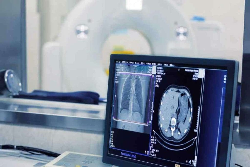
A fluorodeoxyglucose PET scan (FDG-PET) is one of the most important tools in modern medical imaging. It helps detect and manage diseases such as cancer by showing how active cells are inside the body.
During a fluorodeoxyglucose PET scan, doctors use a special tracer called 18F-FDG, a compound that behaves like glucose. Cells that consume a lot of glucose ” such as cancer cells and brain cells ” absorb this tracer. Inside the body, it converts into 18F-FDG-6-phosphate, allowing doctors to visualize areas with high metabolic activity.
At Liv Hospital, we rely on the precision of fluorodeoxyglucose PET scans to detect disease early and guide effective treatment.
Key Takeaways
- 18F-FDG is a radiotracer used in PET scans to measure metabolic activity.
- It is very useful in cancer care for finding tumors and checking how treatments work.
- 18F-FDG helps diagnose many conditions, including cancer and brain disorders.
- These scans give vital info for managing diseases and planning treatments.
- The scan uses a small amount of 18F-FDG, usually 5 to 10 millicuries.
The Fundamentals of Medical Imaging with PET Technology
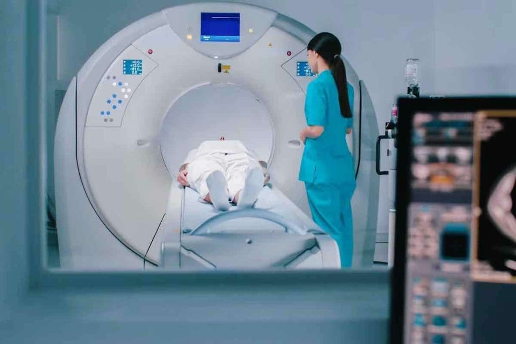
PET scanning is a cutting-edge medical imaging method. It uses radiotracers to measure how active cells are. This tool is key in modern medicine, helping us see and understand the body’s functions and diseases.
How Positron Emission Tomography Works
PET scanning detects gamma rays from a special molecule in the body. This molecule, Fluorodeoxyglucose (FDG), is a glucose molecule with a radioactive tag. It goes to cells that are very active, like cancer cells.
The PET scanner picks up these gamma rays. It makes detailed pictures of how active the body’s cells are. This is super helpful for finding and treating diseases, like cancer.
The Evolution of PET Scanning in Medicine
PET scanning has grown a lot over the years. It started mainly for research but now is a key tool in diagnosing diseases. Thanks to better scanners, tracers, and algorithms, PET scans are more accurate and useful.
Now, PET scanning helps in many areas, like cancer, brain, and heart diseases. It gives us detailed info on how the body works. This info helps doctors understand diseases better, along with CT and MRI scans.
As PET technology keeps getting better, we’ll see even more precise diagnoses. It will also help in more medical areas.
What Is a Fluorodeoxyglucose PET Scan?
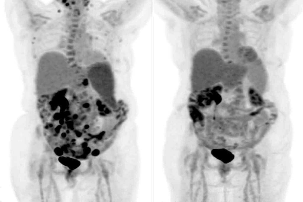
A Fluorodeoxyglucose (FDG) PET scan is a cutting-edge medical imaging method. It shows how the body uses glucose, helping us spot problems. This tool is key for finding and understanding abnormal activity in tissues.
Definition and Basic Principles
An FDG PET scan uses a special tracer called 18F-FDG. This tracer is a glucose molecule with a radioactive fluorine atom. When it’s injected, it goes to cells based on how much glucose they use.
This lets us see and measure how active different tissues are. It’s very helpful for figuring out what’s wrong and how to treat it.
The idea behind FDG PET scans is simple. Cells that use a lot of glucose, like some cancer cells, take up more 18F-FDG. By tracking this radiation, we can make detailed pictures of where glucose is being used in the body.
Key Components of FDG PET Imaging
The main parts of FDG PET imaging are the 18F-FDG tracer, the PET scanner, and special software. The 18F-FDG acts like glucose, getting taken up by cells everywhere. The PET scanner picks up the radiation from 18F-FDG, creating detailed images.
Key elements of FDG PET imaging include:
- The 18F-FDG radiotracer
- The PET scanner
- Image reconstruction software
- Patient preparation and scanning protocols
Comparing FDG PET to Other Imaging Modalities
FDG PET scans have big advantages over other imaging methods. Unlike CT or MRI, which show the body’s structure, FDG PET scans show how tissues are working. This is super useful for finding and tracking diseases like cancer.
When we look at FDG PET compared to other methods, we see it’s better at showing metabolic activity. For example, CT scans are great for seeing the body’s structure, but FDG PET shows how tissues are functioning. This makes FDG PET a valuable tool in medical imaging.
Comparison of FDG PET with other imaging modalities:
| Imaging Modality | Primary Information | Key Applications |
| FDG PET | Metabolic activity | Cancer diagnosis, staging, and monitoring |
| CT | Anatomical detail | Structural abnormalities, trauma assessment |
| MRI | Soft tissue detail | Neurological disorders, soft tissue tumors |
The Science of 18F-FDG as a Radiotracer
Understanding 18F-FDG requires knowledge of its chemical makeup, how it’s made, and its physical traits. It’s a glucose-like substance with a special atom, fluorine-18, added to it. This makes 18F-FDG a key tool in PET imaging.
Chemical Structure of Fludeoxyglucose F 18
18F-FDG is very similar to glucose but has a fluorine-18 atom instead of a hydroxyl group. This change lets it act as a tracer in PET scans without affecting its metabolic path much.
For more details on its chemical structure, check out NCBI’s book on PET imaging.
Production and Synthesis of 18F-FDG
Making 18F-FDG starts with creating fluorine-18, often by hitting oxygen-18 with protons in a cyclotron. This fluorine-18 is then mixed with glucose to create 18F-FDG.
Half-Life and Physical Properties
Fluorine-18 has a half-life of about 110 minutes. This is perfect for PET imaging because it gives enough time for the tracer to be taken up and imaged without too much radiation exposure. The physical traits of 18F-FDG, like its positron emission, are key for clear PET images.
| Property | Value | Description |
| Half-Life | 110 minutes | Ideal for PET imaging, balancing uptake time and radiation safety |
| Positron Emission Energy | 0.633 MeV | Maximum energy of positrons emitted, influencing image resolution |
| Decay Mode | β+ (Positron Emission) | Primary mode of decay, enabling PET imaging |
Metabolic Pathway: How 18F-FDG Mimics Glucose
It’s important to know how 18F-FDG works like glucose in our bodies. We’ll see how it gets into cells, gets changed, and stays in active tissues. This is like how glucose works in our bodies.
Cellular Uptake Mechanisms
The journey starts when cells take in 18F-FDG. Glucose transporters, like GLUT1, help 18F-FDG get into cells. This first step is key because it decides how much 18F-FDG builds up in cells.
Once inside, 18F-FDG goes through the same steps as glucose. But, unlike glucose, 18F-FDG can’t be broken down completely. This lets it be seen with PET scans.
The Phosphorylation Process
Inside the cell, 18F-FDG gets changed by hexokinase into 18F-FDG-6-phosphate. This step is important because it keeps 18F-FDG from leaving the cell. It can’t be broken down further.
This change helps 18F-FDG build up in cells that use a lot of glucose, like cancer cells. This makes it great for PET scans.
Metabolic Trapping in Tissues
18F-FDG gets trapped because 18F-FDG-6-phosphate can’t be used by other enzymes. It can’t go back to its original form to leave the cell. So, it stays in tissues that use a lot of energy, like tumors or areas with lots of activity.
This buildup lets us see these tissues with PET scans. It gives us important info about what’s happening in our bodies.
The Complete FDG PET Scan Procedure
The FDG PET scan procedure is a detailed process. It includes preparation, the scan itself, and care after the scan. Getting ready well, doing the scan right, and taking care after are all key parts.
Pre-Scan Patient Preparation
Before the FDG PET scan, patients need to get ready. They usually fast for 4-6 hours to keep blood sugar steady. They should also avoid hard exercise and sugary foods or drinks.
It’s important to tell the doctor about any medicines being taken. Some might need to be changed or stopped.
The Injection and Uptake Period
Then, patients get an FDG radiotracer injection. The uptake period, lasting 60-90 minutes, lets the FDG absorb into body tissues. During this time, patients rest quietly to avoid extra activity that could mess up the scan.
The Scanning Process
The scanning part has the patient lying on a table that slides into the PET scanner. The scanner picks up the FDG’s radiation to make detailed images of metabolic activity. This process is usually painless and can take 30 minutes to several hours, depending on the area and the scan type.
Post-Scan Care
After the scan, patients can usually go back to their normal day unless told not to by their doctor. Drinking lots of water helps get rid of the radiotracer. Most people don’t have side effects, but some might feel tired or have a bit of soreness at the injection site.
As one doctor said,
“The FDG PET scan has changed how we find and treat cancer and other diseases. It gives us deep insights into how the body works.”
The Warburg Effect: Why Cancer Cells Light Up
Understanding the Warburg effect helps us see why cancer cells show up on PET scans. These cells use glycolysis for energy, even when oxygen is available. This is different from normal cells.
Altered Metabolism in Cancer Cells
Cancer cells have a different way of making energy than normal cells. They rely more on glycolysis, which is less efficient. The Warburg effect is a key sign of this change.
Key aspects of altered metabolism in cancer cells include:
- Increased glucose uptake
- Enhanced glycolytic rate
- Reduced oxidative phosphorylation
Glucose Transporters and Hexokinase Activity
Cancer cells take up more glucose because of special proteins called glucose transporters (GLUT proteins). GLUT1 is very important in many cancers. Inside the cell, glucose is changed into a form that hexokinase can act on.
Hexokinase changes 18F-FDG into a form that gets trapped in cancer cells. This makes them show up on PET scans.
Visual Representation of Tumors on PET Images
The Warburg effect makes cancer cells take up a lot of 18F-FDG. This shows up as bright spots on PET scans. These spots mean there’s cancer.
Characteristics of tumors on PET images include:
- High contrast between tumor and surrounding tissue
- Clear delineation of tumor boundaries
- Ability to detect metabolically active tumors
By understanding the Warburg effect and how it affects 18F-FDG uptake, we can see how PET imaging helps in finding cancer.
Clinical Applications in Oncology
In oncology, FDG PET scans are key for diagnosing, staging, and managing cancer. They help us understand tumor metabolism, which is vital for treatment planning.
Initial Cancer Diagnosis and Staging
FDG PET scans are vital for cancer diagnosis and staging. They show high metabolic activity areas, helping us find tumors and metastases. This info is key for accurate staging and treatment choices.
Accurate staging is critical. It shows how far cancer has spread, guiding treatment. Localized cancer might need surgery or radiation. But widespread cancer might need chemotherapy or immunotherapy.
Treatment Planning and Response Assessment
After diagnosis and staging, FDG PET scans help with treatment planning and response assessment. They show how tumors react to treatment, helping adjust plans if needed.
Being able to see treatment response early is very helpful. It lets us stop ineffective treatments and switch to better ones. This improves patient outcomes and quality of life.
Surveillance and Recurrence Detection
After treatment, FDG PET scans monitor for recurrence. Regular scans help catch recurrence early, when it’s easier to treat.
Early detection of recurrence greatly improves patient outcomes. It allows for timely treatment, potentially leading to better results.
Specific Cancer Types and FDG PET Efficacy
FDG PET scans are effective for many cancers, like lymphoma, lung cancer, colorectal cancer, and melanoma. They provide metabolic information that complements anatomical images.
In lymphoma, FDG PET scans are great for disease extent and treatment response. For lung cancer, they help with staging and treatment monitoring.
Beyond Cancer: Other Applications of FDG PET Imaging
FDG PET scans are not just for cancer. They help in many medical fields, giving doctors important information. This helps in making the right diagnosis and treatment plans.
Neurology and Brain Disorders
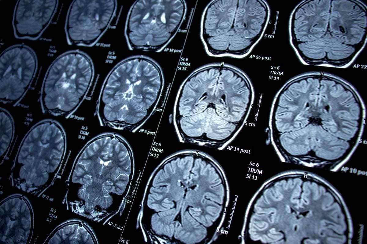
In neurology, FDG PET scans look at brain disorders. They show how brain cells use glucose, which is key for diagnosing diseases like Alzheimer’s and Parkinson’s. NCBI research shows FDG PET’s role in spotting brain activity changes.
FDG PET also helps with seizure disorders and brain injuries. It shows the brain’s metabolic state. This info is key for treatment plans and understanding disease outcomes.
Cardiology Applications
In cardiology, FDG PET scans check if heart muscle is alive but not working. This is important for treating heart disease or after a heart attack.
FDG PET scans find areas of the heart that might work again with treatment. This helps doctors plan treatments that fit each patient’s needs.
Infection and Inflammation Assessment
FDG PET scans are also good for finding infections and inflammation. They help when other tests can’t. They show where glucose uptake is high, which means there’s an infection or inflammation.
FDG PET’s ability to find infections is very useful. It helps doctors choose the right antibiotics and check if treatments are working. This shows how useful FDG PET is in many medical areas.
Radiation Exposure and Safety Considerations
When you get a Fluorodeoxyglucose (FDG) PET scan, you get some radiation. This is something we take very seriously. We work hard to make sure you’re safe from too much radiation.
Typical Radiation Doses from F 18 FDG PET Scans
The amount of radiation from an F 18 FDG PET scan varies. It can be between 7.5 to 30 mSv. This depends on many things like your health, the scanner, and how much tracer you get.
It’s key to find the right dose for good images and less radiation.
A study found the average dose from an FDG PET scan is about 14 mSv. This helps us compare it to other tests.
Comparison with Other Imaging Procedures
Let’s compare the radiation dose from FDG PET scans to other tests. For example, a chest CT scan usually has a dose of about 7 mSv. But, a PET/CT scan can have a dose of up to 25 mSv or more, depending on the CT part.
The International Commission on Radiological Protection says the risk of health problems from radiation depends on the dose. So, knowing these doses helps us choose the best tests.
“The goal is to keep the radiation dose as low as reasonably achievable (ALARA) while getting good images.” –
Radiological Society of North America
Safety Protocols for Patients and Staff
We have many safety steps to follow. For patients, we plan the dose carefully. We make sure it’s enough for good images but not too much. Staff follow strict rules to stay safe, like wearing protective gear and keeping a safe distance from radiation.
- Patient preparation and education
- Optimization of scan protocols
- Use of dose-reducing technologies
- Regular quality control and maintenance of PET scanners
By following these steps, we keep everyone safe from too much radiation.
Advanced Technology and Protocols in Modern FDG PET
The latest in FDG PET technology has made imaging more accurate and detailed. We see big steps forward, like hybrid imaging, better scanner quality, and new protocols.
Hybrid Imaging Systems: PET/CT and PET/MRI
Hybrid imaging mixes PET scans with CT or MRI scans. This mix gives a clearer view of the body’s inner workings.
Systems like PET/CT and PET/MRI have changed how we diagnose. PET/CT shows metabolic activity and CT details, helping pinpoint issues. PET/MRI, with its better soft tissue contrast, is great for brain and cancer studies.
Improvements in Resolution and Sensitivity
Today’s PET scanners can spot smaller problems and track treatment better.
New detector tech and algorithms have led to clearer images and better sensitivity. This helps doctors spot and track conditions more accurately, improving patient care.
LivHospital’s Academic Protocols for F-FDG PET Scanning
LivHospital leads in using the latest F-FDG PET scanning protocols. This ensures top-notch care for patients.
LivHospital’s dedication to excellence shows in its strict F-FDG PET scanning protocols. By keeping up with new research, the hospital offers the latest diagnostic tools to its patients.
Digital PET Technology Advancements
Digital PET technology is a big step up in nuclear medicine. It offers better image quality and quicker scans.
The arrival of digital PET technology has greatly improved image quality and confidence in diagnosis. Its better sensitivity and resolution will make FDG PET even more valuable in medical practice.
Conclusion: The Future of FDG PET in Precision Medicine
Fluorodeoxyglucose (FDG) PET scans are key in today’s medicine, mainly in fighting cancer, studying the brain, and heart health. They have grown a lot, giving clear images that help doctors diagnose and plan treatments better.
We see FDG PET scans staying important in precision medicine. They will help make treatments fit each patient’s needs. With new tech like PET/CT and PET/MRI, they will get even better at finding and treating diseases.
As precision medicine gets better, FDG PET scans will play an even bigger role. They will help us understand how diseases work and grow. We’re excited to use these advances to give top-notch care to patients everywhere, with kindness and thoroughness.
FAQ
What is 18F-FDG and how does it work in PET scans?
18F-FDG, or Fludeoxyglucose F 18, is a special tracer used in PET scans. It looks like glucose and gets taken up by cells, like cancer cells, because they are very active.
How does 18F-FDG PET scanning help in cancer diagnosis?
18F-FDG PET scans help find cancer by showing where cells are very active. This helps doctors spot tumors, figure out how far cancer has spread, and check if treatments are working.
What is the difference between FDG PET and other imaging modalities like CT or MRI?
FDG PET scans show how active tissues are, unlike CT or MRI which show structure. This makes PET scans great for finding and managing cancer.
How is 18F-FDG produced and what are its physical properties?
18F-FDG is made in a cyclotron, where fluorine-18 is attached to glucose. It has a 110-minute half-life, which is important for its use in medical imaging.
What preparation is required before undergoing an FDG PET scan?
Patients need to fast for 4-6 hours before the scan. This helps get clearer images. They should also avoid hard exercise and drink plenty of water.
How does the Warburg effect relate to 18F-FDG uptake in cancer cells?
The Warburg effect says cancer cells use more glucose than normal cells, even with oxygen. This means they take up more 18F-FDG, making them visible on PET scans.
Are there any risks associated with FDG PET scans, particularlly regarding radiation exposure?
Yes, FDG PET scans use a small amount of radiation. But the benefits usually outweigh the risks. Safety steps are taken to protect patients and staff.
Can FDG PET scans be used for conditions other than cancer?
Yes, FDG PET scans are used for more than just cancer. They help in neurology, cardiology, and for checking infections and inflammation.
What advancements have been made in FDG PET technology?
New tech in FDG PET includes hybrid systems like PET/CT and PET/MRI. There are also better resolution and sensitivity, and digital PET tech. These advancements improve PET scan accuracy and usefulness.
How does LivHospital utilize FDG PET scans in their diagnostic protocols?
LivHospital uses the latest in FDG PET scanning for accurate diagnoses. They use advanced PET tech to help patients, focusing on oncology and complex conditions.
Reference
- Wong, T. Z., and Sundaram, M. Positron Emission Tomography (PET). Radiographics. https://radiopaedia.org/articles/positron-emission-tomography


