Last Updated on October 21, 2025 by mcelik

Understanding the difference between MRI scan vs PET scan is essential to making informed health decisions. At Liv Hospital, we guide you through the latest medical imaging technologies to ensure you receive the best and most personalized care. MRI scans use strong magnets and radio waves to create highly detailed images of your body’s structures, making them excellent for detecting issues with the brain, spine, joints, and soft tissues.
In contrast, PET scans use a small amount of radioactive tracer to visualize how your organs and tissues function. This scan shows metabolic activity, helping doctors identify cancers, monitor heart function, and assess brain disorders like Alzheimer’s disease. While MRI focuses on anatomy and physical structures, PET scans highlight how body systems are working at a cellular level. Together, understanding MRI scan vs PET scan empowers patients to know which imaging method best suits their medical needs and how each plays a role in accurate diagnosis and treatment planning.

Medical diagnostics have changed a lot with new imaging tech like MRI and PET scans. These tools help doctors find and treat diseases better.
Medical imaging has grown from simple X-rays to advanced tools like MRI and PET scans. These changes have made medicine better, helping doctors diagnose and treat diseases. The growth of these technologies comes from advances in physics, engineering, and computer science.
Advanced imaging is key in modern medicine. It helps find and treat diseases early. MRI and PET scans are key tools, giving doctors important insights. Knowing the difference between MRI and PET scan helps choose the right test.
The MRI scan is a cutting-edge medical imaging method. It uses strong magnetic fields and radio waves to see inside the body. This non-invasive tech is key in diagnosing diseases, showing detailed images of organs and soft tissues without surgery.
MRI machines align hydrogen atoms in the body with a strong magnetic field. Then, a radiofrequency pulse is sent, making these atoms send signals. The MRI captures these signals to create detailed images.
MRI Technology Key Components:
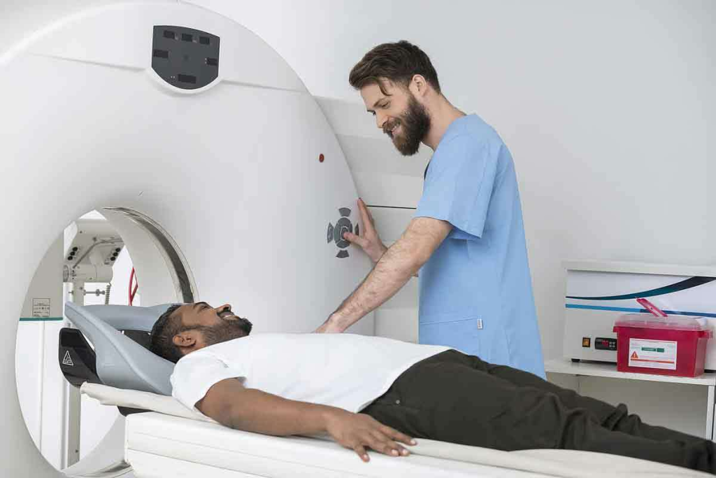
The MRI scanner is a large, tube-shaped machine. It can make loud knocking and tapping sounds during the scan. Patients lie on a movable table that slides into the scanner.
The environment can be scary for some. It requires being inside the machine for 15 to 90 minutes, depending on the scan type. Modern MRI facilities offer earplugs or headphones to reduce noise. Some even provide virtual reality or video entertainment to distract from the scanner’s confines.
Our MRI facilities are designed to be comfortable and safe. Our staff is dedicated to making your experience as pleasant as possible.
PET scans are a top-notch tool in medicine. They use a small amount of radioactive material to see how different parts of the body work. This is key to finding and treating diseases like cancer, brain issues, and heart problems.
Getting a PET scan starts with a small injection of a radioactive tracer. This tracer goes to areas where the body is very active. FDG (Fluorodeoxyglucose) is a common tracer that finds where the body uses a lot of sugar.
As the tracer breaks down, it sends out gamma rays. These rays are caught by the PET scanner. This creates detailed pictures of how the body works.
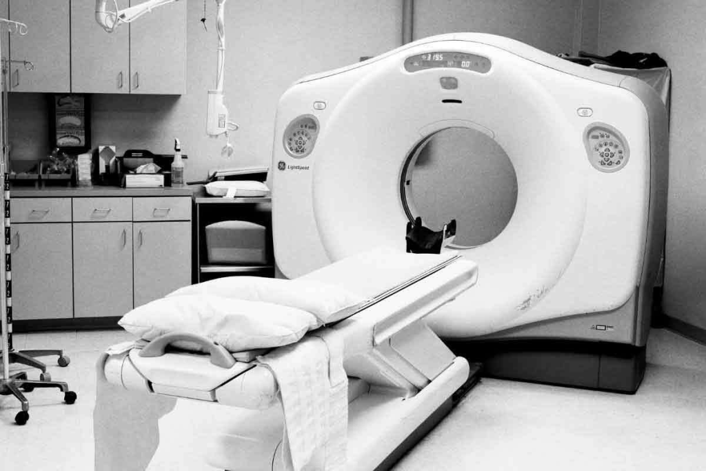
The PET scanner is a big, ring-shaped machine. It wraps around the patient to catch the gamma rays. It then makes images of what’s happening inside the body.
Radioactive tracers are the heart of PET scans. They are made to find specific things in the body. For example, FDG finds cancer because it goes to cells that use a lot of sugar.
Which tracer is used depends on what doctors want to see. Different tracers show different things, like how much oxygen cells use or how fast they grow.
| Tracer | Target | Clinical Use |
| FDG | Glucose Metabolism | Cancer Detection, Neurological Disorders |
| Rubidium-82 | Myocardial Perfusion | Cardiac Function Assessment |
| Florbetapir | Amyloid Plaques | Alzheimer’s Disease Diagnosis |
The table shows how different tracers are used for different things. This shows how flexible PET scans are in helping doctors diagnose and treat many conditions.
Understanding PET scans and tracers shows their importance in medicine. They help doctors find diseases early and accurately. They also help plan treatments and check if they’re working.
MRI and PET scans are both important tools for doctors. They work differently and give different kinds of information. Knowing these differences helps patients and doctors make better choices about tests.
MRI scans use a strong magnetic field and radio waves to show detailed images of the body. PET scans, on the other hand, use a small amount of radioactive tracer. This tracer is absorbed by cells and sends signals to the PET scanner.
MRI technology is great for seeing soft tissues like the brain and spine. PET scans are better for checking how tissues work. They help find cancer, brain problems, and heart diseases.
MRI and PET scans make different kinds of images. MRI scans show detailed pictures of body structures. PET scans show how tissues work by highlighting their metabolic activity.
Here’s a quick look at what MRI and PET scans show:
MRI scans don’t use radiation, using magnetic fields and radio waves instead. PET scans, though, do involve a small amount of radiation from the tracer.
Even though PET scans have low radiation, it’s something to think about, mainly for those needing many scans. Yet, the benefits of PET scans, like finding and tracking cancer, often outweigh the risks.
MRI scans have changed how we diagnose diseases. They show detailed images of what’s inside our bodies. This helps doctors find and track many health issues.
MRI scans are key for brain and spinal cord problems. They spot issues like multiple sclerosis, stroke, and tumors. This is because MRI can see inside the body very well.
Using MRI early can help manage diseases better. For example, it shows brain damage after a stroke. This helps doctors plan the best treatment.
MRI scans are great for looking at muscles, bones, and joints. They help find injuries like sprains and tears. They also track conditions like osteoarthritis.
| Condition | MRI Findings | Clinical Utility |
| Meniscal Tears | Disruption of meniscal tissue | Guiding surgical or conservative management |
| Ligament Sprains | Inflammation and disruption of ligament fibers | Assessing injury severity and guiding rehabilitation |
| Osteoarthritis | Joint space narrowing, cartilage loss, and bone spurs | Monitoring disease progression and response to treatment |
MRI scans also check the heart and blood vessels. They look at blood flow and heart function. This is important for diagnosing heart diseases.
MRI gives clear pictures of the heart and blood vessels. This helps doctors understand how well the heart is working. For example, it shows how much damage there is after a heart attack.
In summary, MRI scans have many uses in medicine. They help doctors see inside the body clearly. As technology improves, MRI will likely play an even bigger role in health care.
PET scans are used in many areas, like oncology, neurology, and cardiology. They help doctors see how the body works. This is key to diagnosing and treating different health issues.
In oncology, PET scans are great for finding and tracking cancer. Cancer cells use more energy than normal cells. So, PET scans can spot tumors and check if treatments are working.
Here are some ways PET scans help in oncology:
The table below shows how PET scans help with cancer:
| Application | Description | Benefits |
| Cancer Detection | Identifying cancerous tissues | Early detection, improved prognosis |
| Treatment Monitoring | Assessing response to therapy | Personalized treatment plans |
| Recurrence Detection | Identifying cancer recurrence | Timely intervention |
PET scans also help with neurological issues like Alzheimer’s and Parkinson’s. They look at brain activity. This helps doctors understand how these diseases progress.
Here are some uses of PET scans in neurology:
In cardiology, PET scans check how well the heart works and find coronary artery disease. They look at blood flow to the heart. This helps doctors find where blood flow is low.
Here are some uses of PET scans in cardiology:
In conclusion, PET scans are used in many areas, from cancer to heart health. They give important information about how the body works. This helps doctors diagnose and treat many diseases.
Doctors have to think carefully when picking between MRI and PET scans for patients. They look at the patient’s condition, what kind of info they need, and the patient’s own situation.
Healthcare pros pick MRI for detailed body images. MRI is great for seeing structural issues like injuries or tumors. But, PET scans are better for showing how tissues work, which is key for cancer checks.
They also think about whether they need contrast agents and the risks they might bring. MRI uses magnetic fields and radio waves. PET scans use tiny amounts of radiation.
PET scans are top for checking cancer because they show how tissues work. MRI is better for soft tissue images. It’s great for injuries or muscle problems.
In brain issues, MRI and PET scans both play a part. MRI shows the brain’s structure. PET scans show brain function, helping with Alzheimer’s diagnosis.
Often, doctors use MRI and PET scans together. For example, in cancer, a PET scan and MRI can give a full picture. This helps doctors make better plans for treatment.
Choosing between MRI and PET scans is complex. It needs a deep understanding of the patient’s needs and the strengths of each scan.
It’s important to know the safety and contraindications of MRI and PET scans. These imaging tools have changed medicine a lot. But they also have safety issues that doctors must think about carefully.
MRI scans are usually safe, but there are some things to watch out for. People with metal implants, like pacemakers, can’t have an MRI. The MRI’s strong magnetic field can harm these devices.
Some big safety worries for MRI include:
PET scans use a little bit of radiation, which is a worry for some, like pregnant women and kids. The radioactive tracers in PET scans are mostly safe. But there are rules to follow to keep exposure low.
Some main safety worries for PET scans are:
Knowing these safety points helps doctors decide when to use MRI and PET scans. This way, patients get the best and safest imaging for their health needs.
It’s important to know the differences between PET scans and MRI for managing cancer. Both are key in finding and treating cancer, but they are used in different ways.
PET scans are great for finding cancer early. They show where cancer cells are active, even if they’re small. This makes them good at spotting tumors that other scans can’t see.
PET Scan Sensitivity: PET scans can find cancer cells, even when they’re tiny or hard to see with other scans.
PET scans and MRI help see how well the treatment is working. PET scans show if a tumor is changing, which means it’s responding to treatment. MRI gives detailed pictures of the tumor’s size and where it is.
Monitoring Treatment: Using both PET scans and MRI gives doctors a full picture of how the cancer is reacting to treatment. This helps them change the treatment plan if needed.
| Imaging Technique | Cancer Detection | Treatment Monitoring |
| PET Scan | High sensitivity for detecting metabolically active cancer cells | Effective for monitoring changes in metabolic activity |
| MRI | Excellent for detailed imaging of tumor size and location | Provides precise measurements of tumor response to treatment |
Even though PET scans and MRI are very helpful, they have their limits. PET scans use radioactive tracers and might not be as clear. MRI shows soft tissue well, but might miss metabolic changes in cancer.
Choosing the Right Tool: The choice between PET scans and MRI depends on the cancer type, stage, and the patient’s health. It’s a decision made by doctors based on the specific situation.
Hybrid imaging technologies combine different imaging methods to offer better diagnostic tools. We’re seeing a big change in medical diagnostics with PET-MRI and PET-CT. These technologies mix PET’s functional info with MRI or CT’s detailed images.
The main benefit of hybrid imaging is getting detailed didiagnostic informationn one go. PET-MRI mixes PET’s functional data with MRI’s soft tissue contrast. This helps diagnose cancer and neurological issues more accurately. PET-CT combines PET’s info with CT’s detailed anatomy, helping in cancer treatment and response checks.
Hybrid imaging is used in many clinical areas. PET-MRI is great for neurological and musculoskeletal imaging. It offers both functional and anatomical info. PET-CT is key in oncology for cancer staging and treatment monitoring.
These technologies boost diagnostic confidence and cut down on extra imaging tests. They also improve patient care by providing accurate staging and treatment plans.
As tech advances, we’ll see more in hybrid imaging. Expect better PET detectors, advanced image algorithms, and AI for better image analysis. These advancements will likely lead to more precise diagnostics and treatments.
The future of hybrid imaging looks bright for better patient care. As these technologies get better and more common, they’ll play a big role in personalized medicine and targeted treatments.
Understanding MRI and PET scans is key to making smart choices in medical imaging. The right choice depends on the condition, the patient’s needs, and what information is needed. This could be about the body’s structure or how it works.
When picking between MRI and PET scans, think about what each offers. MRI scans give detailed pictures of the body’s inside without using radiation. They’re great for looking at the brain, spine, and muscles. PET scans, on the other hand, show how cells are working. This is important for finding and tracking cancer, brain issues, and heart problems.
Knowing the strengths of each scan helps patients and doctors make better choices. This leads to more accurate diagnoses and better treatment plans. As imaging technology grows, staying up-to-date is vital for the best care.
Medical imaging procedures can be tough for patients and doctors to handle. It’s key to have good resources and support to make smart choices.
Many groups and websites have educational materials and support for those going through medical imaging. They help with understanding MRI or PET scans, results, and care plans.
These resources explain medical imaging in detail. This helps patients feel less worried and anxious. Doctors can also use them to learn about new imaging tech and care methods.
Looking for more help? There are many online resources, like patient groups and medical organizations. They offer support and connect people with others who’ve had similar experiences.
MRI scans show the structure of the body. PET scans show how the body works. MRI is great for seeing inside the body. PET scans are good for seeing how different parts work.
Both MRI and PET scans are good for finding cancer. PET scans find cancer by looking at how active cells are. MRI shows tumors and the tissue around them.
MRI uses a strong magnetic field and radio waves. PET scans use radioactive tracers. This means MRI is better for seeing structures. PET is better for seeing how things work.
Yes, PET scans use a little radiation. MRI does not. The radiation from PET scans is safe, but it’s something to think about.
Yes, PET-MRI combines both. It gives detailed information in one scan. This is useful for complex cases or when detailed planning is needed.
MRI can be safe, but there are risks. It can affect metal implants and cause claustrophobia. People with certain implants or severe claustrophobia should not have an MRI.
PET scans are key in cancer care. They help find cancer, see how far it has spread, and check if treatment is working. They show cancer’s activity, which is important for treatment.
Doctors choose based on what they need to know and the patient’s situation. They think about each scan’s strengths and what they can do together.
No PET scans for pregnant women, breastfeeding moms, or those with certain conditions. People with diabetes or those fasting for too long need special care.
Hybrid imaging, like PET-MRI, combines scans for better results. They help doctors make accurate diagnoses and plan treatments. These technologies are improving medical imaging and patient care.
Subscribe to our e-newsletter to stay informed about the latest innovations in the world of health and exclusive offers!
WhatsApp us