Understanding your PSMA PET scan report is key to making smart choices about your prostate cancer care. At LivHospital, we focus on you and set high standards. This helps you grasp these results with confidence and clarity.
Prostate cancer diagnosis and treatment depend a lot on PET scan interpretation. The PSMA PET/CT tech is vital. It spots areas of PSMA uptake, showing prostate cancer cells might be there.
Getting PET scan results right is vital for planning your treatment. We walk you through it. This way, you’ll know your diagnosis and what it means for your care.

PSMA PET scans are key to finding and treating prostate cancer. They are very good at spotting cancer cells. This makes them a vital tool in prostate cancer care.
A PSMA PET scan is a special test that finds prostate cancer cells in the body. It uses a radioactive tracer that sticks to a protein on cancer cells. This protein is called Prostate-Specific Membrane Antigen (PSMA).
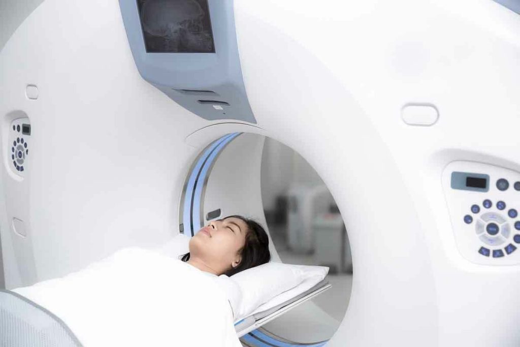
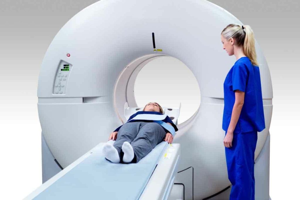
The test starts with a small dose of the tracer being injected into the patient. It goes to areas with lots of PSMA, like prostate cancer cells. A PET scanner then shows where the cancer is by detecting the tracer’s radiation.
PSMA is very important for finding prostate cancer. It’s found more in cancer cells than in healthy cells. This makes it a great target for imaging tests.
Key benefits of PSMA PET scans include:

To understand PSMA PET scans, knowing about PSMA uptake is key. These scans have changed how we find and treat prostate cancer. They are very good at spotting cancer cells.
PSMA tracers stick to the Prostate-Specific Membrane Antigen (PSMA) on prostate cancer cells. This lets us see cancerous tissues through PET imaging. It’s very precise, helping doctors find cancer even when PSA levels are low.
“The use of PSMA tracers has greatly improved PET scans for finding prostate cancer,” say experts. They are very specific for prostate cancer cells, which is why they’re so important for diagnosis and planning treatment.
There are many PSMA tracers, each with its own use. Some are better for finding primary tumors, while others are better for spotting metastatic disease. The right tracer depends on the patient’s situation.
PSMA uptake means the tracer has built up in areas with PSMA. High uptake usually means prostate cancer, but it can also show up in some non-cancerous conditions. It’s important to look at the whole picture when interpreting these scans.
“Understanding the nuances of PSMA uptake is critical for accurate interpretation of PSMA PET scans and for making informed treatment decisions.”
Knowing how PSMA PET scans work and what they show is key for doctors. It helps them diagnose and manage prostate cancer better. This knowledge is vital for giving the best care to patients with prostate cancer.
It’s important to know what a PSMA PET scan report includes. A good report gives details on the scan, the tracer used, and the findings. These are key to diagnosing and managing prostate cancer.
A PSMA PET scan report has several key parts. Patient information and clinical history are listed first. Then, it describes the imaging protocol, like the tracer type and scan timing.
The report then talks about the findings. It points out any abnormal tracer uptake. These findings are matched with CT or MRI images for a full view of the disease.
When looking at a PSMA PET scan report, knowing the technical details is key. Look at the tracer type and dose, the time between injection and scan, and the imaging protocol. These details help judge the scan’s quality and accuracy.
Understanding these technical aspects helps spot any issues in interpreting the scan.
The imaging protocol used in a PSMA PET scan greatly affects the report. It includes the scanning range, slice thickness, and reconstruction algorithms. Knowing these helps grasp the scan’s thoroughness and any report limitations.
For example, a whole-body scan is more detailed than a pelvis-only scan. Thinner slices also help spot smaller lesions better.
Understanding SUV measurements is key to correctly interpreting PSMA PET scan readings. SUV, or Standardized Uptake Value, shows how much tracer is taken up by tissues. In PSMA PET scans, it helps doctors see how much prostate cancer is present.
SUV measurements give a rough idea of tracer uptake in tissues. A high SUV value means more tracer, which might mean cancer in PSMA PET scans. But SUV values can change based on scan timing, tracer dose, and the patient’s metabolism.
When looking at SUV values, we must think about the bigger picture. For example, a high SUV in a lymph node might mean cancer, but a similar value in a salivary gland could be normal.
Different SUV levels mean different things for prostate cancer diagnosis and staging. No one SUV value says cancer is present. Doctors use SUV values, visual checks, and patient history to make decisions.
While SUV values are helpful, they have their limits. Things like partial volume effects, motion artifacts, and scanner calibration can mess with SUV accuracy. Also, SUV values don’t tell us how specific the tracer is to cancer cells. So, SUV values should always be looked at with other diagnostic tools and patient information.
Knowing the details and limits of SUV measurements helps doctors better understand PSMA PET scans. This leads to better care for patients.
Understanding PSMA PET scans means knowing where PSMA is found in the body. PSMA is not just in prostate cancer cells. It’s also in some healthy organs. Knowing where PSMA is normally found helps doctors spot when it’s not where it should be.
PSMA PET scans show that some healthy organs naturally have PSMA. This includes the salivary glands, liver, spleen, and kidneys. These organs usually have more PSMA than others.
The salivary glands and kidneys show a lot of PSMA. This is because PSMA is important in these organs. For example, the kidneys use PSMA for their function.
Other parts of the body also have normal PSMA activity. These include:
Knowing where PSMA is normally found is key. It helps doctors tell the difference between normal and abnormal PSMA activity. This knowledge helps doctors make better decisions for their patients.
Spotting abnormal PSMA uptake patterns is key to diagnosing and treating prostate cancer. It’s important to know what’s normal and what’s not when looking at PSMA PET scans.
Malignant lesions usually have more PSMA uptake than benign ones. Look for these signs on PSMA PET scans:
These signs often point to prostate cancer spreading. But always think about the bigger picture and other test results too.
Telling apart malignant and benign findings is a big part of reading PSMA PET scans. Even conditions like granulomatous disease or osteitis can show up on scans. Here’s how to tell them apart:
| Feature | Malignant Lesions | Benign Lesions |
| Uptake Intensity | Typically high | Variable, often lower |
| Location | May be atypical | Often in typical benign locations |
| SUV Values | Usually higher | Generally lower |
Some PSMA uptake patterns are warning signs for cancer. Watch out for:
Spotting these signs can help find patients who need a closer look or more action.
To get the most from PSMA PET scans, a step-by-step guide is key. This guide helps us understand the scans by looking at different parts of the body. We’ll show you how to read PSMA PET scan reports accurately.
First, we review the patient’s history and the scan details. Knowing the PSMA tracer used is important for correct interpretation. We check the technical parameters like the tracer dose and scan timing.
Then, we assess the image quality and watch for any issues. Good image quality is essential for our analysis. A study on the National Center for Biotechnology Information highlights the importance of technical knowledge.
We systematically check each body part for abnormal PSMA uptake. We start with the prostate gland and nearby areas. Then, we look at lymph nodes, bones, and visceral organs for metastasis signs.
Knowing where PSMA is normally found helps us spot cancer. For example, it’s in the lacrimal glands, the salivary glands, and the liver. Any unusual or intense uptake needs more study.
Clear and detailed reporting is vital for PSMA PET scans. We use a structured reporting template to ensure all important info is included. The report should outline the findings and suggest next steps.
It’s important to be straightforward and avoid complex terms. We summarize the main points and their impact on patient care. This way, our reports are helpful for treatment planning.
PSMA PET scans have many uses in prostate cancer care. They help at different stages of managing the disease. Their role in making treatment choices clearer is growing.
PSMA PET scans have changed how we stage prostate cancer. They give accurate information on how far the disease has spread. This is key to picking the right treatment, like surgery or radiation.
Accurate staging helps doctors decide if a patient needs local or systemic treatments. PSMA PET scans help pinpoint where and how much cancer is present. This makes treatment plans more precise for each patient.
Spotting biochemical recurrence early is vital for prostate cancer patients. PSMA PET scans are very good at finding recurrence, even when PSA levels are low. This means treatments can start sooner.
PSMA PET scans help doctors catch recurrence early. This allows for targeted treatments that could lead to better outcomes. It’s very important for patients with rising PSA levels after the first treatment.
Checking how well treatments work is key in prostate cancer care. PSMA PET scans give valuable insights into treatment success. This helps doctors adjust plans as needed.
By tracking PSMA uptake changes, doctors can see if treatments are working. This helps in making informed decisions about treatment changes.
PSMA PET scan results greatly influence treatment planning. They help pinpoint where and how much cancer is present. This leads to more personalized treatments.
Using PSMA PET scans improves treatment planning. They help define the area for radiation therapy and identify candidates for targeted treatments. This ensures treatments are tailored to each patient’s needs.
As we use PSMA PET scans more, we can offer patients treatments that better match their disease. This leads to better outcomes for patients.
PSMA PET/CT has changed how we diagnose prostate cancer. It offers big advantages over old imaging methods. Knowing about PSMA PET/CT’s benefits is key to fighting prostate cancer.
PSMA PET/CT is better at finding prostate cancer than old methods. It beats traditional imaging in spotting cancer, even when it’s hard to find. This is because prostate cancer cells show up well on PSMA PET/CT scans.
“The high sensitivity and specificity of PSMA PET/CT are big steps forward in prostate cancer care,” say experts. This means doctors can plan treatments more accurately, helping patients more.
PSMA PET/CT can find cancer even when PSA levels are low. This is very helpful when cancer comes back. Early detection can change treatment plans a lot.
The difference between old imaging and PSMA PET/CT is clear. PSMA PET/CT finds cancer better.
PSMA PET/CT is more expensive than some old imaging methods. But its accuracy can save money in the long run. It might mean fewer tests and treatments that don’t work.
In short, PSMA PET/CT is better than old imaging for finding prostate cancer. Its benefits in sensitivity, specificity, and finding cancer at low PSA levels are huge. As we keep improving prostate cancer care, PSMA PET/CT will play a bigger role.
Understanding the common pitfalls in PSMA PET scan interpretation is key to accurate diagnosis and treatment planning. It requires knowledge of prostate cancer and its spread. It also needs awareness of the challenges that can come up during interpretation.
False positives are a big worry in PSMA PET scan interpretation. These happen when the scan shows cancer where there isn’t any. Causes include inflammation, fractures, or certain types of benign tumors. For example, PSMA uptake can be seen in some non-prostatic tissues and benign conditions, leading to possible misinterpretation.
Some specific causes of false positives include:
Knowing these causes can help in making a more accurate interpretation.
False negatives, where the scan misses actual cancerous activity, can also happen. This can be due to small tumor size, low PSMA expression by cancer cells, or technical limitations of the scan. Understanding these limitations is key for clinicians to make informed decisions.
| Cause | Description |
| Small Tumor Size | Tumors that are too small may not be detected. |
| Low PSMA Expression | Cancer cells with low PSMA expression may not be visible. |
| Technical Limitations | Limitations in scanner technology or protocol can affect detection. |
Equivocal findings, where it’s unclear whether the uptake is benign or malignant, are a big challenge. In such cases, additional imaging or clinical correlation may be necessary. For example, correlating the PSMA PET findings with other imaging modalities like MRI or CT scans can provide more clarity.
“The integration of multiple imaging modalities can enhance diagnostic accuracy and confidence, particularly in cases with equivocal findings.” – Society of Nuclear Medicine and Molecular Imaging.
Given the complexities of PSMA PET scan interpretation, seeking a second opinion is very valuable, especially in tough cases. Expert consensus can help in making more accurate diagnoses and treatment plans. We recommend considering a second opinion in cases where the findings are unclear or when there’s a discrepancy between the PSMA PET results and clinical presentation.
By knowing these common pitfalls and challenges, healthcare professionals can improve their interpretation skills and provide better patient care.
Understanding PSMA PET scans is key to managing prostate cancer well. Knowing how to interpret these scans helps doctors make better decisions. This includes diagnosing, staging, and planning treatments.
We’ve seen how PSMA PET scans help find and manage prostate cancer. We talked about the different PSMA tracers and their uses. This knowledge lets doctors see cancer spots, know how far it has spread, and check if treatments are working.
Knowing about PSMA PET scans helps you give better care to patients with prostate cancer. By using what we’ve covered, you can help patients get better and help in finding new ways to fight prostate cancer.
As PSMA PET scan tech gets better, it’s important to keep up with new info and best practices. We urge healthcare pros to use this knowledge in their work. This will help improve care for prostate cancer patients.
PSMA uptake means the PSMA tracer builds up in areas of the body where PSMA is found. In prostate cancer, more PSMA uptake usually means cancer cells are present.
To understand a PSMA PET scan report, look for key parts. These include the imaging details, technical specs, and how PSMA is taken up. Knowing SUV values and normal PSMA spots helps spot unusual uptake.
Normal PSMA distribution shows up in healthy organs like the salivary glands, liver, spleen, and kidneys. Knowing these patterns helps tell normal from abnormal uptake.
To tell malignant from benign, look for intense, irregular uptake and unusual locations. Compare these to normal PSMA patterns.
PSMA PET scan results help stage prostate cancer, find recurrence, check treatment success, and guide treatment. They’re key in managing patient care.
PSMA PET/CT is better at finding and staging prostate cancer, even at low PSA levels. It’s more accurate than traditional imaging.
Common mistakes include false positives from benign conditions or inflammation, and false negatives from small tumors or low PSMA. Being aware helps avoid these errors.
Get a second opinion for unclear findings, if the report is confusing, or if results don’t match the clinical picture. A second opinion can clarify things.
SUV measurements show how much PSMA tracer is taken up. Higher values often mean more aggressive disease. But SUVs have limits and should be seen in context.
PSMA stands for Prostate-Specific Membrane Antigen, a protein found on prostate cancer cells.
Subscribe to our e-newsletter to stay informed about the latest innovations in the world of health and exclusive offers!
WhatsApp us