Last Updated on November 27, 2025 by Bilal Hasdemir
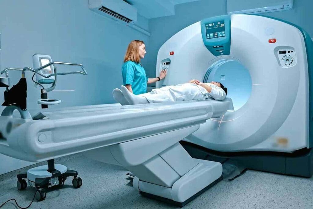
At LivHospital, we use 18F-FDG PET imaging to get deep insights into health. Fluorodeoxyglucose (FDG) is a special sugar that helps us see how active tissues are.
By tracking 18F-FDG in the body, we can see how tissues use sugar. This is key for spotting and tracking diseases, like cancer, brain issues, and heart problems. This fluorodeoxyglucose positron emission tomography tech has changed how we diagnose and treat patients. Get the ultimate answer to what is FDG? This powerful guide explains how 18F-FDG PET imaging works in modern medicine for amazing diagnostics.
Key Takeaways
- 18F-FDG PET imaging assesses metabolic activity in tissues.
- FDG is a glucose analog used in PET scans.
- This technology is key in oncology, neurology, and cardiology.
- 18F-FDG PET scans help diagnose and monitor various conditions.
- Personalized treatment plans are enabled through this technology.
What Is FDG: A Comprehensive Overview
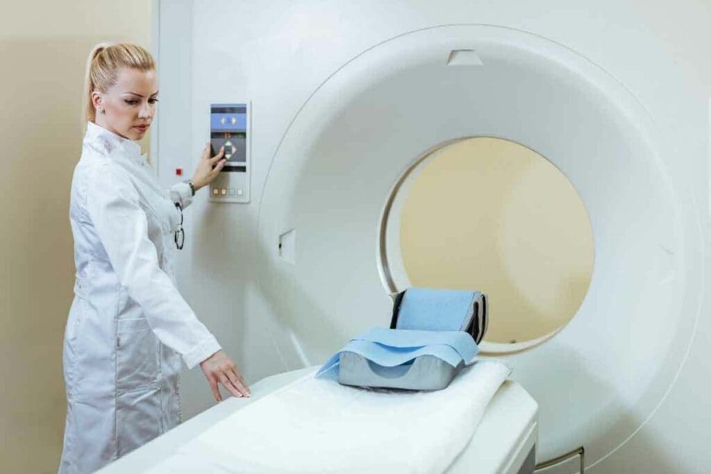
Fluorodeoxyglucose, or FDG, is a glucose-like substance that has greatly helped in medical imaging. It’s used in Positron Emission Tomography (PET) scans. These scans show how active the body’s cells are.
Definition and Development of Fluorodeoxyglucose
FDG is a special glucose molecule with fluorine-18, a radioactive isotope. This makes it useful for seeing how cells work. It was created to help doctors diagnose diseases better.
Making FDG is a complex process. It involves adding fluorine-18 to glucose. This makes a compound that looks like glucose and emits positrons. These positrons are what PET scanners use to create detailed images.
The Glucose Analog Concept in Medical Imaging
Cells use glucose for energy. FDG looks like glucose and is taken up by cells the same way. Inside the cell, it gets stuck because it can’t move forward in the energy process.
This is why FDG is good for PET scans. It shows up in cells that are very active, like cancer cells. This helps doctors see where the body is working too hard.
FDG’s role in showing active cells is key for finding and tracking diseases, like cancer. It helps doctors understand what’s going on inside the body.
The Science of Fluorine-18 in FDG
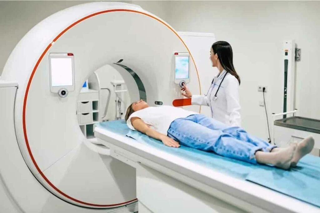
Fluorine-18 is key to understanding FDG PET imaging. It’s a radioactive isotope used in Fluorodeoxyglucose (FDG) for PET scans. This isotope plays a big role in medical diagnostics.
Properties of Fluorine-18 as a Radioisotope
Fluorine-18 is a positron-emitting radioisotope with a short half-life. This makes it perfect for medical imaging. It can be added to glucose molecules, creating FDG, which cells use like glucose.
Key Properties of Fluorine-18:
- Positron emission: F-18 decays by emitting positrons, which are antimatter counterparts of electrons.
- Chemical reactivity: Despite being radioactive, F-18 can be incorporated into biologically active molecules like FDG.
- Half-life: The half-life of F-18 is approximately 110 minutes, which is ideal for PET imaging workflows.
Understanding the 110-Minute Half-Life of F-18
The 110-minute half-life of Fluorine-18 is vital for PET imaging. It allows for the timely production and use of 18F-FDG in clinics.
| Half-Life Characteristics | Implications for PET Imaging |
| Approximately 110 minutes | Sufficient time for production and distribution |
| Relatively short | Minimizes patient radiation exposure |
| Allows for efficient decay | Enables timely imaging with clear results |
We use this property to ensure patients get the imaging they need safely. The half-life is long enough for logistics but short enough for safety. This is a big plus for F-18 in medicine.
Production and Quality Control of 18F-FDG
To make 18F-FDG, a complex radiochemical synthesis is needed. This process has several key steps. These steps make sure the 18F-FDG is of high quality for PET scans.
Radiochemical Synthesis Process
The making of 18F-FDG starts with Fluorine-18 (F-18). This radioactive isotope has a half-life of about 110 minutes. It’s made in a cyclotron by bombarding O-enriched water with protons.
Then, F-18 is added to glucose through chemical reactions. This replaces the hydroxyl group, creating 18F-FDG.
Key Steps in 18F-FDG Synthesis:
- Production of F-18 in a cyclotron
- Synthesis of 18F-FDG through nucleophilic substitution
- Purification of 18F-FDG using techniques like HPLC (High-Performance Liquid Chromatography)
The synthesis of 18F-FDG is complex. It needs precise control to get high yield and purity.
Safety Standards and Quality Assurance
Ensuring 18F-FDG’s safety and effectiveness is key. Quality control is strict at every production stage. This includes from F-18 synthesis to the final 18F-FDG product.
| Quality Control Measure | Description |
| Radionuclidic Purity | Verification that the product contains the correct radionuclide (F-18) |
| Chemical Purity | Assessment of the chemical purity of 18F-FDG, ensuring minimal impurities |
| Sterility and Endotoxin Testing | Testing to ensure the product is sterile and free from bacterial endotoxins |
These quality checks are vital. They make sure 18F-FDG PET scans are accurate and reliable.
By following strict safety and quality standards, we produce top-notch 18F-FDG. This is for use in PET imaging.
Principles of PET Imaging Technology
PET imaging works by detecting metabolic activity in the body. It uses positron-emitting tracers. This tech is key in nuclear medicine, giving insights into living tissues’ biochemical processes.
How PET Scanners Detect Positron Emission
PET scanners find gamma rays from positron-emitting tracers. These gamma rays come from positrons meeting electrons in the body. The detection of these gamma rays is key to making detailed images of metabolic activity.
The PET scanner has a ring of detectors around the patient. When two detectors see gamma rays at the same time, it’s a coincidence event. This shows where the tracer is in the body.
Image Reconstruction and Processing Techniques
Reconstructing images in PET imaging is complex. Advanced algorithms correct for things like attenuation and scatter, improving image quality. These images show where the tracer is, helping to see metabolic activity in different tissues.
There are many ways to reconstruct images, like filtered backprojection and iterative methods. Iterative methods are great because they improve image quality by understanding the data acquisition process. The right algorithm can make PET images better for diagnosis.
After reconstruction, images are processed and analyzed. Experts look at them to find abnormal metabolic activity. This is vital for diagnosing diseases, tracking treatment, and research.
The Metabolic Basis of 18F-FDG PET Imaging
To understand 18F-FDG PET imaging, we need to know how cells take up fluorodeoxyglucose F 18 FDG. This helps us see how the body’s metabolism works through medical imaging.
Cellular Uptake Mechanisms of FDG
Cells use FDG instead of glucose because they’re similar. This happens because of glucose transporters on the cell’s surface. Inside the cell, FDG gets turned into FDG-6-phosphate by hexokinase.
Metabolic Trapping and Signal Generation
Once turned into FDG-6-phosphate, it can’t be broken down further. This is because it gets stuck in the cell. This buildup lets PET scanners see the cell’s activity.
| Process | Description |
| Cellular Uptake | FDG is taken up by cells via glucose transporters |
| Phosphorylation | FDG is phosphorylated by hexokinase to FDG-6-phosphate |
| Metabolic Trapping | FDG-6-phosphate is trapped within the cell, accumulating in proportion to metabolic activity |
To learn more about FDG scans, we can see how they’re used in medicine. They help doctors diagnose and track diseases.
Quantification Methods in FDG-PET Imaging
Measuring FDG uptake in PET imaging is key for accurate diagnosis and treatment tracking. We use different methods to check metabolic activity in the body.
Standardized Uptake Value (SUV) Measurements
The Standardized Uptake Value (SUV) is a common way to measure FDG uptake in PET imaging. It gives a semi-quantitative look at metabolic activity in a specific area.
SUV measurements are figured out by comparing the radioactivity in a certain area to the dose given and the patient’s weight. This makes it easy to compare FDG uptake in different scans and patients.
Here’s an example of how SUV measurements are shown in a clinical setting:
| Region of Interest | SUVmax | SUVmean |
| Tumor | 12.5 | 8.2 |
| Liver | 3.1 | 2.5 |
| Blood Pool | 2.0 | 1.8 |
Dynamic Modeling and Kinetic Analysis
While SUV measurements are useful, dynamic modeling and kinetic analysis give a deeper look at FDG uptake. These advanced methods involve getting PET data over time. This lets us figure out kinetic parameters like Ki (influx rate constant) and k3 (phosphorylation rate constant).
Dynamic modeling helps us understand the metabolic processes behind FDG uptake. This can help better identify tumors and other conditions.
By mixing SUV measurements with dynamic modeling and kinetic analysis, we get a fuller picture of FDG-PET imaging data. This improves diagnostic accuracy and treatment tracking.
Clinical Applications in Oncology
In oncology, 18F-FDG PET imaging is key for checking tumor metabolism and guiding treatment. We use it for many cancer care steps, from first diagnosis to tracking treatment progress and follow-ups.
Cancer Detection and Staging
18F-FDG PET/CT scans are vital for finding and staging cancer. They show where tumors are active, helping spot main tumors and spread. This info is key for the right treatment plan.
This tech is great for cancers like lymphoma, melanoma, and non-small cell lung cancer. It gives us detailed info on how far the disease has spread. This helps us make better treatment choices.
Treatment Response Monitoring
Watching how treatments work is another big use of 18F-FDG PET imaging. It checks if tumors are getting smaller or staying the same. This helps us see if treatments are working and if we need to change them.
Seeing how treatments are working early can stop bad treatments sooner. This helps patients and saves money. We use this info to decide if we should keep or change treatments.
Recurrence Evaluation and Follow-up
Finding cancer early is critical for better treatment and outcomes. 18F-FDG PET imaging is great at spotting cancer coming back, even when other tests don’t show it.
We use 18F-FDG PET scans to watch for cancer coming back. This lets us start new treatments quickly. This is a big part of taking care of cancer patients.
| Application | Description | Benefits |
| Cancer Detection and Staging | Identifying primary tumors and metastases through metabolic activity assessment | Accurate staging, informed treatment planning |
| Treatment Response Monitoring | Evaluating changes in tumor metabolic activity over time | Early assessment of treatment effectiveness, personalized medicine |
| Recurrence Evaluation and Follow-up | Detecting recurrent disease through metabolic activity changes | Timely intervention, improved patient outcomes |
In conclusion, 18F-FDG PET imaging is essential in cancer care. It gives us important info at every stage of cancer management. We keep using this tech to improve care and results for cancer patients.
Neurological Applications of FDG-PET Imaging
FDG-PET is a key tool in neurology for studying brain function and metabolism. It helps us understand various neurological conditions better. This leads to more accurate diagnoses and effective treatment plans.
Alzheimer’s Disease and Dementia Diagnosis
FDG-PET imaging is essential for diagnosing Alzheimer’s disease and other dementias. It shows how different brain areas work by looking at their metabolic activity. Reduced glucose metabolism in certain brain regions is a key sign of Alzheimer’s, helping us catch it early.
It also helps us tell apart different types of dementia. This is important for creating the right treatment for each patient.
Epilepsy and Brain Tumor Assessment
FDG-PET is also key in managing epilepsy and brain tumors. It finds abnormal brain activity that might cause seizures. This info is vital for planning treatments.
For brain tumors, FDG-PET shows how active the tumor is. High-grade tumors typically show increased glucose metabolism. This helps us choose the best treatment.
FDG-PET gives us detailed insights into brain metabolism. It’s essential for diagnosing neurodegenerative diseases, managing epilepsy, and evaluating brain tumors. FDG-PET remains a vital tool in neurology.
Cardiovascular Applications and Inflammation Imaging
18F-FDG PET imaging is key in diagnosing heart conditions. It shows how active the heart’s metabolism is. This helps doctors check heart health, like how well the heart works and if there’s inflammation in blood vessels.
Myocardial Viability Assessment
18F-FDG PET imaging is vital for checking if heart muscle can recover. It looks at how active the heart muscle is. This helps doctors choose the best treatment for heart disease or after a heart attack.
By looking at how much glucose the heart takes up, doctors can tell if parts of the heart are alive. If it takes up a lot of glucose, it’s likely to recover. But if it doesn’t, it might be damaged beyond repair.
Cardiac Inflammation and Infection Detection
18F-FDG PET imaging is also great for finding heart inflammation and infections. It can spot problems like cardiac sarcoidosis, myocarditis, and infections in prosthetic valves. Its high sensitivity means it can catch these issues early.
For example, it can find inflammation in the heart during cardiac sarcoidosis. This helps doctors decide on the right treatment, like medicines to fight inflammation.
Vascular Inflammation and Atherosclerosis Evaluation
18F-FDG PET imaging also helps with vascular inflammation and atherosclerosis. It looks at the activity in atherosclerotic plaques. This helps doctors find plaques that might burst and cause heart problems.
We use 18F-FDG PET to see if treatments for vascular inflammation are working. This is very important for patients with serious atherosclerosis. It helps doctors tailor treatments to each patient’s needs.
Conclusion: The Future of FDG-PET in Medicine
18F-FDG PET imaging is key in today’s medicine, mainly in oncology, neurology, and cardiology. It uses a glucose analog to check metabolic activity. This helps doctors diagnose and track different conditions. The half-life of F-18 in f-fdg is 110 minutes, making it perfect for imaging.
PET technology is getting better, thanks to new tracers and methods. As FDG-PET grows, we’ll see new uses and better treatments. Combining PET with other imaging will make it even more useful. This will lead to better care for patients in many fields.
Knowing about fluorodeoxyglucose and its use in imaging is important. As we go on, f-fdg’s role in medicine will grow. This will lead to more innovation and better healthcare.
FAQ
What is FDG and how is it used in medical imaging?
FDG, or Fluorodeoxyglucose, is a glucose analog used in PET imaging. It shows areas of high metabolic activity in the body. This is important for finding cancer, neurological disorders, and some heart conditions.
What is the role of Fluorine-18 in 18F-FDG PET imaging?
Fluorine-18 is a radioisotope in FDG. It lets PET scanners detect it. Its short half-life of 110 minutes makes it good for clinical use.
How is 18F-FDG produced and what quality control measures are in place?
18F-FDG is made through a radiochemical synthesis process. Quality assurance steps are taken to ensure its safety and effectiveness for use in patients.
What is the principle behind PET imaging technology?
PET scanners use Fluorine-18 to detect annihilation radiation. This radiation happens when positrons collide with electrons. It helps create images showing metabolic activity in the body.
How is FDG taken up by cells and what does it indicate?
FDG enters cells through glucose transporters. It gets trapped after being phosphorylated by hexokinase. This shows areas of high metabolic activity, like in cancer.
What are the common methods for quantifying FDG uptake in PET imaging?
SUV measurements and dynamic modeling with kinetic analysis are common methods. They give insights into metabolic activity. This helps in diagnosing and monitoring various conditions.
What are the clinical applications of 18F-FDG PET imaging in oncology?
In oncology, 18F-FDG PET imaging is used for diagnosis, staging, and assessing treatment response. It’s key in managing cancer patients.
How is FDG-PET used in neurology?
FDG-PET helps diagnose and manage neurodegenerative diseases like Alzheimer’s. It assesses epilepsy and evaluates brain tumors. It provides valuable information on brain metabolic activity.
What are the applications of FDG-PET in cardiology?
In cardiology, FDG-PET assesses myocardial viability and detects cardiac sarcoidosis and other inflammatory conditions. It evaluates vascular inflammation, aiding in diagnosing and managing cardiac diseases.
What is the future of FDG-PET imaging in medicine?
The future of FDG-PET imaging includes emerging applications and technology advancements. It will continue to improve patient outcomes across various medical specialties.
What is the half-life of Fluorine-18 FDG?
The half-life of Fluorine-18 FDG is about 110 minutes.
How does 18F-FDG PET imaging contribute to personalized medicine?
18F-FDG PET imaging helps personalize medicine by providing detailed metabolic activity information. This allows for tailored treatment strategies and better patient outcomes.
Reference
- RadiologyInfo. “PET/CT – Positron Emission Tomography.” 2025., https://www.radiologyinfo.org/en/info/pet


