Can Stress Make Thalassemia Worse? Understanding Stress and Anemia in ThalassemiaAt Liv Hospital, we know getting a PET scan report can be confusing. Terms like FDG avidity and uptake might puzzle you. FDG, or fluorodeoxyglucose, is a special sugar used in PET scans that shows how active tissues are. To help you understand better, here we explain and define FDG clearly.
We use FDG to check how well tissues and organs work. This is key for diagnosing and treating many health issues. Knowing about FDG uptake and activity helps our team give you the right diagnosis and treatment.

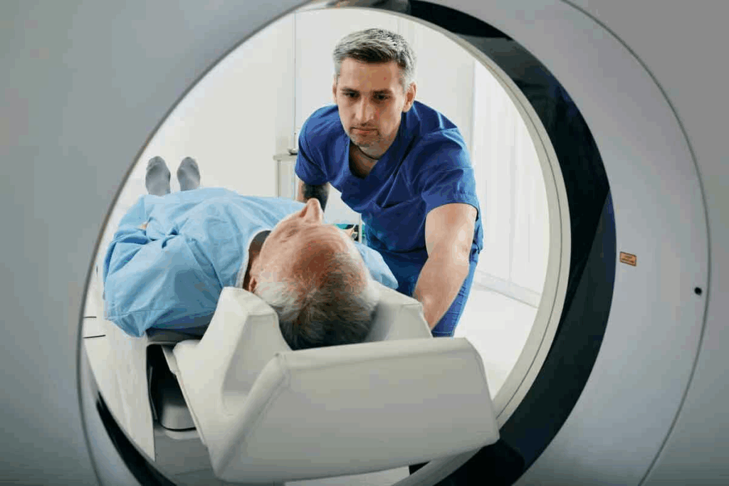
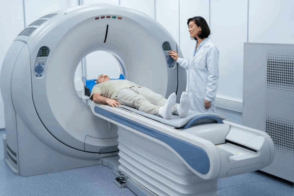
To understand PET scans, we need to know how they work and the role of radiotracers. PET scanning is a complex imaging method. It helps us see and measure how different parts of the body work.
PET scans detect energy from a radiotracer injected into the body. This radiotracer goes to areas with lots of activity, like growing cancer cells. As it decays, it sends out positrons that meet electrons, creating gamma rays.
These gamma rays are caught by the PET scanner. This creates detailed images of the body’s metabolic processes. The data from a PET scan is key for diagnosing and treating many conditions.
Radiotracers are substances with a bit of radioactive material. They are vital for PET imaging. The most used one is FDG (fluorodeoxyglucose), a glucose-like substance. It shows how cells use glucose, helping us study various biological processes.
Fluorodeoxyglucose (FDG) is a key radiotracer in PET scans. It helps check how tissues use glucose. This is very important in medical imaging, like in cancer and brain studies.
To get how FDG works, we look at its structure and how it affects cells. FDG is a glucose-like molecule with a fluorine-18 atom instead of a hydrogen. This makes it special for medical use.
FDG’s structure is close to glucose but has a fluorine-18 atom instead of a hydroxyl group. This lets cells take it up like glucose. But, once inside, FDG gets stuck as FDG-6-phosphate.
FDG acts like glucose in the body by using the same ways cells take it up. This lets PET scans show where glucose is used a lot. This is key for finding tumors, as they use more glucose than normal tissues.
Knowing how FDG works helps doctors use PET scans to see how tissues use glucose. This gives them important info for diagnosis.
FDG uptake is key in PET imaging, showing how active cells are. To grasp what FDG uptake means, we must explore how cells use glucose.
Cells need glucose for energy. The way cells use glucose is controlled by many factors. FDG uptake definition ties to how cells process glucose.
Normal cells use glucose in a controlled way. But cancer cells use more glucose because they are very active. This is why FDG PET scans are used to find and track cancer.
Cancer cells have a different way of making energy, known as the Warburg effect. They use glycolysis even when there’s oxygen, which means they use more glucose. This is why FDG PET scans are good at spotting and tracking cancer.
The Warburg effect is why cancer cells show up high on FDG PET scans. So, knowing what does FDG uptake mean is key for understanding PET scan results in cancer care.
| Cell Type | Glucose Consumption | FDG Uptake |
| Normal Cells | Regulated | Low |
| Cancer Cells | Increased | High |
Understanding FDG uptake’s biological roots helps doctors read PET scans better. This knowledge is vital for diagnosing and treating diseases, like cancer.

Understanding FDG uptake on PET scans is key for accurate diagnosis. FDG, or fluorodeoxyglucose, is a glucose analog that cells with high metabolic rates take up. The body’s FDG distribution is seen through PET scans, giving insights into cellular metabolism.
PET scans show FDG distribution using color scales. Areas with more FDG uptake appear brighter, showing higher metabolic activity. This helps spot tissues or lesions with abnormal glucose metabolism.
Standardized Uptake Values (SUV) measure FDG uptake levels. SUV is calculated by comparing activity in a region to the injected dose and body weight. It gives a semi-quantitative look at glucose metabolism.
The SUV is key for comparing FDG uptake across scans and patients. It helps track disease progression or treatment response.
| SUV Value | Interpretation |
| Low SUV (<2.5) | Typically benign or low metabolic activity |
| Moderate SUV (2.5-4.0) | May indicate inflammation or low-grade malignancy |
| High SUV (>4.0) | Often associated with high-grade malignancy |
PET scan results can be looked at in two ways. Qualitative assessment involves visualizing FDG uptake patterns. Quantitative assessment uses SUV to give a more objective measure of FDG uptake.
Both methods are used together for a full understanding of PET scan results. SUV is very useful for tracking changes over time or in response to treatment.
In PET imaging, FDG activity shows how much fluorodeoxyglucose is taken up by tissues. This is key to knowing how much glucose they use.
FDG activity is when cells take in fluorodeoxyglucose, showing how much glucose they use. This is seen through PET scans. It gives a clear picture of how active the cells are.
In medical reports, FDG activity helps check how tissues are doing. High levels often mean tissues are using more glucose, like in cancer. A study on the National Center for Biotechnology Information shows it’s key for cancer diagnosis and staging.
Many things can change how much FDG is taken up. For example, diabetes can affect it because high blood sugar levels can block FDG from getting into cells.
| Condition | FDG Uptake Pattern | Clinical Significance |
| Malignant Tumors | High FDG uptake | Indicates aggressive tumor metabolism |
| Inflammatory Processes | Moderate FDG uptake | Suggests active inflammation |
| Benign Lesions | Low FDG uptake | Typically indicates non-malignant conditions |
In summary, knowing about FDG activity is vital for reading PET scan results right. It helps doctors understand tissue health. This helps in making diagnoses, planning treatments, and caring for patients.
The term FDG avidity shows how much fluorodeoxyglucose (FDG) lesions take up during a PET scan. It’s key in imaging because it helps spot and understand tissues by their glucose use.
FDG avidity matters because it shows how active tissues are metabolically. Areas with high glucose use, like many cancers, show high FDG avidity. Knowing this helps doctors read PET scans right.
Lesions with high FDG uptake are called FDG-avid lesions. These spots use more glucose than usual, often meaning cancer is present. The more glucose they use, the faster they grow.
But, not all high FDG uptake means cancer. Some infections or inflammation can also show high uptake. So, doctors must look at the whole picture when seeing FDG avidity.
FDG avidity ranges from low to high. Low avidity might mean the tissue is less active, possibly benign or slow-growing. High avidity, though, usually means the tissue is more aggressive or cancerous.
Knowing the range of FDG avidity is key for doctors. It helps them tell different types of lesions apart and figure out how serious they are. For example, a high avidity lesion might need quicker or more intense treatment than a low one.
Doctors use FDG avidity to make better care plans. This includes choosing treatments, tracking disease, and seeing how well treatments work.
It’s key to know the difference between normal and abnormal FDG uptake patterns. PET scans help us understand many health issues. Getting FDG uptake right is very important.
FDG uptake changes a lot in different body parts. It’s vital to know these changes to spot normal versus abnormal conditions.
In a normal PET scan, we see certain FDG uptake patterns. This is because of how our organs and tissues work. For example, the brain uses a lot of glucose, so it shows high FDG uptake.
The heart, liver, and muscles also have different FDG uptake levels. This depends on how active we’ve been and our metabolism. The kidneys and urinary tract also take up FDG as they filter and remove it.
Abnormal FDG uptake can mean many things, like cancer. Tumors use more glucose, so they take up more FDG. But, it’s not just cancer. It can also show up in infections and inflammation.
The way FDG uptake looks can tell us a lot. A bright spot might mean a tumor. But, a spread-out uptake could mean inflammation or infection.
We need to look at all the information together. This helps us make the right diagnosis and treatment plan.
Understanding mild FDG uptake in PET scans is complex. It involves knowing both benign and malignant processes. This slight increase in fluorodeoxyglucose (FDG) by cells can show up in many medical conditions.
When looking at low-grade or mild FDG uptake, several factors are important. These include the patient’s medical history, where and how the uptake is seen, and other test results. Mild FDG uptake can be seen in both benign and malignant conditions. It’s key to link PET scan results with other tests and the patient’s overall health.
When checking mild FDG uptake, doctors look at a few things:
It’s hard to tell if mild FDG uptake is from a benign or early malignant process. Some benign conditions, like inflammation or infection, can show mild FDG uptake. On the other hand, some cancers might only show a little FDG uptake, even in the early stages.
To help tell them apart, doctors use more information, like:
| Diagnostic Factor | Benign Conditions | Malignant Conditions |
| FDG Uptake Pattern | Typically diffuse or homogeneous | Often focal or heterogeneous |
| Clinical Context | Presence of inflammatory or infectious symptoms | History of malignancy or risk factors |
| Correlative Imaging | CT or MRI findings consistent with benign etiology | Imaging features suggestive of malignancy |
By combining PET scan results with other diagnostic info and the patient’s situation, doctors can make better decisions about treatment.
PET FDG uptake has many uses in healthcare and is growing. It helps in many areas of medicine, not just one. This shows how important it is for health care today.
PET FDG uptake is key in finding and checking cancer. It spots tumors by seeing where cells are most active. This helps doctors know how big the tumor is and if it has spread.
Experts say FDG PET/CT is essential for cancer care. It helps in planning and checking treatment. It can find small tumors that other tests miss.
PET FDG uptake helps in many ways for cancer:
PET FDG uptake is also important for checking how well cancer treatment works. It looks at how tumors change with treatment. This helps doctors know if the treatment is working.
A study found that early checks with FDG PET/CT can help predict treatment success. This lets doctors tailor treatment for each patient.
PET FDG uptake is also useful for diseases like inflammation and infections. It spots where cells are very active, which helps find and check these diseases.
“FDG PET/CT is a great tool for finding and managing diseases like vasculitis and other inflammatory conditions. It’s very good at spotting inflammation in blood vessels.”
PET FDG uptake helps in many ways for diseases like inflammation and infections:
| Disease Category | Specific Conditions | Role of PET FDG Uptake |
| Inflammatory Diseases | Vasculitis, Sarcoidosis | Detecting and assessing disease activity |
| Infectious Diseases | Abscesses, Osteomyelitis | Identifying sites of infection and guiding treatment |
FDG uptake in PET scans is not just about disease. Many factors influence it. Knowing these is key for correct scan results.
Getting ready for a PET scan is important. Things like fasting, blood sugar, and recent exercise can change FDG uptake.
Some medical issues can change where FDG goes, making scan results tricky. Diabetes, infections, and inflammatory diseases can impact FDG uptake.
For example, diabetes can change how glucose is used, affecting FDG. It’s important to think about these conditions when looking at PET scans.
Medications can also change FDG uptake. They can alter how glucose is used or affect the scan itself. Here are a few examples:
Knowing what medications a patient takes is essential for understanding PET scan results.
FDG PET scans are a key tool in diagnosing diseases. Yet, they come with challenges. We must look at different factors that can impact their accuracy to get reliable results.
False positives happen when FDG uptake is seen as cancer when it’s not. This can occur due to inflammation, infection, or normal body processes. For example, FDG uptake in brown adipose tissue might be mistaken for cancer. It’s important to know the patient’s situation and use other imaging methods when needed.
Some reasons for false positives include:
False negatives happen when cancer is hard to find because it doesn’t show up well on FDG PET scans. Some cancers, like certain types of prostate cancer or neuroendocrine tumors, might not show up because they don’t use much glucose.
To avoid false negatives, we need to know when FDG PET might not work. We should think about using other imaging methods or different tracers when needed.
By knowing these challenges, we can make FDG PET scans more accurate. This helps us give better care to our patients.
FDG is key in medical imaging, mainly for cancer diagnosis and tracking treatment. It has changed oncology by helping doctors find and understand cancer better.
FDG highlights areas with high glucose use, common in cancers. This helps doctors make better choices for their patients.
As medical imaging gets better, FDG’s importance grows. It helps see how treatments work and spot infections. This shows its wide use in medicine.
In short, FDG’s role in medical imaging is clear. We must keep learning about it to help patients more and improve imaging technology.
FDG avidity shows how much Fluorodeoxyglucose (FDG) cells or tissues take up. It measures their metabolic activity.
In a PET scan, FDG uptake shows how much Fluorodeoxyglucose cells or tissues take up. It helps check glucose metabolism and diagnose conditions like cancer.
FDG activity shows how much Fluorodeoxyglucose cells or tissues take up. It’s a way to check glucose metabolism and diagnose medical conditions.
Mild FDG uptake means a low amount of Fluorodeoxyglucose in cells or tissues. It might show a benign or early cancer. The exact meaning depends on the clinical context.
FDG is key in PET imaging for checking glucose metabolism in tissues and organs. It’s used to diagnose, stage, and check cancer treatment. It also helps with inflammatory and infectious diseases.
FDG helps diagnose cancer by checking glucose metabolism in tumors. Cancer cells use more glucose, making FDG useful for finding and staging cancer.
Several things can change FDG uptake, like patient preparation, medical conditions, and medications. For example, high blood sugar can lower FDG uptake, and some medicines can change glucose metabolism.
Interpreting FDG PET scans has limitations, like false positives and negatives. False positives can happen with inflammation or infections. False negatives can occur in tumors with low glucose use.
FDG avidity helps check the metabolic activity of cells or tissues. It’s a valuable tool for detecting and characterizing medical conditions, including cancer.
Radiotracers, like FDG, are vital in medical imaging. They help assess physiological processes, like glucose metabolism. This visualization is key for diagnosing and managing medical conditions.
Subscribe to our e-newsletter to stay informed about the latest innovations in the world of health and exclusive offers!
WhatsApp us