Last Updated on October 21, 2025 by mcelik

A CT scan of the abdomen is a top-notch tool for seeing inside the body. It uses X-rays and computers to make detailed pictures. Doctors can then spot many health issues with these images.
At Liv Hospital, they focus on the patient. An abdominal CT scan helps doctors make sure diagnoses are right and fast. Knowing how a CT scan works helps patients understand their health better.

Abdominal CT scans use advanced X-ray tech to take images from many angles. This gives a full view of the abdomen. It’s key for spotting many abdominal problems, making it a must-have in medical imaging.
CT scans combine X-rays and computers to show body parts in detail. They spin an X-ray source and detectors around the body. This captures images from all sides.
Then, a computer puts these images together into detailed views or slices of the abdomen.
Key components of CT imaging technology include:
Abdominal CT scans beat ultrasound and X-ray in many ways. They show detailed images of the abdomen’s complex parts. This includes organs, blood vessels, and lymph nodes.
| Imaging Modality | Advantages | Limitations |
| CT Scan | Detailed cross-sectional images, excellent for complex structures | Exposure to radiation, need for contrast agents |
| Ultrasound | No radiation, real-time, cost-effective | Limited depth, depends on the operator |
| X-ray | Quick, widely available, low cost | Limited soft tissue detail, radiation |
The table shows the good and bad of CT scans, ultrasound, and X-ray. CT scans stand out for their detailed images of the abdomen. They help diagnose many conditions.

Getting ready for an abdominal CT scan involves several steps. These steps make the process smooth and effective. Knowing what to expect can help you feel more comfortable.
To get ready for your scan, take off any metal jewelry. Wear loose, comfy clothes. You might need to change into a hospital gown. Always follow the instructions from your doctor or the radiology team.
Important Preparation Steps:
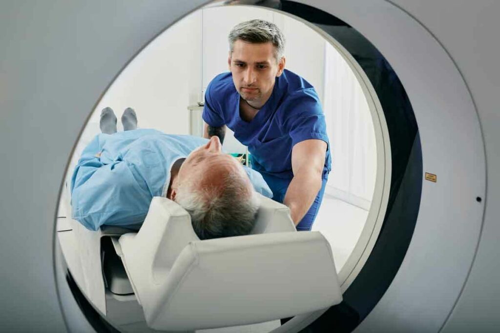
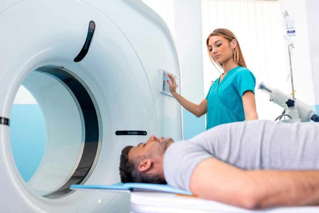
During the scan, you’ll lie on a table that slides into a big, doughnut-shaped machine. The machine will move around you, taking X-rays from different angles. It’s usually painless and takes a few minutes to half an hour.
The CT scan machine is designed to be open and spacious, reducing feelings of claustrophobia. You can talk to the radiology technician through an intercom system.
Contrast material, or “dye,” might be used to make images clearer. It can be given orally, intravenously, or through an enema. This helps doctors see specific structures or problems better.
Contrast enhancement is key in the abdomen CT scan procedure. It helps doctors make accurate diagnoses. Your healthcare provider will explain how and why contrast is used during your preparation.
By knowing what happens during an abdomen CT scan, patients can prepare better. This reduces anxiety and makes the exam more effective.
An abdominal CT scan is a powerful tool for diagnosing many conditions in the abdomen. It gives doctors detailed images of the abdomen. This helps them find and treat various problems accurately.
An abdominal CT scan can show many organs and structures in the abdomen. It can see the liver, spleen, pancreas, kidneys, adrenal glands, and parts of the digestive system. It also shows blood vessels, lymph nodes, and other tissues.
CT scans are very clear. They help doctors spot problems like tumors, cysts, abscesses, and inflammation. For example, they can find liver lesions, pancreatic cancer, and kidney stones very accurately.
Doctors order abdominal CT scans for many reasons. They use them for abdominal pain, trauma, suspected tumors or infections, and to monitor known conditions. They are very useful in emergencies when quick diagnosis is needed.
“CT scans have revolutionized the field of diagnostic medicine, providing unparalleled insights into the abdominal cavity.” – A Radiologist
Recent studies show that abdominal CT scans are very accurate. They are a trusted tool for doctors. The detailed images help in planning treatments, whether surgery or not.
In summary, an abdominal CT scan is a versatile tool for diagnosing many conditions. Its ability to show detailed images of abdominal organs and structures makes it essential for doctors.
Liver diseases often go unnoticed until they are advanced. But, CT imaging can spot issues like tumors and cirrhosis early. This can lead to better treatment outcomes. The liver is key for metabolism, detoxification, and digestion, making it important to monitor.
CT scans are great at showing the liver’s structure. They can find problems that other scans might miss. This is key for diagnosing and treating liver diseases.
Hepatocellular carcinoma (HCC) is the most common liver cancer. CT scans are vital in finding it. They show tumors’ size, location, and how they relate to other parts of the liver. This info is important for treatment planning.
CT scans can also find other liver tumors and metastatic lesions. They can tell different types of lesions apart. This is a big help in diagnosis.
Cirrhosis is a late stage of liver scarring. It’s caused by many diseases and conditions, like hepatitis and alcoholism. CT scans can see how much cirrhosis there is. They show changes in the liver, like atrophy and hypertrophy.
CT imaging can also stage cirrhosis. This helps doctors decide the best treatment and predict how well it will work.
Hepatic abscesses are pockets of pus in the liver. They can come from infections. CT scans are key in finding these abscesses. They show where they are, how big they are, and how many there are.
The use of contrast enhancement in CT scans helps tell abscesses apart from other liver problems. This guides the right treatment, like antibiotics or drainage.
The CT scan of the upper abdomen is a key tool for spotting problems with the gallbladder and pancreas. It gives clear images that help doctors diagnose many issues. This makes it a must-have in gastroenterology.
Gallstones are small, hard deposits in the gallbladder. CT scans can spot these stones, mainly if they are calcified. Cholecystitis, or inflammation of the gallbladder, often comes with gallstones.
CT scans show signs of cholecystitis like a swollen gallbladder, thickened walls, and inflammation around it.
A study in the Journal of Clinical Gastroenterology found CT scans are very good at spotting acute cholecystitis. This makes them a reliable tool for diagnosing this condition.
“CT is a valuable tool in the diagnosis of acute cholecystitis, particular in patients with atypical presentations or when ultrasound findings are equivocal.”
Journal of Clinical Gastroenterology
Pancreatic adenocarcinoma is a cancer that starts in the pancreas. CT scans are key for finding and figuring out how far this cancer has spread. They show the tumor’s size, location, and if it has spread to other areas.
| CT Findings | Clinical Significance |
| Hypoattenuating mass | Indicative of pancreatic adenocarcinoma |
| Vascular invasion | Suggests advanced disease |
| Lymph node enlargement | May indicate metastasis |
Pancreatitis is inflammation of the pancreas, which can be acute or chronic. CT scans help diagnose pancreatitis, see how severe it is, and find any complications. Acute pancreatitis shows as swelling and inflammation of the pancreas. Chronic pancreatitis leads to atrophy and calcifications of the pancreas.
In conclusion, CT scans of the upper abdomen are essential for diagnosing many gallbladder and pancreatic disorders. Their detailed images make them a critical tool in medical practice.
CT imaging is key for spotting many kidney and urinary tract issues. It gives clear images that doctors use to find problems. This part will look at the different kidney and urinary tract issues that CT scans can find.
Nephrolithiasis, or kidney stones, are small, hard deposits in the kidneys. CT scans are great at finding these stones, even the small ones. The National Institute of Diabetes and Digestive and Kidney says CT scans are top for checking the urinary tract, including stones.
CT scans help see how big the stones are, where they are, and if they’re causing problems. This info is key for picking the right treatment.
Renal cell carcinoma is the main kidney cancer in adults. CT scans are vital for finding this cancer and telling it apart from other kidney growths. The contrast-enhanced CT scan is best for looking at kidney masses, like their size and how they react to contrast.
Contrast enhancement is important for telling apart bad tumors from harmless growths. By looking at how things enhance, doctors can help figure out what’s going on and how to treat it.
Pyelonephritis is an infection of the kidney, which can be either acute or chronic. CT scans help find this infection and see how bad it is. They show signs like a bigger kidney, inflammation, and possible abscesses.
If pyelonephritis turns into a renal abscess, CT scans are key for spotting it and planning how to drain it. The detailed pictures from CT scans help doctors manage these issues well.
CT scans of the abdomen help find and treat gastrointestinal problems. These issues can really affect a person’s life. They give a detailed look at the gut, helping spot important conditions.
Appendicitis is a common cause of belly pain that needs quick surgery. CT scans are now the top choice for finding appendicitis. They show an enlarged appendix and inflammation around it.
Using CT scans has cut down on wrong surgeries for appendicitis. CT imaging lets doctors see the appendix and nearby areas. This helps confirm the diagnosis and rule out other pains.
Diverticulitis happens when the colon’s pouches get inflamed. CT scans are great at spotting diverticulitis and its problems. They show thickened colon walls and inflammation.
CT scans also help figure out how bad diverticulitis is and if there are abscesses or holes. This info is key for deciding treatment, like antibiotics or surgery.
Intestinal obstruction blocks the normal flow of gut contents. CT scans are key in finding the cause and where the blockage is. Common reasons include adhesions, hernias, and tumors.
On a CT scan, you see swollen gut parts before the blockage and shrunk parts after. CT imaging can also find the blockage’s cause and check for serious issues like ischemia or holes.
Abdominal vascular problems, like aneurysms and ischemia, are serious. CT scans can spot these issues accurately. If not treated quickly, they can be deadly.
An abdominal aortic aneurysm (AAA) is when the aorta gets too big. It’s bigger than 3 cm or 50% larger than usual. CT scans are great at finding and watching AAAs. They give clear images to check size and risk of rupture.
CT imaging helps with:
| Characteristics | Normal Aorta | Abdominal Aortic Aneurysm |
| Diameter | Typically < 2 cm | > 3 cm or 50% larger than normal |
| Wall Thickness | Normal wall thickness | May be thinned or irregular |
| Risk Factors | Low risk | High risk of rupture |
Mesenteric ischemia happens when blood flow to the intestines drops. This can cause damage and death if not treated fast. CT scans are key in finding mesenteric ischemia by showing blocked or narrowed mesenteric arteries.
CT scans show signs of mesenteric ischemia like:
CT scans are vital for spotting vascular problems in the abdomen. They help with both emergency and diagnostic care.
CT scans are key in emergency care for upper abdomen injuries. These injuries can come from car accidents, falls, or fights. CT scans quickly show how bad these injuries are, helping doctors decide on treatment right away.
Injuries to organs like the liver, spleen, or kidneys are common in the abdomen. CT scans can spot these injuries well, like cuts or bleeding. Blood in the belly, called hemoperitoneum, often goes with these injuries.
CT scans are great at seeing how much blood is in the belly and where it’s coming from. This helps doctors decide the best treatment for patients. A study in the Journal of Trauma and Acute Care Surgery shows CT scans help doctors make better diagnoses. They also cut down on the need for surgery to check for injuries.
There are grading systems for describing abdominal injuries. The Organ Injury Scale (OIS) is the most used. It rates injuries from I (minor) to VI (very severe).
“The use of CT scans in evaluating abdominal trauma has become the standard of care, providing critical information that guides treatment decisions and improves patient outcomes.”Trauma Surgeon
These systems help predict how patients will do and guide treatment. They also help doctors talk clearly with each other. This ensures patients get the right care every time.
Understanding CT abdomen results needs a good grasp of radiology reports. When a CT scan is done, radiologists look at the images. They then write a detailed report based on what they see.
These reports are key for spotting problems in the abdomen. Knowing the terms in these reports helps doctors make the right choices for patients.
Radiology reports use special words to talk about what the CT scan shows. Words like “hypodense,” “hyperdense,” and “isodense” describe how lesions or problems look compared to the rest of the body.
A hypodense spot might mean a cyst or tumor. A hyperdense spot could mean bleeding or calcification. It’s important to know these terms to understand the scan results well.
Common Terms Used in Radiology Reports:
When a CT scan shows something odd, more tests are often needed. These might include MRI or PET scans.
In some cases, a biopsy might be suggested to get tissue for lab tests. The right next step depends on what the first scan shows and the patient’s situation.
| Initial Finding | Possible Follow-up Study | Clinical Context |
| Mass or Tumor | MRI or PET Scan | Suspected malignancy |
| Liver Lesion | Contrast-enhanced MRI | Characterization of liver lesions |
| Pancreatic Abnormality | Endoscopic Ultrasound (EUS) | Suspected pancreatic pathology |
It’s important to understand what abnormal CT abdomen results mean and what comes next. This helps both doctors and patients make good choices and plan the best care.
Abdominal CT scans have changed how we diagnose diseases. They give quick and accurate results that help doctors treat patients better. These scans show detailed images of the abdomen’s organs.
They help find many health issues, like liver and pancreatic problems, blood vessel issues, and injuries. This makes them very important in medicine today.
CT scans help doctors make quick decisions without needing surgery or more tests. This shows how important they are in modern medicine. They improve patient care and make doctor’s work easier.
In short, abdominal CT scans are a big step forward in diagnosing diseases. They are key to giving patients the best care possible.
A CT scan of the abdomen can reveal many conditions. This includes tumors, infections, and problems with blood vessels. It can also show injuries to organs like the liver, gallbladder, pancreas, kidneys, and intestines.
A CT scan gives detailed cross-sections of the body. This is more detailed than an X-ray, which shows a flat image.
A CT scan uses X-rays and computer tech to make detailed images. These images show what’s inside the body.
Contrast material makes certain parts of the body stand out. It helps doctors see different tissues and organs more clearly.
Abdominal CT scans are used to find many conditions. This includes appendicitis, diverticulitis, kidney stones, and liver disease. They also help spot injuries and track disease progress.
Yes, a CT scan can find liver problems. This includes cancer, cirrhosis, and abscesses.
A CT scan can spot gallstones, cholecystitis, and other issues. It gives clear images of the gallbladder and pancreas.
A CT scan can find kidney stones, cancer, and infections. It helps identify problems in the kidneys and urinary tract.
Yes, a CT scan can find issues like appendicitis, diverticulitis, and blockages. It provides important information for diagnosis and treatment.
A CT scan helps check for injuries in the upper abdomen. It spots organ damage and bleeding, guiding treatment.
An abnormal CT abdomen result means there’s a problem or disease. More tests are needed to figure out what it is and how serious it is.
After an abnormal result, more tests or studies might be needed. This helps confirm the diagnosis and plan treatment.
Subscribe to our e-newsletter to stay informed about the latest innovations in the world of health and exclusive offers!
WhatsApp us