Last Updated on November 27, 2025 by Bilal Hasdemir
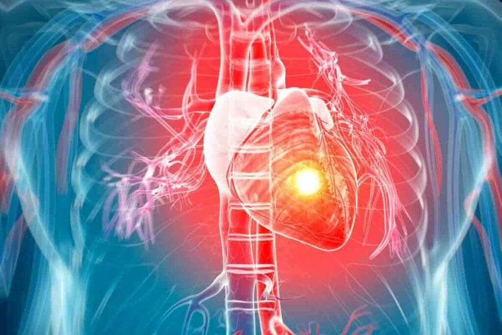
Diagnosing coronary artery disease is key for top-notch patient care. We use advanced tools like myocardial perfusion imaging (MPI) tests to check blood flow to the heart.
Cardiac SPECT is a mainstay in non-invasive MPI. It gives us vital info for making treatment plans. At LivHospital, we focus on trust, innovation, and putting patients first. We use these advanced imaging methods to give precise care.
Key Takeaways
- Myocardial perfusion imaging tests are vital for diagnosing coronary artery disease.
- Cardiac SPECT is a key non-invasive diagnostic tool.
- Advanced imaging techniques improve patient outcomes.
- LivHospital prioritizes patient care with innovative diagnostic approaches.
- Effective diagnosis is crucial for guiding treatment decisions.
Understanding Cardiac SPECT Technology
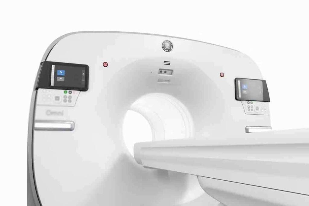
We use Cardiac SPECT technology to understand the heart better. This advanced imaging is key in diagnosing and managing heart disease.
What is Single-Photon Emission Computed Tomography?
Single-Photon Emission Computed Tomography (SPECT) is a way to see the heart in detail. It uses tiny amounts of radioactive tracers. These tracers send out gamma rays that a camera catches.
The Role of Radioisotopes in Cardiac Imaging
Radioisotopes are vital in Cardiac SPECT. Isotopes like Technetium-99m are used as tracers. They show how blood flows in the heart muscle.
How SPECT Creates Three-Dimensional Images
The SPECT scanner moves around the patient. It takes many two-dimensional images from different angles. Then, a computer turns these into a three-dimensional image of the heart. This 3D view helps doctors see the heart’s structure and function more clearly.
The Fundamentals of Myocardial Perfusion Imaging
Myocardial perfusion imaging (MPI) is a detailed method to see how blood flows through the heart muscle. It’s key for checking heart health and spotting problems linked to coronary artery disease (CAD).
We’ll dive into MPI basics, like what it is, why blood flow matters, and its role in finding CAD. Knowing these points helps us see why MPI is so important in medical care.
Defining Myocardial Perfusion
Myocardial perfusion is about blood moving through the heart muscle. Good blood flow is vital for the heart to get oxygen and nutrients and work right. MPI, like Single-Photon Emission Computed Tomography (SPECT), shows this flow.
A tiny amount of radioactive tracer is given, which the heart muscle absorbs based on blood flow. Where the tracer is less, it might show heart muscle problems.
The Importance of Blood Flow Assessment
Checking blood flow to the heart is key for spotting and treating CAD. Less blood flow can cause ischemia, where the heart muscle doesn’t get enough oxygen. This can lead to a heart attack if not treated. MPI spots these issues and helps decide how to treat them.
- Identifies regions of reduced blood flow
- Helps in risk stratification for cardiac events
- Guides therapeutic decisions, including revascularization procedures
Detecting Coronary Artery Disease Through Perfusion
MPI is great for finding CAD, where arteries get blocked by plaque. It looks at blood flow when the heart is stressed and at rest. This helps find ischemia and scar tissue.
Finding CAD early and accurately is key to stopping heart problems and helping patients. MPI gives doctors the info they need to make good treatment plans.
In summary, myocardial perfusion imaging is a vital tool for checking heart health and managing CAD. Its ability to show blood flow to the heart muscle makes it essential in cardiology.
How MPI Stress Tests Work
MPI stress tests are key in checking how the heart works under stress. They help find coronary artery disease and see how the heart reacts to stress or exercise.
Exercise vs. Pharmacologic Stress Testing
There are two main ways to do MPI stress tests: exercise and pharmacologic stress testing. Exercise stress testing uses physical activity, like on a treadmill, to stress the heart. It’s best for those who can exercise, showing how well the heart works under stress.
Pharmacologic stress testing is for those who can’t exercise, like because of mobility issues. It uses medicine to mimic exercise, letting doctors check the heart’s function under stress.
- Exercise stress testing is ideal for patients who can physically exert themselves.
- Pharmacologic stress testing is used for patients unable to undergo physical exercise.
The Two-Phase Testing Protocol
The MPI stress test has two parts: a resting phase and a stress phase. In the resting phase, images of the heart are taken when the patient is calm. This gives a starting point for heart function.
The stress phase comes next, where the heart is stressed, either through exercise or medicine. More images are taken. By comparing these images, doctors can spot heart problems.
- The resting phase establishes a baseline for myocardial perfusion.
- The stress phase assesses the heart’s response to stress.
- Comparing both phases helps in diagnosing coronary artery disease.
Patient Preparation and Experience
Before the test, patients should avoid caffeine and some medicines. They should wear comfy clothes and be ready for the test, including pharmacologic stress if needed.
During the test, patients are watched closely, and their health signs are checked. Most people do okay, but might feel some side effects from the stress agent or exercise.
Understanding MPI stress tests helps doctors better diagnose and treat heart disease. This improves patient care and outcomes.
Interpreting Cardiac SPECT Results
Understanding Cardiac SPECT results is key for healthcare providers. It helps them make better decisions for patient care. The process involves looking at different parameters, with the Summed Stress Score (SSS) being a major one.
Understanding the Summed Stress Score (SSS)
The SSS is a way to measure how bad myocardial perfusion defects are. It scores images from 0 to 4. A score of 0 means normal perfusion, and 4 means no perfusion at all.
We use the SSS to sort patients by risk. It shows how bad the ischemia or scar tissue is. This helps us understand the extent and severity of the problem.
| SSS Range | Interpretation |
| ≤3 | Normal or Equivocal |
| 4-8 | Mild Defects |
| 9-12 | Moderate Defects |
| >12 | Severe Defects |
Normal vs. Abnormal Findings
Normal Cardiac SPECT results mean the heart is well-perfused at rest and stress. But, abnormal results show ischemia or scar tissue. This can be seen through visual or quantitative analysis.
Abnormal results can be reversible or fixed. Reversible defects mean ischemia, while fixed defects mean scar tissue.
Visual and Quantitative Analysis Methods
Visual analysis looks at the SPECT images directly for reduced or absent perfusion. Quantitative analysis uses software to measure perfusion, like the SSS. This gives a more precise look.
Both methods are important. They help make the diagnosis more accurate. Quantitative analysis makes the readings more consistent, reducing variability.
The Clinical Significance of SSS Scoring
Understanding the Summed Stress Score (SSS) is key to reading cardiac SPECT results well. The SSS shows how bad the heart’s blood flow problems are.
Normal or Equivocal Results (SSS ≤3)
Patients with an SSS of 3 or less are at low risk for heart problems. This includes both normal and unclear results. These results show little to no heart blood flow issues.
Mild Defects (SSS 4-8)
An SSS of 4 to 8 means there are mild heart blood flow problems. These patients face a bit higher risk of heart issues than those with normal results. Doctors often need to check these results more closely.
Moderate Defects (SSS 9-12)
SSS scores of 9 to 12 show more serious heart blood flow issues. This means a higher chance of heart problems. Patients in this group usually need more careful checking and treatment.
Severe Defects (SSS >12)
SSS scores over 12 show very bad heart blood flow problems. This means a big risk of heart issues. These patients need quick and strong treatment.
SSS scoring is important for figuring out heart risk and treatment plans. It helps doctors decide who needs stronger treatments.
| SSS Range | Category | Risk Level | Clinical Implication |
| ≤3 | Normal/Equivocal | Low | Conservative management |
| 4-8 | Mild Defects | Mildly Increased | Clinical correlation recommended |
| 9-12 | Moderate Defects | Moderately Increased | Further evaluation and potential intervention |
| >12 | Severe Defects | High | Prompt and intensive management |
Left Ventricular Function Assessment
Assessing the left ventricle is key to understanding heart health. It helps decide the best treatment. The left ventricle pumps blood to the body, showing how well the heart works.
Measuring Left Ventricular Ejection Fraction
Measuring LVEF is important for checking the left ventricle’s function. LVEF shows how much blood the ventricle pumps out with each beat. It’s vital for diagnosing and treating heart issues.
We use Cardiac SPECT to accurately measure LVEF. This technology shows how well the heart pumps, helping doctors make better decisions for patients.
Normal LVEF Values and Significance
LVEF values between 55% and 70% are normal. This means the left ventricle is working right. Values below this might show heart problems.
Knowing LVEF values helps doctors plan treatments. For example, patients with low LVEF might need special medicines or treatments to improve their heart function.
Gated SPECT for Combined Perfusion and Function Analysis
Gated SPECT is a new imaging method. It checks both how well blood flows to the heart and how well the left ventricle works. This is done by matching the SPECT images with the patient’s heart rhythm.
This method gives a full picture of the heart’s health. It helps find patients at risk and plan their treatment better.
| LVEF Range (%) | Interpretation | Clinical Significance |
| 55-70 | Normal | Left ventricle is functioning properly |
| 40-54 | Mildly reduced | May indicate early stages of heart failure |
| 30-39 | Moderately reduced | Suggests significant impairment of left ventricular function |
| <30 | Severely reduced | Indicates severe heart failure or significant cardiac dysfunction |
Recent Advancements in Cardiac SPECT Technology
Cardiac SPECT technology has seen big changes lately. We’ve moved from old SPECT systems to newer, better ones. These updates have made Cardiac SPECT more accurate and useful in hospitals.
Evolution from Traditional to Modern SPECT Systems
Old SPECT systems had problems with image quality and how long it took to get images. New SPECT systems have fixed these issues. Improved detector materials and designs have made images clearer, helping doctors make better diagnoses.
Full-Ring Solid-State CZT Detectors
One big step forward is the use of full-ring solid-state Cadmium Zinc Telluride (CZT) detectors. These detectors are more sensitive and have better resolution than old ones. They also let doctors get images faster and use less radiation on patients.
The Veriton-CT System and Its Capabilities
The Veriton-CT system is a top-notch SPECT/CT scanner. It combines advanced SPECT tech with CT features. This system gives complete cardiac images, showing both how the heart works and its structure. It has better image quality, helps doctors be more sure in their diagnoses, and can do many cardiac imaging tasks.
| Feature | Traditional SPECT | Modern SPECT (e.g., Veriton-CT) |
| Detector Material | NaI(Tl) | CZT |
| Image Resolution | Lower | Higher |
| Acquisition Time | Longer | Shorter |
| Radiation Dose | Higher | Lower |
In summary, recent changes in Cardiac SPECT technology have greatly improved cardiac imaging. Moving from old to new SPECT systems, using CZT detectors, and the Veriton-CT’s features have all helped make diagnoses better and care for patients better.
Benefits of Advanced SPECT Imaging Systems
Advanced SPECT imaging systems have changed how we diagnose heart problems. They offer better images, fewer errors, and more accurate diagnoses than old SPECT tech.
Improved Image Quality and Resolution
These new systems make high-quality images with better detail. This is key for spotting heart issues and seeing how much damage there is. The clear images help doctors make better choices for their patients.
Reduced Attenuation Artifacts
Old SPECT scans often had errors that made it hard to read them right. But, the new systems cut down on these mistakes. This means doctors get a clearer view of the heart’s health.
Enhanced Visualization of the Inferior Myocardial Wall
The bottom part of the heart is hard to see with old SPECT scans. But, the new systems can see it better. This helps doctors find problems and check how well the heart is working.
Correlation with Coronary Angiography Findings
These new SPECT scans match up well with what coronary angiography shows. This is a big plus for figuring out and treating heart artery disease. A study on PMC shows how important this match is for getting a correct diagnosis.
| Benefits | Description | Clinical Impact |
| Improved Image Quality | High-resolution images | Better diagnosis and patient care |
| Reduced Attenuation Artifacts | Minimized image distortion | More accurate diagnoses |
| Enhanced Visualization | Better imaging of the inferior myocardial wall | Improved detection of abnormalities |
| Correlation with Angiography | Strong correlation with coronary angiography findings | Enhanced diagnostic confidence |
Clinical Applications of Myocardial Perfusion Imaging
MPI is used in many ways to help manage heart diseases. It’s a key tool in cardiology, helping doctors understand and treat coronary artery disease (CAD).
Diagnosis of Coronary Artery Disease
MPI is key for finding CAD because it shows how blood flows to the heart. It spots problems like ischemia or infarction, helping doctors decide what to do next. It’s especially helpful for people who might have CAD but aren’t sure.
- Finds signs of CAD
- Shows how bad the ischemia is
- Helps decide on next steps for testing or treatment
Risk Stratification in Known or Suspected CAD
Knowing the risk is important for managing CAD. MPI helps by showing how bad the heart’s blood flow problems are. People with normal MPI results are usually at low risk for heart problems.
- Normal MPI means a good outlook
- Abnormal results show who’s at high risk
- Helps decide how closely to follow up and what preventive steps to take
Pre-operative Cardiac Assessment
MPI is also important before non-heart surgery. It finds out who’s at risk for heart problems during surgery. It helps doctors decide if more heart tests or treatments are needed before surgery.
Evaluation After Revascularization Procedures
After heart surgeries like CABG or PCI, MPI checks if the surgery worked. It shows if there’s still heart problems or if the surgery fixed them.
| Application | Description |
| Diagnosis of CAD | Detects perfusion defects and assesses ischemia |
| Risk Stratification | Provides prognostic information based on perfusion defects |
| Pre-operative Assessment | Identifies patients at risk for perioperative cardiac complications |
| Post-revascularization Evaluation | Assesses the success of revascularization and detects residual ischemia |
Comparing SPECT with Other Cardiac Imaging Modalities
There are many ways to diagnose coronary artery disease. Each method has its own strengths and weaknesses. The right choice depends on the patient’s health, the suspected disease, and what technology is available.
PET vs. SPECT Myocardial Perfusion Imaging
PET and SPECT are both used for imaging the heart. But they work differently. PET MPI gives clearer images and is more sensitive than SPECT. Yet, PET is less common and costs more.
- PET MPI offers better image quality and quantification.
- SPECT is more established and widely available.
- PET has a lower radiation dose in some protocols.
SPECT vs. Cardiac CT Angiography
Cardiac CT Angiography (CCTA) shows detailed pictures of the heart’s arteries. SPECT looks at how well the heart is working. CCTA is great for finding blockages, but it doesn’t always show how serious they are.
- CCTA is ideal for assessing coronary artery anatomy.
- SPECT is better for evaluating the functional impact of CAD.
- Combining CCTA and SPECT can provide a comprehensive assessment.
SPECT vs. Stress Echocardiography
Stress Echocardiography is another tool for checking the heart. It looks at how the heart moves under stress. Unlike SPECT, it doesn’t use radiation.
- Stress Echocardiography is free from radiation.
- SPECT provides direct visualization of myocardial perfusion.
- The choice between them may depend on patient characteristics and local expertise.
Choosing the Right Test for Different Clinical Scenarios
Choosing the right test depends on the patient’s situation and what information is needed. For example, patients with known CAD might get SPECT or PET MPI for more details. On the other hand, patients with suspected CAD might have CCTA to see the heart’s structure.
It’s important to know the good and bad of each test to help patients. By understanding each method’s benefits, we can make better choices. This helps us get the best results for our patients.
Conclusion: The Future of Cardiac SPECT in Clinical Practice
Cardiac SPECT is key in diagnosing and managing heart disease. New technology and imaging methods are making it even better. It’s becoming a must-have for patient care.
Looking ahead, Cardiac SPECT will keep being a big part of healthcare. It helps doctors understand heart function and make better treatment plans. As cardiology advances, so will Cardiac SPECT, helping more patients globally.
FAQ
What is Cardiac SPECT and how does it work?
Cardiac SPECT is a test that uses tiny amounts of radioactive tracers. It creates detailed 3D images of the heart. This helps doctors see how well blood flows to the heart muscle, spot coronary artery disease, and decide on treatments.
What is the difference between exercise and pharmacologic stress testing in MPI?
Exercise stress testing makes you move to stress your heart. Pharmacologic stress testing uses medicine to mimic exercise. The choice depends on your ability to exercise and other health factors.
What is the Summed Stress Score (SSS) in Cardiac SPECT?
The Summed Stress Score (SSS) measures how severe heart muscle problems are. It’s based on SPECT images. The score shows how likely you are to have heart problems.
How is Left Ventricular Ejection Fraction (LVEF) measured using Cardiac SPECT?
LVEF is measured with gated SPECT. This test syncs with your heart’s rhythm. It shows how well your heart pumps, giving important info on its function.
What are the advantages of advanced SPECT imaging systems?
New SPECT systems, like those with full-ring solid-state CZT detectors, improve image quality. They reduce errors and match better with coronary angiography. This makes Cardiac SPECT more accurate and useful.
How does Cardiac SPECT compare to other cardiac imaging modalities like PET or cardiac CT angiography?
Cardiac SPECT, PET, and cardiac CT angiography each offer unique views. SPECT is great for seeing how blood flows and if heart muscle is alive. PET gives detailed images and exact measurements. The right choice depends on what you need to know and the patient’s situation.
What are the clinical applications of Myocardial Perfusion Imaging (MPI)?
MPI helps find coronary artery disease, figure out risk, check the heart before surgery, and see how well treatments work. It shows how well the heart muscle gets blood and works, helping doctors make better choices for treatment.
What is the role of radioisotopes in Cardiac SPECT?
Radioisotopes, like Technetium-99m, help see blood flow and heart function in Cardiac SPECT. They are injected into the blood and build up in the heart muscle. This lets doctors make detailed images of how well the heart muscle gets blood.
What is myocardial perfusion imaging (MPI) stress test?
An MPI stress test checks blood flow to the heart muscle when it’s stressed, usually with exercise or medicine. It finds areas of heart damage or blockages, helping doctors diagnose and treat heart disease.
What is a PET scan for cardiology?
A PET scan for cardiology is a test that uses tiny amounts of radioactive tracers to see the heart. It checks how well the heart muscle gets blood, works, and is alive. This helps doctors diagnose and manage heart disease.
References
- Bateman, T. M., Dilsizian, V., Beanlands, R. S., DePuey, E. G., Heller, G. V., & Wolinsky, D. A. (2016). American Society of Nuclear Cardiology and Society of Nuclear Medicine and Molecular Imaging joint position statement on the clinical indications for myocardial perfusion PET. Journal of Nuclear Cardiology, *23*(5), 1227–1231. https://pubmed.ncbi.nlm.nih.gov/27495328/


