Last Updated on November 25, 2025 by
When you have a medical imaging exam, you might get a contrast dye. This helps doctors see inside your body better. It’s key for diagnosing and treating many health issues.

We use contrast materials to make imaging exams like x-rays and CT scans clearer. These substances change how imaging tools see the body. This leads to better images and more accurate diagnoses.
Most people don’t have serious reactions to contrast dye. Knowing how it works helps us see its value in healthcare. It’s important for our health.
In medical imaging, contrast dye changes how x-rays or other tools see the body. This makes images clearer, helping doctors make better diagnoses.
Contrast agents, or media, make internal body structures more visible during imaging. The National Center for Biotechnology Information explains they change the contrast of body parts. Their main goal is to help doctors see inside the body better, leading to more accurate diagnoses.

Contrast agents make internal structures stand out by changing how imaging tools see the body. For example, in a CT scan with contrast, the dye highlights specific areas. This makes it easier to see different tissues or structures.
This clear view is key for spotting many health issues, like vascular diseases or cancers.
Contrast and non-contrast studies differ mainly in their use of agents. Non-contrast studies use natural body tissue contrast. Contrast studies, on the other hand, use agents to boost contrast, giving clearer images.
Non-contrast studies are good for some diagnoses, but contrast studies offer more detailed info. The choice between them depends on the health issue and the doctor’s judgment.
Contrast media are key in medical imaging. They come in different types for various imaging methods. Each imaging technique needs a specific contrast agent for clear images.
Iodinated contrast agents are used in CT scans. They contain iodine, which absorbs x-rays well. This makes it easier to see different body parts and structures.
Iodinated contrast is great for imaging blood vessels and organs. It helps doctors see more clearly during CT scans. This is helpful for diagnosing injuries and cancers.

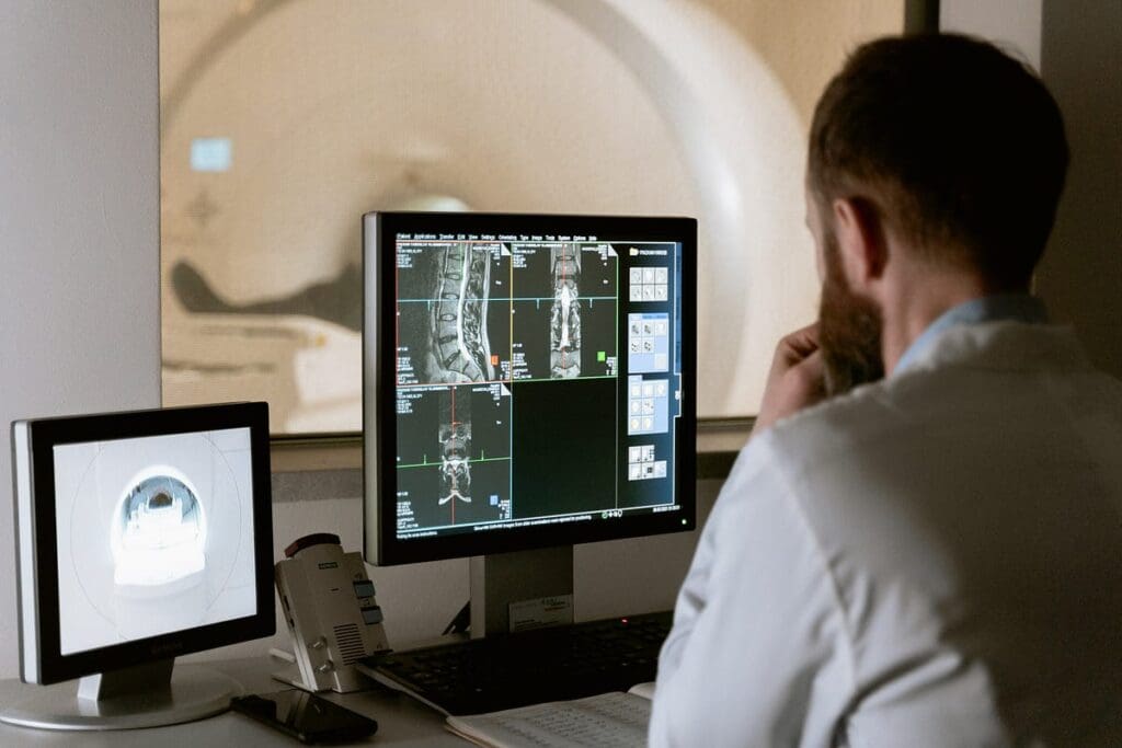
Gadolinium-based contrast agents are for MRI. Gadolinium changes the magnetic properties of hydrogen nuclei. This improves the contrast between tissues on MRI images.
Gadolinium contrast is excellent for seeing tumors, inflammation, and blood vessels. It’s very useful for MRI scans of the brain and spine.
Barium sulfate is used in x-rays of the digestive tract. When swallowed, it coats the digestive tract. This allows for detailed x-ray images of the esophagus, stomach, and intestines.
We also have specialized contrast agents for ultrasound and fluoroscopy. Each agent is chosen for its unique properties and the imaging procedure’s needs.
In summary, the contrast media type depends on the imaging method and procedure needs. Knowing these differences is important for healthcare providers and patients. It helps ensure the best diagnostic results.
Contrast dye starts a journey through the body when it’s introduced. This journey is key for the dye to work in medical imaging.
Contrast materials can get into the body in different ways. They might be swallowed, given by enema, injected into a blood vessel, or injected into body spaces. The choice depends on the imaging procedure.
After being given, contrast agents start moving through the body. If taken by mouth or given by enema, they are absorbed in the gut. Injected into a blood vessel, they spread through the body via blood.
The path it takes depends on the contrast agent. For example, iodinated agents in CT scans quickly move through blood vessels. This makes blood vessels and organs more visible.
Contrast dye meets various organs and tissues as it moves. The interaction depends on the dye’s chemical makeup and the organs’ or tissues’ characteristics.
Gadolinium-based agents in MRI scans tend to gather in specific tissues. This makes these tissues more visible during imaging. It helps in spotting conditions affecting them.
The body naturally gets rid of contrast agents once they’re done. The kidneys are key in filtering out many agents, which then leave through urine.
Other agents, like barium sulfate for X-rays of the gut, are removed through feces. How fast it’s eliminated can vary based on the agent and kidney function.
Knowing how contrast dye moves through and is removed from the body is important. It helps both patients and healthcare providers understand medical imaging’s complexity. It also shows why choosing the right contrast agent is critical for each test.
Contrast dye is usually safe, but it’s good to know about possible side effects. Contrast agents help make internal structures visible during medical scans. But, they can cause reactions in some people.
Mild side effects from contrast dye are rare, happening in less than 1% of patients. Symptoms include nausea, vomiting, and feeling warm or flushed. These usually go away on their own without needing medical help.
More serious side effects from contrast dye are very rare, affecting fewer than 0.05% of people. Symptoms can be hives, itching, and trouble breathing. In rare cases, severe reactions like anaphylaxis might happen, needing quick medical care.
Allergic-like reactions to contrast dye are the most common, making up about 90% of all reactions. These reactions can seem like true allergies but aren’t always caused by one. Symptoms can be mild to severe, like skin rashes, and in rare cases, anaphylaxis.
It’s important for patients to tell their doctors about any allergies or past reactions to contrast dye before a procedure. Knowing the possible side effects and taking steps to prevent them helps keep contrast dye safe for medical imaging.
Medical imaging keeps getting better, and we must understand how contrast dye affects kidney health. We’re digging deep into how contrast media and kidneys interact. This ensures patients get the best care possible.
Contrast-induced nephropathy (CIN) has long worried doctors. It’s when kidney function drops suddenly after contrast use. It was thought to be a big cause of kidney problems in hospitals.
But new studies show the risk might be lower than we thought. This is true for people with normal kidneys. Yet, those with kidney issues or diabetes are at higher risk.
Recent research aims to understand acute kidney injury (AKI) risks from contrast. A major study found AKI rates after CT scans with contrast were similar to non-contrast scans.
This means contrast might stress kidneys, but the AKI risk is low. But, we must be careful, mainly with patients at higher risk.
We’re also looking into how often using contrast affects kidneys over time. Some research checks if it can cause long-term kidney damage or worsen existing conditions.
More research is needed, but it’s clear that those with kidney issues or needing many scans should be watched closely. Doctors should think hard about the benefits and risks of using contrast for each patient.
In summary, while old worries about CIN are real, new studies offer a clearer picture. By keeping up with the latest research, we can better care for our patients.
Getting ready for your contrast-enhanced imaging appointment is key. We’ll show you how to prepare for a smooth process. You’ll learn what to do before, during, and after the exam.
In some cases, you might need to fast before your MRI with contrast. Fasting before MRI with contrast helps the contrast agent work better. It also reduces side effects. The fasting time depends on your healthcare provider’s instructions.
It’s important to follow these instructions closely. This avoids any last-minute changes to your appointment. If you’re unsure, always ask your healthcare provider.
Before your imaging appointment, share important information with your healthcare provider. This includes:
Sharing this information ensures your safety during the procedure. It also reduces the risk of adverse reactions to the contrast agent.
During your contrast-enhanced imaging procedure, you can expect the following:
After the procedure, you’ll be monitored for a short time. This is to check for any immediate reactions to the contrast agent. Usually, you can go back to your normal activities right away. But, it’s best to follow any specific instructions from your healthcare provider.
Understanding what to expect and how to prepare makes your imaging experience better. If you have any concerns or questions, don’t hesitate to reach out to your healthcare provider for guidance.
We’ve looked into how contrast media help in medical imaging and its effects on the body. While it’s mostly safe, there’s a chance for bad reactions, some serious. The American College of Radiology says iodinated contrast media is okay for pregnant women in certain cases. It’s also safe for breastfeeding after using iodine-based contrast media.
The good news is that the benefits of using contrast often outweigh the risks. But, it’s important to think about the risks, like for those who’ve had bad reactions before. Studies, like those on PubMed Central, show knowing the risks and benefits is key to safe use.
Knowing the possible side effects and taking steps to avoid them helps patients safely get imaging done. This includes MRI with or without contrast. Our talk shows how vital it is to weigh the benefits against the risks of contrast media. This ensures patients get the best care possible.
Contrast dye, also known as contrast media, is used in medical imaging. It makes internal structures more visible. This is done by changing how X-rays or magnetic fields interact with the body.
There are many types of contrast media. Iodinated contrast is used for CT scans. Gadolinium-based contrast is for MRI. Barium sulfate is used in other imaging procedures.
Contrast dye is given through an IV. It goes into the bloodstream and spreads through the body. The kidneys remove it, mostly in the urine, within hours.
Side effects can range from mild to severe. Mild reactions include nausea and vomiting. More serious reactions include hives and anaphylaxis.
Fasting is needed for some procedures, like MRI with contrast. It helps prevent aspiration and ensures proper absorption. Your doctor will tell you what to do.
Tell your doctor about any health conditions, allergies, or medications. This helps them decide if contrast dye is safe for you.
You’ll get the contrast agent through an IV during the procedure. Afterward, you’ll be watched for a bit. Your doctor will tell you what to do next.
Studies have looked at the long-term effects of contrast dye on kidneys. While there were once concerns, newer research suggests the risk might be lower. Always talk to your doctor about your risks.
A CT scan with contrast uses dye to show more details. A CT scan without contrast uses natural differences in tissues. The choice depends on what the doctor needs to see.
Gadolinium-based contrast is used in MRI scans. It changes the magnetic properties of hydrogen nuclei. This makes it easier to see blood vessels, tumors, and other structures.
Subscribe to our e-newsletter to stay informed about the latest innovations in the world of health and exclusive offers!