


What Is Radiology? Understanding Patient Imaging and Diagnosis

What Does Radiology Do? How Imaging Guides Care
- Detecting injuries and infections quickly so treatment can begin without delay.
- Clarifying the cause of pain, swelling, or fever when a physical exam alone can’t provide the full picture.
- Mapping anatomy before procedures or surgery to improve precision and safety.
- Monitoring known conditions over time to see if they are stable, improving, or changing.
- Guiding minimally invasive treatments using live imaging so therapies can be delivered precisely where needed.
What Is Diagnostic Radiology? Common Tests and When They’re Used
Diagnostic radiology includes imaging tests that help find, diagnose, and follow health problems. Here’s a look at the main types from a patient’s perspective.
X ray (Radiography)

* Best for: bones, joints, chest and lungs, and certain abdominal assessments.
* Experience: quick and painless; you may be asked to hold your breath briefly. Uses a low dose of ionizing radiation.
* Common reasons: fractures, arthritis, pneumonia checks, line and device placement.
Ultrasound
* Best for: soft tissues and fluids; blood flow; thyroid; kidneys; gallbladder; pelvis; pregnancy imaging.
* Experience: a clear gel on the skin and a handheld probe; no radiation; images in real time.
* Common reasons include abdominal pain, suspected gallstones, vascular evaluations, and guidance on fluid drainage.
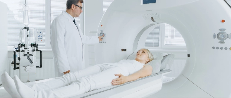
CT (Computed Tomography)
* Best for: rapid, detailed cross sectional images of the chest, abdomen, pelvis, and head.
* Experience: a quick scan through a donut shaped machine. Sometimes, contrast is used to highlight organs or blood vessels. This test uses ionizing radiation, but modern machines use lower doses.
* Common reasons: trauma, appendicitis, kidney stones, suspected clots, or detailed chest or abdominal evaluation.
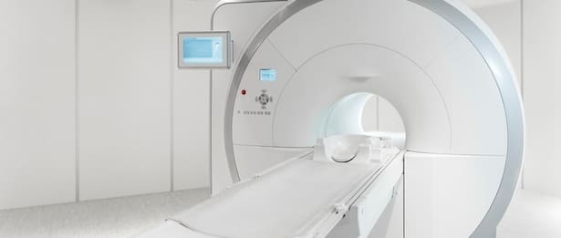
MRI (Magnetic Resonance Imaging)

* Best for: brain, spine, joints, muscles, abdominal and pelvic organs, certain heart and vascular studies.
* Experience: No ionizing radiation. You will hear tapping sounds and need to stay still. Most exams take 15 to 45 minutes. Sometimes, contrast is used to highlight certain tissues.
* Common reasons: neurologic symptoms, back and joint pain, liver or pelvic evaluations, soft‑tissue assessment.
Mammography

* Best for: breast imaging for screening and problem‑focused evaluation.
* Experience: brief compression for clear pictures; low radiation dose; often combined with ultrasound when needed.
Nuclear Medicine and PET/CT
* Best for: evaluating function and cellular activity rather than just structure.
* Experience: A small amount of radiotracer is used to highlight target tissues; cameras detect the signal to create images.
* Common reasons: infection or inflammation scans, cardiac perfusion studies, cancer staging, and treatment response.
The best exam is the one that answers your health question clearly and safely. Your care team reviews your symptoms, medical history, and prior images to determine which test is right for you.
What Is Interventional Radiology? Minimally Invasive Image Guided Treatments
Interventional radiology (IR) uses imaging, such as ultrasound, X ray video (fluoroscopy), CT, or MRI, to perform treatments through tiny skin punctures rather than large incisions. This usually means less pain, lower risk, and a quicker recovery.:
• Vascular care: opening narrowed vessels, stopping internal bleeding, treating certain varicose veins, or placing targeted medications.
• Oncology support: image guided biopsies for diagnosis, localized tumor therapies, and procedures that relieve pain or symptoms.
• Organ and drainage procedures: relieving blocked bile ducts or kidneys, draining abscesses, and placing feeding or access tubes when needed.
• Pain and spine care: guided injections to reduce inflammation and improve mobility.
Many IR treatments use local anesthesia and mild sedation, and most patients go home the same day. You will get clear instructions about eating, medicines, and activity to help you recover smoothly.
Is Ultrasound Considered Radiology? How Ultrasound, X ray, CT, and MRI Are Different
- Ultrasound works well for soft tissues and blood flow. It is safe during pregnancy, portable, and provides real time images.
- X ray is quick and especially good for looking at bones and the chest. It uses a small amount of radiation.
- CT scans provide fast, detailed 3D images, which are helpful in emergencies and for complex anatomy. They use more radiation than X rays, but modern machines keep the dose as low as possible.
- MRI does not use radiation and is excellent for viewing soft tissues. It takes longer than other tests but gives very detailed images.
Is MRI Radiology? What to Expect From an MRI Scan

- Remove all metal items and inform the team of any implants or prior surgeries.
- If you have claustrophobia, options include eye masks, breathing techniques, a call button for reassurance, and, when appropriate, mild calming medication.
- Contrast agents may be used to highlight specific tissues; you’ll be screened carefully to ensure it’s safe for you.
- After most MRI exams, you can immediately resume normal activities.
Is Radiology Hard to Undergo? Comfort, Safety, and Preparation Tips
Most imaging tests are straightforward. A few simple steps can make them even smoother:
• Clothing: wear comfortable, metal free clothing if possible; lockers are usually available.
• Food and drink: follow any fasting instructions, especially if contrast is planned for CT or MRI.
• Motion: staying still is key to clarity; technologists use cushions and brief breath holds to help.
• Contrast: Some studies use contrast to make structures stand out. You might feel a warm sensation during CT contrast or a cool feeling for MRI contrast both pass quickly. Tell the team if you feel unwell.
• Allergies and conditions: always share your medical history, prior reactions, and medications so the team can plan safely.
Radiology departments use the lowest possible doses for X ray and CT, have special protocols for children, and choose ultrasound or MRI when possible to avoid radiation. Your safety, privacy, and comfort are always the top priorities.
Radiology Workflow in a Hospital Setting: From Referral to Results

A clear process helps your visit go smoothly and makes sure the results answer your doctor’s question.
Referral and Scheduling
Your clinician places an order describing your symptoms and the question to be answered. The radiology team reviews the request, assesses safety considerations (such as metal implants or kidney function for contrast), and shares any preparation instructions with you.
Check In and Safety Screening
On the day of your exam, staff confirm your identity and complete screening forms, including allergy, medication, and relevant medical history questions. For MRI, you’ll answer questions about metal and implanted devices; for contrast studies, kidney function and prior reactions are reviewed.
Image Acquisition
Technologists help position you for comfort and to get clear images, adjusting the settings for your body and the test. For X ray and CT, modern machines use lower doses but still give good images. For ultrasound, the technologist uses a handheld probe. For MRI, you get ear protection and comfort items.
Image Interpretation
A radiologist reviews your images, compares them with any previous scans, and considers the details from your doctor’s referral. The radiologist then writes a report that highlights the key findings and main impression.
Results and Next Steps
Your clinician receives the report and discusses it with you. Urgent or unexpected findings are communicated rapidly. Follow up recommendations might include additional imaging at a specific interval or a referral for treatment. You’ll be given clear guidance on what to watch for and how to proceed.
This step by step process keeps you safe, improves accuracy, and helps you get answers faster.

Will Radiology Be Replaced by AI? How Technology Supports, Not Replaces, Doctors
Safety in Radiology: Dose Awareness and Contrast Considerations
Safety is built into every imaging decision. Key elements include:
• Dose optimization: scanners use smart protocols to minimize X ray and CT radiation while preserving clarity.
• Ultrasound/MRI first: radiation free options are chosen when they can fully answer the clinical question.
• Pediatric care: child specific settings and techniques help keep dose as low as possible.
• Pregnancy: imaging choices and shielding are tailored; ultrasound and MRI are often preferred when appropriate.
• Contrast safety: screening for kidney function and allergy risk is standard. Teams are prepared to promptly manage rare reactions.
If you have concerns about radiation or contrast, your radiology team can explain the risks and benefits in simple terms and adjust the plan if possible.
Preparing for Common Exams: Quick Checklists
Chest X ray : Remove jewelry and metal objects; you may take a deep breath and hold it briefly.
Abdominal Ultrasound : Follow fasting instructions if provided; staying hydrated can improve visibility.
CT Abdomen/Pelvis : You may receive oral or IV contrast; expect a brief warm sensation with IV contrast. Follow any fasting guidance.
Brain MRI : Remove all metal items; share any implants or prior surgeries. Eye masks, music, and cushions can improve comfort.
Mammography : Avoid deodorant or powders on exam day, as they can appear on images. Schedule when breasts are least tender if possible.
Understanding Your Radiology Report

• Technique: the type of study and whether contrast was used.
• Findings: detailed, objective observations from the images.
• Impression: the bottom line, with key conclusions and, if needed, suggested next steps.
Why Radiology Matters Today ?
Radiology helps doctors diagnose faster, avoid unnecessary surgeries, target treatments more precisely, and track how well treatments are working. New technology keeps improving image quality, lowering radiation doses, shortening exam times, and helping doctors with advanced analysis. For patients, this means quicker answers, safer tests, and care plans based on the best information, turning uncertainty into a clear path forward
Liv Hospital Radiology
Liv Hospital Radiology Clinic, equipped with high tech devices, provides healthcare services with a multidisciplinary approach with its specialist physician staff and physicians from different disciplines.
Liv Hospital Radiology Clinic has a 3 Tesla MRI device with a large magnet (70 cm) that is found very rarely in our country, 256 slice CT device, 2 digital x ray devices, digital fluoroscopy device, 2 portable digital x ray devices, a digital mammography device, a bone densitometer device, 3 USG and Doppler devices, and an angiography device that guides neuroradiological and vascular interventions.
For more information about our academic and training initiatives, visit Liv Hospital Academy .
Frequently Asked Questions for Radiology
Is X‑Ray Part of Radiology?
Absolutely. X ray is one of the most common radiology exams, ideal for bones and chest imaging, and modern systems use low radiation doses.
Is MRI Radiology?
Yes. MRI uses a magnet and radio waves no ionizing radiation to create detailed images, especially of the brain, spine, joints, and organs.
Is Ultrasound Radiology?
Yes. Ultrasound uses sound waves (no radiation) to create real‑time images of soft tissues and blood flow.
What Is Interventional Radiology?
Interventional radiology performs treatments through tiny punctures using imaging guidance for example, biopsies, drainages, vascular procedures, pain interventions, and targeted tumor therapies.
What Is Diagnostic Radiology?
It includes tests such as X ray, ultrasound, CT, MRI, mammography, nuclear medicine, and PET/CT to detect and track disease, with each exam selected for a specific clinical question.
What Does Radiology Do?
It finds the cause of symptoms, checks injuries, monitors known conditions, plans procedures, and guides minimally invasive treatments using real time imaging.
What Is Radiology?
Radiology is the medical specialty that uses imaging such as X ray, ultrasound, CT, MRI, mammography, nuclear medicine, and PET/CT to diagnose conditions and guide treatments safely and non invasively.