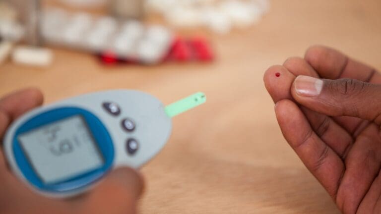
Doing a kidney examination is key to finding kidney problems. At Liv Hospital, we stress the need for a detailed physical check-up. This is along with lab and imaging tests, like ultrasound and serum creatinine level.
We’ll show you how to do a renal exam, including kidney palpation and assessment. A good physical exam is vital for checking the kidneys. When you add urinalysis and imaging tests, you get a full view of kidney issues.
Key Takeaways
- Understanding the importance of a thorough physical examination in kidney assessment.
- Learning the steps involved in performing a renal exam.
- Recognizing the role of laboratory and imaging tests in evaluating kidney disorders.
- Appreciating the significance of kidney palpation techniques.
- Combining physical exam findings with diagnostic test results for a complete kidney check.
The Fundamentals of Kidney Assessment
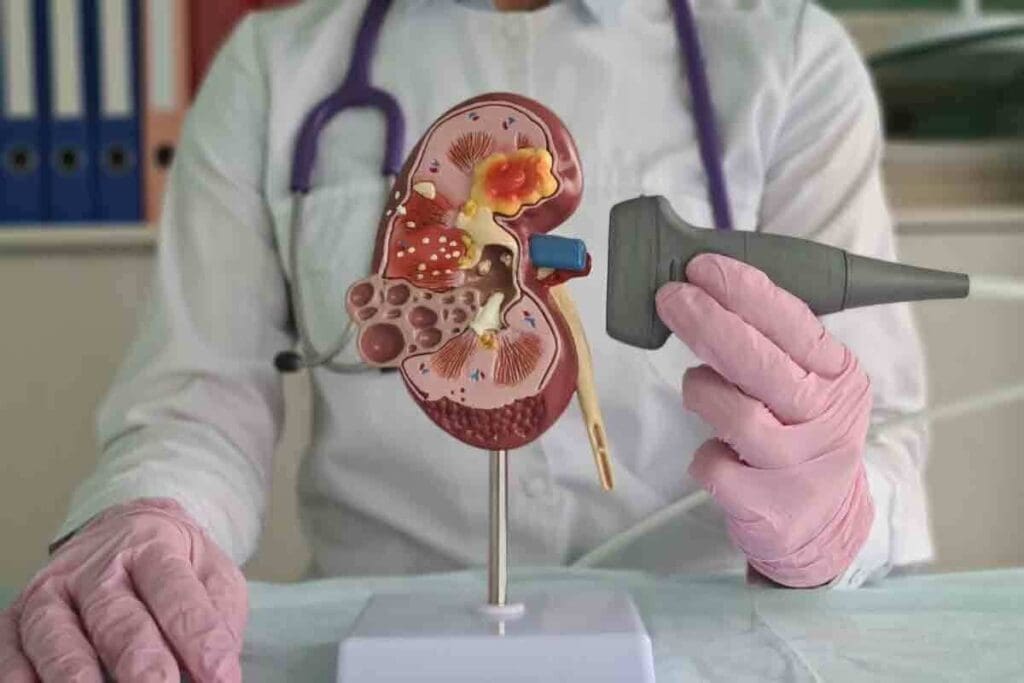
Kidney assessment is key for healthcare providers to diagnose and manage kidney issues. Knowing the renal system well helps spot problems early and treat them right.
Definition and Clinical Importance
Kidney assessment is a vital part of checking patients. It helps doctors find and diagnose kidney problems. This early detection is key to timely treatment.
Understanding the clinical importance of kidney exam helps doctors see its role in patient care. It’s not just for kidney diseases but also for checking overall health.
Anatomy and Location of the Kidneys
The kidneys sit on each side of the spine, just below the ribs. They are about the size of a palm. Knowing the anatomy of the kidneys is essential for a detailed check-up.
They filter waste and excess fluids, balance electrolytes, and make hormones. This knowledge helps doctors use techniques like palpation and percussion to check kidney function.
When to Conduct a Renal Exam

Knowing when to do a renal exam is key for better patient care, mainly for those at risk of kidney disease. We’ll cover the main times a renal exam is needed. This helps doctors make the best choices for their patients.
Common Clinical Indications
Some signs mean a renal exam is needed. These include symptoms of kidney disease, like blood in the urine, too much protein in the urine, or abnormal kidney tests. People with diabetes or high blood pressure should also get regular kidney checks because they’re at higher risk.
Also, if someone has side pain or pain in the area where the kidney is, a renal exam is a good idea.
Risk Factors Requiring Kidney Assessment
Some risk factors mean a kidney check is needed. These include a family history of kidney disease, being older, and certain ethnic backgrounds that face a higher risk. Lifestyle choices like smoking and being overweight also raise the risk of kidney disease.
Patients with a history of kidney stones, urinary tract blockages, or kidney surgery should get regular kidney exams. By spotting these risk factors and signs, doctors can decide when to do a renal exam. This helps improve patient care.
Preparation for a Complete Renal Exam
Getting ready for a renal exam is all about careful planning and the right steps. It’s important to prepare well to check how our kidneys are working and find any problems early.
Optimal Patient Positioning
Patient positioning is the first step for a detailed renal exam. We make sure patients are comfy to get the best results.
- Make sure the patient is relaxed and comfy on the table.
- They can sit or lie down, depending on the exam’s needs.
- It’s also key to keep the patient covered to respect their privacy and comfort.
Required Equipment and Environment
Having the right equipment and a good environment is essential for a renal exam. You’ll need:
- A stethoscope for listening to sounds in the body.
- A percussion hammer to check for pain in the back.
- A bright room for a clear view of the body.
The room should also be quiet, private, and at a good temperature. This helps the patient relax and makes the exam better.
By paying attention to these details, we can make sure the renal exam goes well. This leads to better diagnoses and treatment plans.
Collecting a Thorough Kidney-Related History
Starting a successful renal exam begins with a detailed kidney-related history. This step helps healthcare providers grasp the patient’s situation. It also helps spot kidney problems early and plan further tests.
Essential Questions for Patient Assessment
To assess a patient for kidney disease, we must ask key questions. These questions help us understand the patient’s health better.
- Medical History: We ask about past kidney issues, diabetes, high blood pressure, and other health problems.
- Family History: We look into the patient’s family history of kidney disease. Some kidney problems run in families.
- Symptoms: We note any symptoms like changes in urination, pain in the flank, or swelling.
- Lifestyle Factors: We talk about how diet, staying hydrated, and medications might affect the kidneys.
Documenting Relevant Symptoms and Complaints
It’s important to document the patient’s symptoms and complaints accurately. This helps us understand their condition fully.
- We record when symptoms started and how long they’ve lasted.
- We note how severe the symptoms are and how they affect daily life.
- We look at what makes symptoms better or worse.
By gathering a detailed kidney-related history, we can create a better treatment plan. This approach improves patient care and helps healthcare providers make better decisions.
Visual Inspection in the Renal Exam
The renal exam starts with a detailed visual check. This step is key to spotting kidney problems. We look for any signs of illness or discomfort that might point to kidney issues.
Systematic Observation Techniques
We use a methodical approach for the visual check. First, we examine the patient’s overall look. We search for signs like edema, pallor, or fluid retention that could hint at kidney problems.
Then, we check the belly area for any oddities. This includes looking for masses or scars that might suggest kidney issues or past surgeries.
Recognizing Visual Abnormalities
Spotting visual signs is key in the renal exam. We watch for periorbital edema or swelling in the legs and feet. These can be signs of fluid buildup due to kidney issues.
We also look for skin color or texture changes, like pallor or uremic frost in severe kidney disease. Checking the patient’s urine output is also important. Changes in volume, color, or consistency can tell us a lot about kidney health.
By using these observation methods and knowing what visual signs to look for, we can do a detailed renal exam. This helps us make a good diagnosis and treatment plan.
Kidney Palpation Techniques
Checking kidney health through palpation needs precision and the right method. Kidney palpation is a key tool for doctors to spot issues like masses or kidney growth.
Proper Hand Positioning for Bimanual Palpation
To do bimanual palpation well, correct hand placement is key. We put one hand in front on the patient’s belly, just under the ribs. The other hand goes behind the ribs.
This setup lets us feel the kidney area gently but firmly. The front hand should be relaxed, fingers together. The back hand applies light pressure to move the kidney forward for better feeling.
Anterior Approach to Kidney Palpation
The anterior method means feeling the belly to locate the kidney. We tell the patient to breathe deeply, which moves the kidney down for us to feel better.
As they inhale, we press our fingers into the belly wall. We try to feel the kidney between our hands.
Detecting Masses and Enlargement
While palpating, we look for any oddities like masses or growth. Finding these signs can show problems that need more checking. We check the size, softness, and how easily it moves of any found masses or growth.
Getting good at kidney palpation takes time and effort. By learning bimanual palpation and the anterior method, doctors can better find and treat kidney problems.
Performing Kidney Percussion
Kidney percussion is a method to check for tenderness by gently tapping the costovertebral angle. It’s key for spotting kidney problems. Healthcare pros find it very useful.
Correct Percussion Methodology
To do kidney percussion right, the patient sits or stands. This makes it easy to reach the costovertebral angle. We start by finding the 12th rib and the spine. Then, we tap the area just below the rib gently.
Proper hand positioning is key; one hand taps while the other supports the back. The tap should be firm but not too hard. This helps us see if there’s tenderness.
Assessing Costovertebral Angle Tenderness
Costovertebral angle (CVA) tenderness shows kidney problems, like infections. If tapping hurts, it might mean there’s a kidney issue.
We check for CVA tenderness by watching the patient’s face. If they show pain, we need to do more tests. This could include blood work or scans to find out why it hurts.
In short, kidney percussion is a simple yet helpful way to check on kidney health. By using the right method and watching the patient’s signs, doctors can get vital info.
Additional Physical Assessment Techniques
Assessing kidney function involves more than just tests. Physical exams are key to understanding a patient’s health fully. These exams help us see how well the kidneys are working and any effects on the body.
Evaluating for Edema and Fluid Retention
Checking for edema and fluid retention is vital. Edema is swelling from too much fluid in the body’s tissues. We look for swelling in the legs, warmth, and tenderness. A simple test is pressing on the skin. If it stays indented, it’s pitting edema.
Fluid retention can also show up as puffiness around the eyes or swelling in the hands and feet. These signs are important to notice. For example, people with nephrotic syndrome often have a lot of edema because of lose too much protein in their urine.
Assessing Related Abdominal Structures
Examining the abdomen is also important. It helps us find any problems that might affect the kidneys. We start by looking for any unusual shapes or sizes in the abdomen.
When we touch the abdomen, we check for tenderness, lumps, or if organs are bigger than usual. We pay special attention to the liver, spleen, and bladder. Problems with these organs can be linked to kidney issues. For example, a big liver can be a sign of polycystic kidney disease.
- Check for abdominal tenderness or guarding
- Assess for masses or organ enlargement
- Evaluate for ascites, which can be associated with fluid retention
By using these extra physical checks, we get a better picture of a patient’s health. This way, we can spot problems early, track how the disease is progressing, and create a treatment plan that fits the patient’s needs.
Laboratory Evaluation in Renal Assessment
Renal assessment is not complete without laboratory evaluations. These tests give us key insights into kidney function. They are vital for diagnosing and managing kidney disease.
Blood Tests for Kidney Function
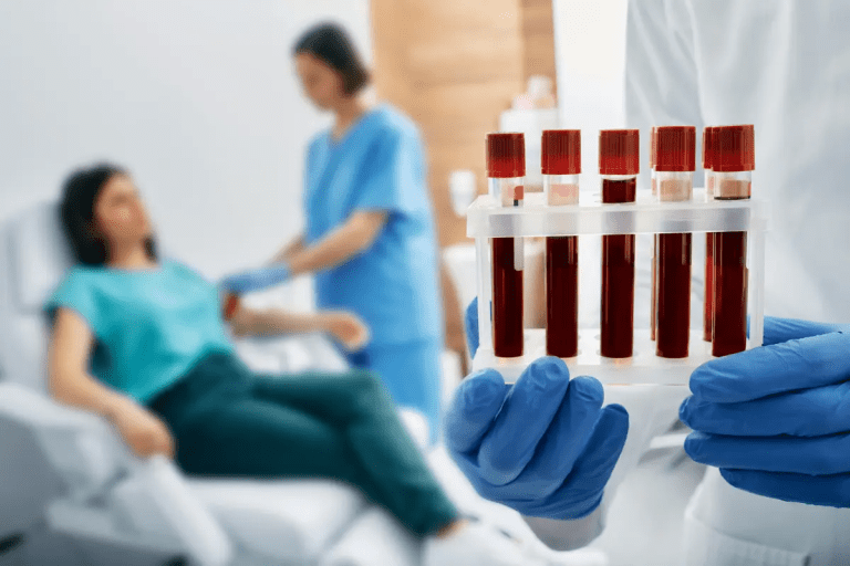
Blood tests are key in renal assessment. They tell us a lot about how well our kidneys are working. Serum creatinine is a common test. It checks the level of creatinine in our blood, a waste product from muscle wear and tear.
High levels of serum creatinine can mean our kidneys are not working properly. Another important test is blood urea nitrogen (BUN). It measures urea, a waste product our kidneys filter out. While BUN is not as specific, it helps when used with other tests.
Urine Studies and Their Clinical Significance
Urine studies are also vital in renal assessment. Urinalysis looks at urine’s physical, chemical, and microscopic properties. It can spot problems like proteinuria, hematuria, and casts, signs of kidney damage.
A leading nephrology expert says, “Urinalysis is key in diagnosing and managing kidney disease. It helps guide further testing and treatment.”
“The presence of certain elements in the urine, such as red blood cells or casts, can be indicative of specific kidney disorders.”
Urine culture and sensitivity tests can find urinary tract infections. These infections can harm our kidneys if not treated.
By looking at blood tests and urine studies together, doctors can understand a patient’s kidney health. They can then create a proper treatment plan.
Imaging Modalities in Kidney Evaluation
Imaging technologies have changed how we check and manage kidney problems. Different imaging methods are key in diagnosing and treating kidney diseases.
Renal Ultrasound: The Gold Standard
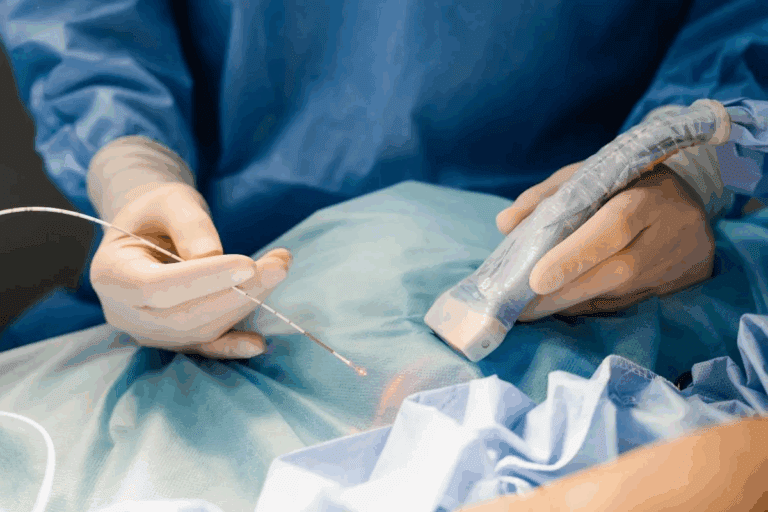
Renal ultrasound is seen as the top choice for checking kidney size, shape, and any issues. It’s safe, doesn’t use radiation, and is affordable. This makes it a great first choice for imaging.
A study in the Journal of the American Society of Nephrology found renal ultrasound is very good at spotting kidney problems. It can find cysts, tumors, and blockages well.
“Renal ultrasound is the first-line imaging modality for the evaluation of kidney disease, providing valuable information on kidney size, echogenicity, and the presence of masses or obstruction.”
| Imaging Modality | Advantages | Limitations |
| Renal Ultrasound | Non-invasive, no radiation, low cost | Operator-dependent, limited detail |
| CT Scan | High detail, fast scanning | Radiation exposure, contrast required |
| MRI | No radiation, high soft tissue detail | High cost, claustrophobia, contraindicated in some patients |
Advanced Imaging Techniques
CT scans and MRI are also important for looking closely at kidney diseases. They help see the kidney’s structure and how it works.
CT scans are great for finding kidney stones, tumors, and blood vessel problems. MRI is best for seeing soft tissues clearly, helping with complex kidney issues.
Interpreting Imaging Results
Understanding kidney anatomy, disease, and each imaging method’s strengths is key. Accurate interpretation helps doctors make the right treatment plans.
Doctors must look at the whole picture when reading imaging results. They consider symptoms, lab tests, and medical history. This ensures imaging findings match the patient’s condition.
Clinical Interpretation of Renal Exam Findings
It’s key to understand renal exam findings to spot kidney problems and know what to do next. We’ll show you how to tell normal from abnormal results. We’ll also talk about common issues and when to send patients to nephrology.
Normal vs. Abnormal Kidney Assessment Results
Knowing the difference in kidney test results is vital for right diagnosis and treatment. Normal results mean the kidneys are working well and don’t have big issues. But, abnormal results might point to kidney disease or damage, needing more checks.
Signs of abnormal kidney test results include:
- Kidney enlargement or masses found during touch
- Costovertebral angle tenderness when tapping
- Edema or fluid buildup
- High creatinine or urea levels in blood tests
Common Pathological Findings
Many kidney problems can be found with a detailed renal exam. We’ll cover some common ones and what they mean.
| Pathological Condition | Clinical Findings | Implications |
| Kidney Stones | Severe pain, costovertebral angle tenderness | May need surgery or lithotripsy |
| Chronic Kidney Disease | Edema, abnormal blood tests | Needs ongoing care and checks |
| Pyelonephritis | Fever, flank pain, costovertebral angle tenderness | Usually treated with antibiotics |
When to Refer to Nephrology
It’s important to know when to send a patient to a nephrologist for the best care. Refer when there’s:
- Significant kidney dysfunction or failure
- A complex or unclear diagnosis
- The need for specialized treatment, like dialysis or transplant
By understanding renal exam findings and knowing when to get specialist help, healthcare can greatly improve patient care.
Conclusion
We’ve shown how to do a complete renal exam. It’s key to use history, physical check-ups, lab tests, and imaging. A detailed kidney check is vital for spotting and treating kidney disease.
Healthcare pros can make patients better by learning these methods. Our guide shows the importance of a step-by-step approach. This includes looking and feeling, lab work, and scans.
In the end, a good renal exam is critical for finding problems early. This guide helps doctors get better at checking kidneys. It’s all about improving care for patients.
FAQ
What is a renal examination?
A renal examination checks the kidneys thoroughly. It includes physical checks, lab tests, and imaging to see how well the kidneys work. It helps find kidney problems early.
Why is kidney palpation important in a renal exam?
Kidney palpation is key to finding issues like masses or swelling. It’s done by feeling the kidneys with both hands. This helps spot kidney problems.
What is the purpose of kidney percussion in a renal exam?
Kidney percussion checks for tenderness in the back. It’s a way to see if there’s a kidney issue. The method used is specific.
What are the common clinical indications for a renal exam?
Reasons for a renal exam include symptoms like blood in the urine or pain in the side. Risk factors like diabetes or a family history of kidney disease also matter.
How is a renal ultrasound used in kidney evaluation?
Renal ultrasound is the top choice for checking the kidneys. It shows the size and structure of the kidneys. It’s often used with other tests.
What laboratory tests are used to assess kidney function?
Tests like blood creatinine and urea levels check the kidneys. Urine tests, like urinalysis, also help. They find kidney disease early.
When should a patient be referred to nephrology?
If a renal exam finds serious kidney damage, a nephrology referral is needed. Nephrologists handle further evaluation and treatment.
What is the significance of assessing related abdominal structures during a renal exam?
Checking the liver and spleen gives important health info. It helps find conditions linked to kidney disease.
How is costovertebral angle tenderness assessed during a renal exam?
Costovertebral angle tenderness is checked by tapping the back. The patient is in a certain position. It looks for kidney problems.
What is the role of visual inspection in a renal exam?
Visual inspection looks for signs like swelling or a belly bulge. These can mean kidney disease.
How is a thorough kidney-related history collected?
A detailed history is gathered by asking key questions. It includes symptoms and medical history. This helps plan the next steps in diagnosis.
References
- Chronic Kidney Disease Diagnosis and Management: A Review. (n.d.). PLOS ONE / PMC. https://pmc.ncbi.nlm.nih.gov/articles/PMC7015670/


