Last Updated on November 27, 2025 by Bilal Hasdemir

Knowing how our kidneys work is key to spotting and treating kidney problems. We use the MAG 3 renal scan, a high-tech test, to check kidney function, blood flow, and urine flow from the kidneys to the bladder.
The Tc-99m MAG3 is the main agent in this scan. Sometimes, a Lasix (furosemide) challenge is added to find out about any blockages or issues with urine flow. A study on NCBI shows this test helps check the kidney’s tissue and how urine moves through the system and bladder.
At Liv Hospital, we focus on top-notch, patient-focused kidney care. Our team is here to support and guide you every step of the way.
Key Takeaways
- The MAG 3 renal scan checks kidney function, blood flow, and urine flow.
- Tc-99m MAG3 is mainly secreted by the kidney’s tubules.
- This scan helps find and track kidney issues.
- Diuretic renography with MAG3 checks for urinary tract blockages.
- It gives semi-quantitative data for a detailed look.
Understanding MAG 3 Renal Scan for Adults: A Complete Overview

The MAG 3 renal scan is a detailed test for checking how well the kidneys work in adults. It uses nuclear medicine to see how the kidneys function and if the urinary tract is open.
Renography, which includes the MAG 3 scan, is a moving picture test. It looks at how kidneys work and drain over time. This is different from static tests that just show what kidneys look like.
Definition and Clinical Applications
The MAG 3 renal scan, or renal scintigraphy, uses a special dye called Technetium-99m MAG3. This dye is mainly cleared by the kidneys’ tubules. It’s great for checking how well the kidneys work and drain.
This scan is used for many things. It helps find blockages in the urinary tract, check how well transplanted kidneys work, and see how each kidney is doing. It’s even better when done with a Lasix (furosemide) challenge. This is called a Lasix renogram or renal scan. It helps find problems with how urine flows.
Renography vs. Static Kidney Imaging
Renography, like the MAG 3 scan, is very different from static kidney tests. Static tests show what kidneys look like. But, renography looks at how kidneys work, like blood flow and how they drain.
This moving picture test lets doctors find problems that static tests can’t. For example, a Lasix renogram can tell if a blockage is causing urine to back up or not.
The Science Behind Nuclear Medicine Renal Scans

Nuclear medicine renal scans use radiopharmaceuticals and advanced imaging. They help doctors understand how the kidneys work and drain. This is key for diagnosing and treating kidney problems.
Radiopharmaceuticals in Kidney Imaging
Radiopharmaceuticals are vital in these scans. Tc-99m MAG3 is a top choice for checking kidney function. Experts say it’s great because it’s quickly taken up and cleared by the kidneys.
After it’s given to the patient, Tc-99m MAG3 goes to the kidneys and then out. This lets doctors see how well the kidneys are working. It also helps spot any issues in the urinary system.
Gamma Camera Technology and Image Acquisition
Gamma camera tech is key for these scans. It picks up gamma rays from the radiopharmaceuticals. This creates detailed images of the kidneys.
The patient lies under the gamma camera for the scan. It captures both static and dynamic images. These images show how well the kidneys are working and their structure.
Together, advanced radiopharmaceuticals and gamma camera tech make nuclear medicine renal scans very useful. They help doctors make better diagnoses and treatments. As we keep improving, we’ll see even better care for patients with kidney issues.
Medical Indications for MAG 3 Renal Scans
MAG 3 renal scans are used for two main reasons. They help find urinary tract blockages and check how well a transplanted kidney is working. These scans look at how well the kidneys function and how they drain.
Suspected Urinary Tract Obstruction
When we think a urinary tract blockage might be present, a MAG 3 scan is very helpful. It shows how bad the blockage is and how it affects the kidney. It looks at blood flow, how the tubes in the kidney work, and how well the kidney drains.
If someone has symptoms like flank pain or if an ultrasound shows hydronephrosis, we might suggest a MAG 3 scan. This test helps us figure out if there’s a blockage and how serious it is. It helps us decide what to do next.
Renal Transplant Evaluation
MAG 3 scans are also key when checking on a kidney transplant. They help us see how well the new kidney is working. They can spot problems and keep an eye on the kidney’s health.
For transplant patients, these scans help find issues like damage to the kidney’s tubes, rejection, or blockages. By watching how the kidney changes over time, we can make sure it’s working as well as it can. This helps the kidneys last longer and improves the patient’s health.
| Indication | Clinical Use | Information Provided |
| Suspected Urinary Tract Obstruction | Diagnose and manage obstruction | Renal blood flow, tubular function, and drainage |
| Renal Transplant Evaluation | Assess graft function, detect complications | Graft function, possible complications, and overall kidney health |
The Lasix Renogram: Enhancing Diagnostic Accuracy
The Lasix renogram is a key tool for better kidney function tests. It uses furosemide, a diuretic, to stress the kidneys. This helps find blockages or poor drainage in the urinary tract.
Purpose of the Furosemide Challenge
The furosemide challenge is a big part of the Lasix renogram. It tests how the kidneys handle diuretic stress. This is great for spotting functional blockages in the urinary tract.
The main goal of the furosemide challenge is to distinguish true blockages from other issues. It helps figure out if a blockage is real or not.
Protocols for Lasix Renal Scan Administration
Administering Lasix during a scan follows strict rules for the best results. The furosemide is given intravenously 15-20 minutes after the radiopharmaceutical.
- The furosemide dose is usually 0.5 to 1 mg/kg of body weight.
- When to give furosemide is very important. It’s done when the radiotracer peaks in the kidneys.
- After furosemide, images are taken to see how the kidneys react.
By sticking to these steps, we get important information on kidney function and drainage. This is key for accurate diagnosis and treatment plans.
Renal Scan Results Interpretation: Normal Patterns
Understanding renal scan results is key to spotting normal kidney function versus possible issues. We’ll explore how to read these results, focusing on what’s normal and what’s expected for each kidney’s function.
Renal Scan with Lasix Normal Results
A normal scan with Lasix shows the kidneys take in and release the tracer quickly, showing they’re working right. The Lasix test checks how well the kidneys handle a diuretic, looking for any blockages.
Here’s what normal results look like:
- Both kidneys take in the tracer evenly and fast.
- The tracer leaves quickly after Lasix is given.
- The tracer doesn’t stay in the kidney’s tubes or sacs too long.
Normal Split Renal Function Parameters
Split renal function means how much each kidney does compared to the other. It’s checked by looking at how each kidney works.
| Parameter | Normal Value |
| Differential Renal Function (DRF) | 45-55% for each kidney |
| Time to Peak Activity (Tmax) | <5 minutes |
| Half-time (T1/2) after Lasix | <10 minutes |
These numbers help doctors see how well each kidney is doing. A normal split renal function means both kidneys are working together well.
Knowing these normal patterns and numbers is vital for correctly reading renal scan results. It helps doctors tell if the kidneys are working right or if there’s a problem, guiding treatment choices.
Identifying Abnormal Renogram Patterns
Abnormal renogram patterns can show different kidney problems. It’s key to know what they mean. When we check MAG 3 renal scans, we look for any patterns that don’t seem right.
Abnormal Renal Scan with Lasix Findings
An abnormal renal scan with Lasix can show issues like urinary tract blockages or kidney problems. The Lasix test, which uses furosemide, helps figure out if the problem is a blockage or not.
After the Lasix test, we check the scan for changes. A normal response means the radiopharmaceutical washes out quickly. This shows the kidneys are working well and there’s no big blockage.
Interpreting Obstructive vs. Non-Obstructive Patterns
Telling apart obstructive and non-obstructive patterns is key to finding the right treatment. Obstructive patterns show a steady increase in the renogram curve, even with Lasix. This might mean there’s a blockage in the urinary tract.
Non-obstructive patterns might show other issues, like delayed or less uptake of the radiopharmaceutical. This could mean kidney damage or problems, not just a blockage.
By studying these patterns closely, we can learn a lot about kidney function and possible issues. This helps us decide what tests to do next and how to treat the problem.
DTPA Scanning vs. MAG 3: Choosing the Right Nuclear Renal Scan
DTPA scanning and MAG 3 renal scans are used in nuclear medicine. They help check how well the kidneys work. The right choice depends on what information is needed for patient care.
Glomerular Filtration Rate Assessment with DTPA
DTPA scans mainly check the glomerular filtration rate (GFR). GFR shows how well the kidneys filter blood. DTPA is great for looking at glomerular function.
We use DTPA scans for many reasons. This includes checking for kidney disease or tracking kidney problems. It helps diagnose and track kidney disease.
Tubular Function Evaluation with MAG 3
MAG 3 (Mercaptoacetyltriglycine) scans look at tubular function. MAG 3 is secreted by the tubules. It’s good for checking tubular secretion and kidney function, even when glomerular filtration is low.
MAG 3 scans are best when we need to know about tubular function. This is true for checking transplanted kidneys or finding out about blockages in the urinary tract.
Knowing the benefits of DTPA and MAG 3 scans helps doctors choose the best test. This ensures accurate diagnosis and effective treatment plans for patients.
Patient Preparation and Procedure for MAG 3 Renal Scintigraphy
To get accurate results, patients must follow certain steps before a MAG 3 renal scintigraphy. This test, also known as a MAG 3 renal scan, checks how well the kidneys work and finds any problems.
Pre-Scan Instructions and Requirements
Before the test, patients need to follow some important steps. Tell your doctor about any medicines you’re taking, as some might need to stop before the test. Also, be ready to:
- Arrive with a full bladder, as this helps in the accurate assessment of the kidneys.
- Wear comfortable, loose-fitting clothing to facilitate the imaging process.
- Avoid wearing jewelry or clothing with metal parts that could interfere with the scan.
A leading nuclear medicine specialist says,
“Proper hydration is key before undergoing a MAG 3 renal scan. Patients should drink plenty of water to ensure they are well-hydrated.”
This makes sure the images are clear and the results are accurate.
The Imaging Process: What to Expect
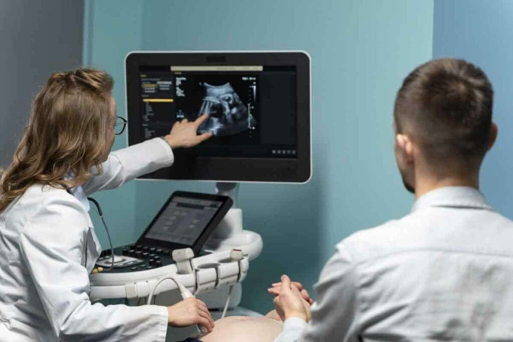
During the MAG 3 renal scintigraphy, patients lie on their backs on an imaging table. A small dose of MAG 3 is given through a vein in the arm. The gamma camera then takes pictures of the kidneys as the tracer moves through the urinary system.
The procedure is generally well-tolerated, with most patients experiencing no discomfort. It usually takes 30 to 60 minutes, and patients must stay very quiet to get clear images.
After the scan, patients can usually go back to their normal activities. The test results will be reviewed by a nuclear medicine specialist. They will talk about the results with the patient during a follow-up appointment.
Potential Limitations and Considerations in Renal Scan Interpretation
Interpreting renal scans needs a deep understanding of technical aspects and the patient’s situation. We must look at several limitations that can affect how accurate the results are.
Technical Factors Affecting Accuracy
Many technical factors can change how accurate renal scan results are. Image quality is key; bad images or artifacts can cause mistakes. Patient positioning is also important; wrong alignment can hide important details. The radiopharmaceutical used and its dose also play a big role in the scan’s accuracy.
To deal with these issues, we follow strict rules for taking and processing images. We do regular checks on our equipment and prepare patients carefully. By focusing on these technical details, we can make our renal scan interpretations more reliable.
Clinical Scenarios Requiring Additional Testing
Some situations need more tests than just a standard renal scan. For example, if someone might have a urinary tract blockage, we might do a Lasix renogram. For renal transplant evaluations, we often use a mix of scans to check how well the transplant is working.
If the scan results are unclear or don’t match what the patient is feeling, we might suggest more imaging studies. This could be ultrasound or CT scans to get a better look at the kidneys. By using information from different tests, we can make better choices for patient care.
Knowing the limits and things to consider in renal scan interpretation helps us give more precise diagnoses. This way, we can create treatment plans that really fit each patient’s needs.
Conclusion: The Clinical Value of MAG 3 Renal Scans in Modern Nephrology
The MAG 3 renal scan is a key tool in modern nephrology. It gives important insights into how the kidneys work and how urine moves. We’ve looked at how this nuclear medicine works, its uses, and what it can show doctors.
It helps find and track kidney problems like blockages and damage. The Lasix renogram makes it even better by checking how the kidneys react to a certain drug.
Its main benefit is helping doctors make better treatment plans and track how well patients are doing. Knowing its strengths and weaknesses helps doctors give better care and improve health outcomes.
As we move forward in nephrology, MAG 3 scans will likely play an even bigger role. They might use new tech to get even more accurate results and help patients even more.
FAQ
What is a MAG 3 renal scan, and how does it work?
A MAG 3 renal scan is a test that checks how well your kidneys work. It uses a special dye called Tc-99m MAG3. This dye is injected into your blood and goes to your kidneys.
Then, it’s passed out in your urine. A camera captures images of your kidneys. This helps doctors see how well they’re working.
What is the difference between renography and static kidney imaging?
Renography shows how the kidneys work over time. Static imaging takes a snapshot of the kidneys at one moment. MAG 3 scans are a type of renography.
What are the medical indications for a MAG 3 renal scan?
MAG 3 scans help find and track kidney problems. They’re used for things like checking for blockages, evaluating transplants, and spotting damage.
What is a Lasix renogram, and how does it enhance diagnostic accuracy?
A Lasix renogram tests how the kidneys handle a diuretic. It helps tell if there’s a blockage or not. This makes it easier to diagnose problems.
How are renal scan results interpreted, and what are normal patterns?
Doctors look at the images and data from the scan to understand the results. Normal scans show good kidney function. Abnormal scans might show blockages or damage.
What is the difference between DTPA scanning and MAG 3 scanning?
DTPA scans check how well the kidneys filter waste. MAG 3 scans look at how well the kidneys handle substances. The right scan depends on what the doctor needs to know.
How should I prepare for a MAG 3 renal scintigraphy, and what can I expect during the procedure?
Before the scan, you’ll need to follow some steps. You might need to arrive with a full bladder and stay very quiet. The scan is done in a special room where a dye is injected and images are taken.
What are the limitations and considerations in renal scan interpretation?
Things like image quality and how the patient is positioned can affect the scan’s accuracy. Sometimes, more tests are needed to confirm certain conditions.
What is a normal split renal function parameter, and what does it signify?
A normal split renal function means both kidneys are working about the same. If there’s a big difference, it could mean one kidney isn’t working as well.
How do I know if my renal scan results are normal or abnormal?
A doctor will look at your scan and tell you if it’s normal or not. They’ll explain what it means for your health.
What is the clinical value of MAG 3 renal scans in modern nephrology?
MAG 3 scans are a key tool in diagnosing and managing kidney diseases. They provide important information for patient care.
References
- Taylor, A. T., Nally, J., Aurell, M., et al. (2018). SNMMI Procedure Standard / EANM Practice Guideline for Diuretic Renal Scintigraphy in Adults With Suspected Upper Urinary Tract Obstruction. Seminars in Nuclear Medicine, 48(4), S1–S35. https://pmc.ncbi.nlm.nih.gov/articles/PMC6020824/
- O’Dell, M., et al. (2024). Effects of Renal Scintigraphy Processing With and Without Kidney Depth Correction. Journal of Nuclear Medicine, 65(supplement). https://jnm.snmjournals.org/content/65/supplement_2/2417
- “99mTc-MAG3 Diuretic Renography: Intra- and Inter-Observer Reproducibility.” (2020). European Journal of Nuclear Medicine and Molecular Imaging. https://pmc.ncbi.nlm.nih.gov/articles/PMC7554833/


